Causes of candidiasis
Thrush in children is caused by yeast-like fungi of the genus Candida, class Fungi Imperfecti. They belong to opportunistic microorganisms, that is, they can provoke disease only if they are directly in the child’s body in sufficient quantities and under favorable conditions. These mushrooms can retain their properties in a dried state for three years, easily tolerate freezing, but when boiled and under the influence of disinfectants they die instantly.
Yeast infection most often occurs in newborns of both sexes, as well as in teenage girls. The main reason infants become infected with candidiasis is infection from a sick mother during the birth process. When a child passes through the birth canal, Candida yeast-like fungi settle on his skin and mucous membranes. In addition, infection can occur while bathing after an infected family member. In teenagers, thrush usually develops due to hormonal changes.
There are a number of factors contributing to the formation of candidiasis in children:
- period of teething;
- long-term and unsystematic use of antibiotics;
- weakened immunity;
- frequent colds;
- endocrine pathology in children (diabetes mellitus, obesity, bronchial asthma);
- diseases of the digestive tract;
- prematurity;
- dry and hot climate in the room where the child is;
- unfavorable sanitary and hygienic conditions.
Candidiasis of smooth skin
Candidiasis is a common disease classified as mycoses caused by opportunistic fungi. In our country, at the end of the 18th century, this fungal disease was known, but numerous reports of candidiasis, which developed as a complication after antibacterial therapy, began to appear after the introduction of broad-spectrum antibiotics into practice.
According to G. A. Samsygina (1997), there is an increase in various clinical forms of candidiasis in newborns, including children infected in the early neonatal period. When using antibacterial drugs in children of somatic departments, damage to the oral mucosa caused by yeast fungi of the genus Candida is observed in 6.6%, oral mucosa and skin - in 15%, intestinal mucosa - in 2.5%, candidiasis in the intestines - in 9.2% of patients (Zh.V. Stepanova, L.L. Smolyakova, 1999).
The increase in the incidence of candidiasis is associated with the use of modern means of therapy (antibiotics, hormonal drugs, cytostatics, immunosuppressants), as well as with an increase in the number of diseases that create a favorable background for its development: immunodeficiency states, dysfunction of the endocrine glands, malignant neoplasms, blood diseases, etc. In addition, the increase in the number of cases of candidiasis is influenced by increased background radiation and other factors that weaken the immune system. The increasing detection of this disease in patients after surgical treatment, as well as with gynecological, urological, hematological and other disorders in recent years, has increased interest in the problem of candidiasis among doctors of various specialties.
Candidiasis is caused by yeast fungi of the genus Candida, widespread in nature and classified as opportunistic pathogens. There are more than 130 species, but a little more than 10 species can be pathogenic for humans, such as C. albicans, C. tropicalis, C. krusei, etc.
The causative agents of candidiasis are isolated on average from every third person from the intestines, genitals, and bronchial secretions. Primary colonization of the body occurs in the mother's birth canal, and after birth - through contact and nutrition. Infection of a child can occur due to candidiasis of the mother’s nipples, through contact with service personnel, through household items, etc.
The leading factors in the development of candidiasis are the background condition or diseases of the body, in which opportunistic pathogens acquire pathogenic properties. In recent years, many researchers have come to the conclusion that the main predisposing factor for the occurrence of superficial forms of candidiasis, including in HIV infection and AIDS, is a violation of cellular immunity. A certain role in the development of candidiasis is played by the frequent and not always justified prescription of broad-spectrum antibiotics, including for prophylactic purposes, as well as the widespread use of drugs that have an immunosuppressive effect - glucocorticoid hormones and cytostatics.
Candidal paronychia and onychia on the fingers, as a rule, develops in women who have frequent contact with water, while separation of the nail skin (eponychion) from the nail plate is observed, creating favorable conditions for fungal infection in the matrix area. The disease can occur in people with diabetes. In recent years, women have begun to use false nails, which has created another risk factor for the development of fungal and bacterial infections.
Yeast fungi of the genus Candida can cause damage to the mucous membranes, skin and its appendages, and internal organs. The most common forms of mycosis are superficial.
| Figure 1. Interdigital candidiasis erosion |
On smooth skin, large (inguinal-femoral, intergluteal, under the mammary glands, axillary cavities) and small (interdigital) folds are affected, but rashes can also occur outside the folds on the smooth skin of the trunk and extremities, including the palms and soles. Outside the folds, foci of candidiasis develop mainly in infants or in adults suffering from diabetes mellitus or severe somatic illness. In large folds, small bubbles the size of millet grains appear, sometimes pustules, which quickly open with the formation of erosions. Due to peripheral growth, erosions quickly increase in size and merge with each other, forming large areas of damage. The lesions are dark red in color with a burgundy tint, shiny, with a moist surface, irregular in shape, with a strip of exfoliating epidermis along the periphery. Fresh small erosions (foci of elimination) form around large foci. In children, especially weakened ones, from the folds the lesion spreads to the skin of the thighs, buttocks, abdomen, and sometimes to the entire skin. There may be painful cracks deep in the folds. Candidiasis of smooth skin outside the folds (chest, abdomen, shoulders, forearms, legs, etc.) in children has a clinical picture of seborrheic dermatitis in the form of foci of erythematous-squamous nature or hyperemia. In adults, the disease can manifest itself as erythematous spots with peeling in the center and small blisters along the periphery. In small folds of the skin, the lesion usually occurs between the 3rd and 4th, 4th and 5th fingers of the hands, less often the feet, and is characterized by the formation of eroded lesions of a deep red color with a smooth, shiny, as if varnished surface, clear boundaries, with peeling of the stratum corneum of the epidermis along the periphery (Fig. 1). The disease can begin with the appearance of small blisters on the lateral touching hyperemic surfaces of the skin. Then it spreads to the area of the interdigital fold, swelling, maceration, and peeling appear.
Interdigital candidiasis erosion is observed mainly in individuals who have prolonged contact with water, which contributes to the development of skin maceration, and, as a result, favorable conditions are created for the development of candidiasis infection. In addition to the folds between the 3rd and 4th, 4th and 5th fingers, others can be affected, not only on one, but also on both hands. Patients are concerned about itching, a burning sensation, and if there are cracks, pain. The course of the disease is chronic, with frequent relapses. Breastfeeding women may develop candidiasis of the smooth skin of the nipples. Clinical manifestations can be different: in the form of slight hyperemia in the area of the isola; in the form of a lesion near the nipple with maceration, clear boundaries; in the form of a crack with maceration along the periphery and small bubbles between the nipple and the isola.
Candidiasis of the palms and soles is rare. On the palms, the disease can occur as dry lamellar dyshidrosis (superficial lamellar, ring-shaped or garland-shaped peeling); may have a vesicular-pustular form (vesicles and pustules against the background of hyperemic and edematous skin); can occur as hyperkeratotic eczema (against the background of diffuse hyperkeratosis or individual areas of keratinized skin, sharply limited wide skin furrows with a dirty brown color are observed). Candidiasis of the skin of the soles is observed mainly in children and is characterized by the presence of small blisters and pustules, hyperemic spots with peeling and exfoliating macerated epidermis along the periphery.
| Figure 2. Candidal paronychia and onychia |
With candidiasis of the nail folds (candidal paronychia), the disease begins with the posterior fold, often in the area where it transitions to the lateral fold, with the appearance of hyperemia, swelling and swelling of the skin. Then the inflammatory phenomena spread to the entire roller, which becomes thicker and seems to hang over the nail, and peeling is observed along the edge of the roller. The skin of the cushion becomes thin, shiny, and the eponychion disappears. When pressing on the roller, ichor, a lump of white crumbly mass or a drop of pus may be released (due to the addition of a secondary infection). Later, the nail plate changes: it becomes dull, in the area of the lunula it is separated from the bed, destroyed as onycholysis, or transverse grooves and elevations appear. These changes are associated with impaired blood supply in the area of the matrix, i.e. they are trophic in nature and caused by inflammation in the area of the cushion. Nail damage caused by a yeast fungus of the genus Candida begins from the lateral edges, the nails become thinner, separate from the bed, acquire a yellow-brown color and look as if they are trimmed on the sides (Fig. 2). In young children, inflammatory phenomena in the area of the cushion are more pronounced, and the nail plate changes from the distal edge. There is candidiasis of the nails without inflammation of the ridge. In this case, the change in the plate begins from the distal edge and develops according to the type of onycholysis: the plate becomes thinner, does not adhere to the bed, and multiple lesions of the nails may occur.
In a special type of the disease - chronic generalized (granulomatous) candidiasis - clinical manifestations on the skin can be varied.
- Lesions with clear boundaries, round and oval in shape, ranging in size from 1.5 to 5 cm in diameter, of an erythematous-squamous nature develop on any part of the skin; they often merge with each other and form large areas; in some cases, the lesion takes on the character of erythroderma.
- Lesions in the form of plaques ranging in size from 1 to 5 cm in diameter, with hyperemia, a bluish or bright red rim, sometimes with papillomatous or verrucous growths on the surface, covered with horn-like formations, can occur on the scalp, face, trunk and limbs ; when the crusts are rejected, an easily bleeding surface opens; after resolution of such elements, cicatricial atrophy of the skin is formed (Fig. 3).
- Foci with clear boundaries are surrounded by a continuous infiltration ridge, consisting of tubercles, vesicles, with layering of yellow crusts and pronounced exudation; lesions are located predominantly on the dorsum of the hands and (or) feet with transition to the palmar and plantar surfaces; some patients experience dry skin on the palms and (or) soles, often spreading to the dorsum of the fingers and (or) feet (Fig. 4 ).
Candida balanoposthitis is quite common. The disease can occur in a mild form with minor lamellar ring-shaped peeling or be more pronounced. In this case, hyperemia, maceration, erosions appear on the skin of the glans penis and the inner layer of the preputial sac, as well as in the coronary groove on the contacting surfaces. Skin looks damp. Erosions can merge, and foci with clear boundaries, polycyclic outlines, with a shiny, varnished surface are formed; a fringe of exfoliating epidermis is observed along the periphery. Subjectively, patients are bothered by itching and a burning sensation. The disease can be complicated by ulceration and the development of phimosis.
| Figure 3. Lesions of the scalp, face and nails in a patient with chronic generalized granulomatous candidiasis |
In superficial forms of candidiasis, the diagnosis is based on the presence of a characteristic clinical picture in the patient and the detection of the fungus in pathological material (skin flakes, scrapings from nails) during microscopic examination. The diagnosis can be considered reliable if pseudomycelium or true mycelium and budding cells are detected. Sowing on a nutrient medium is carried out to identify the type of yeast fungus of the genus Candida and to determine its sensitivity to antimycotic drugs. Isolating only a culture of the fungus has no diagnostic value, since it can be obtained by culturing scrapings from the skin and nails of healthy people.
With candidiasis of smooth skin of large folds and outside the folds, the disease should be differentiated from seborrheic eczema, psoriasis, other mycoses - inguinal epidermophytosis, superficial trichophytosis, pseudomycosis - erythrasma (complicated form); with interdigital candidiasis erosion on the hands - from dyshidrotic eczema, on the feet - from mycosis caused by Trichophyton interdigitale and Trichophyton rubrum; for damage to nails and ridges - from onychia and paronychia of bacterial etiology, eczema and psoriasis; with balanoposthitis - from a similar disease of bacterial etiology.
Limited, sometimes widespread, forms of smooth skin lesions, especially those that developed during treatment with antibacterial drugs, as a rule, are easily treated with local antimycotic agents and can resolve without treatment after discontinuation of antibiotics. Local and systemic antimycotics are prescribed as etiotropic therapy. In recent years, azole drugs with a broad spectrum of action, as well as polyene antibiotics, have been widely used in the treatment of candidiasis.
For candidiasis of smooth skin of large folds with acute inflammatory phenomena, treatment should begin with the use of an aqueous solution of brilliant green (1–2%) in combination with powder and continue for 2–3 days, then antifungal drugs are prescribed until the clinical manifestations resolve.
| Figure 4. Lesions of the skin of the feet and nails in a patient with generalized granulomatous candidiasis |
For candidiasis of smooth skin of large, small folds and other areas of the skin, antifungal agents are used in the form of cream, ointment and solution: ketoconazole (ketoconazole, mycozoral, nizoral), clotrimazole (clotrimazole, canizon, canestene, candide), oxyconazole (mifungar), bifonazole ( mycospor, bifosin), sertaconazole (zalain), natamycin (pimafucin). Cream or ointment is applied in a thin layer to the affected areas and rubbed in 1-2 times a day. The duration of treatment is until clinical manifestations resolve, then continue to use the cream for another 7-10 days to prevent relapse. In case of common skin processes and the ineffectiveness of local therapy, systemic antimycotics are prescribed: fluconazole (Diflucan, Flumicon, Mycosist, Flucostat, etc.) for adults at a dose of 100–200 mg 1 time per day, for children at a rate of 5 mg/kg body weight 1 time per day; itraconazole (orungal, irunin, rumicosis) for adults, 100–200 mg 1–2 times a day; ketoconazole (nizoral, mycozoral) for adults 200 mg once a day daily, as well as the polyene antibiotic natamycin (pimafucin) for adults 100 mg 4 times a day, children 50 mg 2–4 times a day. The duration of therapy is 2–4 weeks.
In case of candidiasis of the skin of the nail folds, anti-inflammatory treatment of the nail folds is first carried out using applications with pure ichthyol, which are prescribed once a day, until the inflammatory phenomena subside. Then use antimycotic agents (ketoconazole, mifungar, travogen, clotrimazole, zalain, etc.) for topical use, rubbing them under and around the roller. The procedures are carried out 2 times a day, in the evening they can be applied under an occlusive dressing. It is effective to prescribe a combination of ointment (cream) and solution, alternating them. If local therapy is ineffective and if the nail plates are affected, systemic antimycotics are indicated: fluconazole (Diflucan, Flumicon, Mycosist, Flucostat, etc.) according to an intermittent regimen (adults 150 mg once a week, children 5–7 mg/kg body weight); itraconazole (orungal, irunin, rumicosis) for adults using the pulse therapy method (200 mg 2 times a day for 7 days, then a 3-week break) for 2–3 months; ketoconazole (nizoral, mycozoral) for adults, 200 mg per day daily for 2–4 months.
Considering the fact that candidiasis is an opportunistic mycosis, it is first necessary to identify and, if possible, eliminate the pathogenetic factors of the disease (a study of the immune and endocrine status, the gastrointestinal tract and corrective therapy are indicated).
Prevention of candidiasis includes the following measures.
- Prevention of the development of candidiasis in patients with severe somatic and endocrine diseases, as well as immunodeficiency. They should be tested repeatedly for fungi.
- To prevent the development of candida infection in children who are in somatic departments and receiving antibacterial drugs, it is necessary to prescribe fluconazole (Diflucan, Flumicon, Mikosist, Flucostat, etc.) at a rate of 3–5 mg/kg body weight, 1 time per day. The daily dose of the drug depends on the degree of risk, treatment is carried out in parallel with therapy for the underlying disease.
- Prevention of dysbiosis.
- Prevention of the development of candidiasis in newborns.
Literature
- Stepanova Zh. V. Fungal diseases: diagnosis and treatment. M.: Miklos, 2005. 124 p.
- Stepanova Zh. V., Smolyakova L. L. Candida infection as a complication of antibiotic therapy in children // Bulletin of Dermatology and Venereology. 1999. No. 1. P. 55–56.
Zh. V. Stepanova , Doctor of Medical Sciences, Professor TsNIKVI, Moscow
Types of candidiasis
In children, the following main types of thrush are distinguished:
- candidiasis of the skin and its appendages;
- candidiasis of the mucous membranes (oral cavity, corners of the mouth, tongue, pharynx, larynx, trachea, gastrointestinal tract, genitals, urethra);
- visceral or systemic candidiasis (yeast endocarditis, meningitis, sepsis, etc.).
Depending on the duration of the inflammatory process and the manifestations of the disease, acute and chronic forms of thrush are distinguished. Depending on the extent of the lesions, candidiasis can be focal or generalized. Based on the depth of spread, thrush is divided into visceral and superficial (smooth skin, mucous membranes, and nails are affected).
A separate type of disease includes secondary allergic rashes that appear as a result of additional sensitization of the skin to the proteolytic enzymes of the fungal pathogen.
Candidiasis in children. Diaper dermatitis and candidiasis of skin folds
In infants, candidiasis of large folds often occurs simultaneously with oral thrush.
Skin candidiasis in children
The skin in the perianal area, intergluteal and inguinal-femoral folds is affected.
The process then spreads to the skin of the inner and back surfaces of the thighs, genitals, buttocks, and abdomen. In rare advanced cases, almost the entire skin is involved, even outside the folds.
Skin lesions with candidiasis in young children
Candidiasis in a child
The disease begins with the appearance of pink, slightly itchy lesions, possibly with small blisters and pustules, which then turn into red, weeping and erosive painful rashes.
As a result of the merging of several lesions, large, clearly demarcated areas are formed, surrounded by a whitish border of weeping skin. After the process resolves, peeling may remain.
Symptoms of candidiasis
The clinical symptoms of candidiasis vary due to the different localization of the inflammatory process in the body.
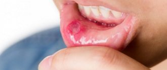
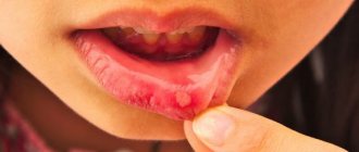
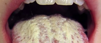
Oral thrush in children is characterized by:
- A whitish, cheesy coating in the mouth that is easily removed with a finger or a spatula. It is localized mainly on the mucous membrane of the cheeks, tongue, tonsils, hard and soft palate, red border of the lips. At first, candidal plaque is present in the form of small white dots, reminiscent of semolina, then merges into larger plaques with the characteristic appearance of “curd residue” or milky films in the mouth. In the absence of proper treatment and when the process becomes chronic, plaque transforms into a dense yellow film, which is difficult to separate from the mucous membranes with the formation of bleeding erosions.
- Hyperemia of the mucous membrane (overflow of blood vessels), erosion.
- Pain and burning in the mouth.
- Seizures, maceration of the skin in the corners of the mouth.
- Child's moodiness, loss of appetite up to complete refusal to eat.
Genital candidiasis is characterized by the following symptoms:
- Redness and swelling of the genitals, severe itching.
- A cheesy, thick discharge from the genitals with an unpleasant sour odor.
- Pain during urination.
- White milky films on the skin of the external genitalia, in the chronic process - congestive hyperemia, tissue thickening.
Candidiasis of the esophagus , stomach and intestines is characterized by the presence of the following symptoms:
- Colic and flatulence, frequent regurgitation.
- Pain when swallowing.
- Pain in the stomach and intestines.
- Decreased appetite, refusal to eat.
- Unshaped stool with an admixture of white inclusions, reminiscent of curd mass.
Skin candidiasis is characterized by the formation of a rash in the form of papules or vesicles, which eventually open and form weeping erosions with a white coating. In infants, the elements of the rash are most often localized in the area of large folds on the abdomen. In older children, yeast infections usually appear between the fingers.
Fisenko Karina Olegovna
Specialty: Adult gynecologist, gynecologist-endocrinologist Work experience: more than 25 years Cost of appointment: 1600 rubles Cost of online appointment: 1200 rubles
Make an appointment
- Clinical picture of the disease
- The main causes of inflammation of the external genitalia and vagina in children
- Types of vulvovaginitis in girls
- Diagnosis of the disease
- Treatment of the disease
Diagnosis of candidiasis
The first step in diagnosing thrush is an examination by a pediatrician. If the doctor detects signs of fungal infection (white cheesy coating on inflamed mucous membranes, the appearance of eroded surfaces after removing the milk film), a set of additional laboratory and instrumental studies is prescribed to confirm the diagnosis of candidiasis and determine the cause of its appearance:
- General blood and urine analysis.
- Blood chemistry.
- Microbiological examination of stool.
- Serological study.
- Microscopy of a scraping or smear from the affected organ to determine the presence of Candida fungus.
- Cultural culture of blood, urine, feces, biopsy of the mucous membrane.
- Ultrasound examination of the abdominal and pelvic organs.
- Endoscopy of the digestive organs.
Treatment methods for candidiasis
Therapy for yeast infections in children involves an integrated approach to treatment. Pediatricians are faced with the task of not only destroying the fungus that causes lesions, but also eliminating pathogenetic mechanisms, restoring normal microflora and increasing immunological reactivity.
When choosing tactics for drug treatment of thrush in children, they try to refrain from prescribing antibiotics. Affected mucous membranes and skin are usually treated with antifungal creams, ointments and solutions. For the visceral form of candidiasis, systemic antifungal drugs are used.
During treatment, the patient needs a healthy diet, in which proteins predominate and carbohydrates are limited. In addition, as prescribed by your doctor, you need to take vitamins, anti-dysbacteriosis medications and immunostimulating drugs.
Since children’s immunity is still quite weak, it is necessary to take a responsible approach to the prevention of thrush and follow simple rules:
- regularly sterilize all pacifiers and pacifiers given to the child;
- bathe in clean water in an individual bath;
- carefully care for the skin and mucous membranes;
- observe the correct diet, monitor the state of the child’s gastrointestinal tract;
- completely cure candidal colpitis and vulvitis before or during pregnancy, so as not to infect the child during childbirth.
Clinical picture of the disease
Vulvovaginitis in girls is accompanied by complaints of severe itching, pain, burning and discomfort in the external genitalia and perineum. Additionally, pathological vaginal discharge of a reddish, yellow or green color, leucorrhoea or curdled discharge may appear. The girl’s genitals were hyperemic and very red, and a rash and scratches appeared on the skin.
The baby constantly pulls her hands to the crotch area and scratches. Vulvovaginitis may not seriously affect the general health, except for the deterioration of the child’s mental state - tearfulness, irritability, anxiety, etc. As the disease progresses, when the inflammatory process affects other organs, additional symptoms may appear - difficulty urinating, pain in the lower abdomen, fever, etc.
Consequences of candidiasis
If a timely diagnosis is made and rational treatment is prescribed, thrush in children is completely cured and leaves no consequences. With improper therapy and late seeking medical help, the disease can become chronic and become resistant to further treatment measures. In addition, against the background of a chronic yeast infection, candidosepsis can develop, a dangerous disease that can be fatal. Intestinal candidiasis is fraught with complications such as heavy bleeding, perforation of the intestinal wall, and ulcers.