Enamel is the hardest tissue in the human body and a natural protective barrier for teeth. However, some people are diagnosed with its deficiency. Hypoplasia of dental enamel - as this condition is professionally called - can have different causes, but appropriate treatment methods must be used each time.
The main function of enamel in the oral cavity is to protect teeth from harmful factors: the action of bacteria, thermal and chemical irritants, as well as abrasion during chewing. Although there are proven ways to strengthen enamel, its gradual erosion is associated with various reasons. Some patients, even children, are also diagnosed with underdevelopment of this tissue. Dental hypoplasia can have many causes and each time requires appropriate treatment.
What is tooth enamel hypoplasia?
Literally, the term “hypoplasia” can be translated as “little enamel.” This formulation fully reflects the situation, since the disease is characterized by underdevelopment or absence of areas of enamel. The opposite of hypoplasia is enamel hyperplasia, when pathological growth of tooth tissue occurs, most often manifested in the form of enamel drops. Due to phonetic similarity, confusion often arises. Hypoplasia often develops on baby teeth, which, by default, have thinner and more vulnerable enamel, but the disease can also affect molars.
Attention!
The risk of hypoplasia of the enamel of permanent teeth increases many times if at the milk stage the pathology was not corrected in time and transferred to the rudiments of the molars. Enamel hypoplasia in adults occurs much less frequently and, as a rule, is a consequence of pathologies that developed in childhood.
Causes of abnormal tooth shapes
Abnormalities such as excessive tooth wear or enamel hyperplasia are often genetic disorders. But sometimes they can be caused by traumatic, inflammatory, chemical or allergic processes in the oral cavity, which can be easily eliminated using classical dentistry methods. Causes also include intrauterine diseases, such as syphilis or endocrine system disorders.
In the milk and permanent teeth of children and adolescents, various deviations from normal development may occur, which causes the formation of tartar on the teeth more actively, or pathological abrasion of the teeth occurs. These developmental disorders can affect the number, shape, or color of teeth.
Our job is to recognize these changes so that we can assess the consequences at an early stage.
Causes of hypoplasia
Treatment of dental enamel hypoplasia in children is complicated by the fact that this disease has a very extensive etiology. Enamel hypoplasia occurs under the influence of a number of factors that are formed both at the stage of intrauterine development and after birth.
Causes of enamel hypoplasia before birth
- Heredity is one of the main factors. Enamel hypoplasia is inherited as a dominant (X-linked) trait. Women have two X chromosomes, so a mother's hypoplasia can be passed on to her children, regardless of their gender. In men, hypoplasia is inherited only by daughters. If both parents have hypoplasia, the chances of transmitting the pathology to children increase.
- Viral and chronic diseases in the mother.
- Severe forms of toxicosis during pregnancy.
- Lack of vitamins and microelements.
- Bad habits, drug addiction and alcoholism.
Reasons for the development of hypoplasia after birth
- Prematurity and traumatic birth.
- Metabolic disease.
- Diseases of the cardiovascular system in children.
- Hemolytic disease.
- Infectious diseases.
- Anemia.
- Complications after taking antibiotics.
- Dental diseases.
- Damage to the rudiments of permanent teeth. This factor provokes the development of hypoplasia of the enamel of molars in children.
- Vitamin deficiency and unhealthy diet.
Not all of the above factors are the root cause of hypoplasia, but each of them contributes to the development of the disease to varying degrees.
Hypoplasia of permanent teeth in children: treatment
Sometimes the process of hypoplasia cannot be stopped by applying mineral and fluorine-containing compounds. In such cases, pediatric dentists offer options for reconstructing tooth enamel or even the crowns of permanent teeth themselves.
Restoration is carried out using one of the following methods:
Filling children's teeth
Using grinding, dentists clean the affected area and then restore the surface using high-quality, reflective filling compounds. Installing fillings makes teeth straight, white and neat, and also reduces the rate of spread of the lesion and reduces the risk of complications.
Prosthetics
Installation of full crowns on affected permanent teeth or production of veneers. For children, prosthetic options are selected by the doctor according to the indications and the current clinical situation.
Plaksina Margarita
Unfortunately, children with hypoplasia are sometimes brought to the appointment too late. It is not always possible to save a tooth; it has to be removed. In the case of a primary malocclusion, the child is given an orthodontic ring to maintain space for the permanent unit. This is necessary so that neighboring teeth do not take up free space and spoil the bite.
Forms of enamel hypoplasia
Hypoplasia is divided into two types: systemic and local. They differ in both symptoms and etiology.
Systemic enamel hypoplasia
With systemic enamel hypoplasia in children, a large area of the dentition is affected. It is often called congenital and genetic, since it occurs immediately with the appearance of baby teeth. May cause changes in tooth shape, especially in the incisal area. The disease has several forms and varieties.
- Spotted.
Light spots appear on the enamel, but its structure is not damaged. - Erosive enamel hypoplasia.
A more severe form, in which round-shaped depressions appear on the surface of the enamel. - Furrowed.
Depressions in the form of large or small grooves, in some cases may resemble waves. - Mixed.
The simultaneous manifestation of two or more forms is observed.
Local enamel hypoplasia
Local enamel hypoplasia is localized, that is, it affects one or two teeth. Most often acquired and occurs against the background of inflammation and damage to the buds of the teeth. It appears in the form of spots of various shapes, pits and grooves. If left untreated, the disease affects the deeper tissues of the tooth and provokes the development of caries.
Aplasia or absence of enamel
The most severe form of the disease, which some experts distinguish separately. It can be either local or systemic (less commonly).
Treatment
First you need to determine the cause of tooth enamel destruction. The dentist will conduct an examination and interview. For example, if it turns out that the patient is not eating properly, the doctor will give recommendations regarding the diet, tell you which foods should be avoided and which should be included in the menu.
Therapy for mild enamel damage
Next, they begin the treatment itself. In the early stages, damage to tooth enamel is reversible. Even if the teeth become thinner or worn out, with timely treatment the enamel is restored.
First, the dentist will perform a professional cleaning of the oral cavity, removing plaque and tartar. The next stage is remineralization. To restore weak, damaged enamel, it is saturated with calcium and phosphorus.
The doctor uses specialized solutions and gels containing minerals and vitamins. Enamel absorbs beneficial microelements like a sponge. To consolidate the effect, the teeth must be coated with fluoride-containing varnish. The coating creates a thin, dense film on the surface that prevents demineralization.
Treatment of severe enamel damage
Due to untimely consultation with a doctor, the destruction progresses: the enamel is erased, chips, pronounced wedge-shaped defects and carious cavities appear on the surface of thinned teeth. The dentist recommends filling or artistic restoration. First, the doctor cleans the teeth of the affected tissues, then you can put a filling or restore it. In some cases, inlays or microprostheses are used.
Diagnosis of enamel hypoplasia
- Visual inspection. Hypoplasia always has external manifestations, so the first conclusions can be drawn.
- Anamnesis collection.
- Analyzes and studies to identify the root cause.
Differential diagnostics are carried out separately in order to make the correct diagnosis and exclude diseases with similar symptoms. For example, to exclude suspicion of caries, vital staining with methylene blue is performed (areas with hypoplasia are not stained, unlike carious ones). Fluorosis and enamel hypoplasia can also have similar symptoms, but the nature of these diseases is different. Fluorosis occurs due to excess fluoride in the body. Spots may appear on the surface of the enamel, but unlike hypoplasia, they have a characteristic brown tint.
Diagnostics
To conduct a diagnosis, the doctor must conduct an initial examination, interview the patient, identify increased sensitivity, and the presence of pain. Externally, the pathology differs from other types of hypoplasia by local damage. That is, a small number of units are affected, and asymmetry of placement appears. In case of a systemic disorder, the anomaly manifests itself on the right and left sides, since the causes are anomalies in the functioning of the glands. During the examination, it is necessary to determine what exactly is the cause of the disease, to isolate the pathology from caries, fluorosis, and pigmentation disorders. To do this, the doctor uses the presence or absence of the following signs:
- caries is manifested by the development of white spots in the cervical area, hypoplasia is characterized by spots in different areas of the unit;
- the enamel has a rough surface, which is determined by a special probe; in this disease, the tissues are smooth;
- caries is stained (methylene blue is used), hypoplasia does not imply a change in tissue color.
To clarify the diagnosis, radiography is performed to identify points and depressions. Teleradiography, orthopantomography, and MGE are used to diagnose and identify malocclusions.
Treatment regimen
Therapy depends on the severity of the problem. If single spots appear on the surface of the tissue, treatment is not prescribed. To pigment the frontal part, composite fillings are installed, restoring aesthetics. If structural changes in dentin and enamel are detected, the use of filling materials such as compomers and composites is indicated. If there is a large area of damage, prosthetics and the use of ceramic and other crowns are recommended.
Treatment of the problem is necessary, since hypoplasia over time causes tissue deterioration and caries damage. Without treatment, malocclusion is diagnosed and there is a high risk of tooth loss.
Treatment of enamel hypoplasia
Treatment of dental enamel hypoplasia in children is carried out in two directions. Firstly, it is necessary to eliminate or correct the main factor that provoked the development of the pathology. Of course, if the disease was inherited, it is difficult to do anything about it, but in most other cases it is necessary to minimize the influence of concomitant diseases and pathologies. The treatment method for manifestations of hypoplasia depends on the form and severity of the disease.
- Hypoplasia of the enamel of primary and molar teeth in the stained stage, when the enamel structure is not damaged, is treated conservatively. Remineralization, fluoridation, resurfacing and whitening are the main procedures that restore the aesthetics of a smile.
- Erosive and grooved hypoplasia affecting dentin is treated with filling with composite materials.
- Severe cases of hypoplasia, in particular aplasia, require orthopedic treatment. The most effective methods for treating enamel hypoplasia of permanent teeth are the installation of crowns and veneers.
Hypoplasia of primary teeth: treatment
Since one of the reasons for the abnormal development of enamel is low tissue mineralization, doctors have found an opportunity to help young patients. During treatment, two goals are set - to stop pathological changes, reduce or eliminate the severity of existing defects.
The following methods are used in pediatric dentistry in Moscow:
Enamel remineralization
The procedure allows you to replenish the deficiency of missing minerals and strengthen the child’s teeth. At the appointment, the doctor sequentially treats healthy and damaged teeth with a special gel. Treatment is continued at subsequent appointments or even at home. For home procedures, the doctor prescribes the child the optimal paste composition.
Fluoridation of teeth
Treatment of hypoplasia differs in the choice of drug. When fluoridating, the dentist uses a gel with a high fluoride content - up to 65-70%.
The method of restoring baby teeth with hypoplasia is chosen by the dentist after assessing the condition of the enamel and the indications.
Symptoms of dysplasia
The main manifestations of dentin and enamel dysplasia are observed in such dental diseases as:
- dentinogenesis imperfecta;
- Stanton-Capdepont syndrome;
- imperfect amelogenesis.
Stanton-Capdepont syndrome
Stanton-Capdepont disease or syndrome is a hereditary disorder of the formation of enamel and dentin tissue. The main symptoms of this pathology are:
- eruption of teeth having a natural shade and shape, and the subsequent change in the color of their enamel to gray;
- gradual destruction and chipping of tooth enamel;
- early, rapidly progressing abrasion of dentin;
- illumination of the contours of the dental cavity through dentin defects;
- decreased sensitivity of the pulp.
Molars with Stanton-Capdepont disease are subject to destruction and abrasion to a lesser extent than milk teeth. In some cases, the disease is completely asymptomatic, while the teeth are in excellent condition and look practically healthy.
Dentinogenesis imperfecta
Dentinogenesis imperfecta is a pathology characterized by a disruption in the formation of a layer of hard dental tissue located under the enamel (dentin). According to statistics, this disease is found in one out of 8,000 babies, with girls being diagnosed more often.
Depending on the symptoms, there are 4 forms of dentinogenesis imperfecta:
- Type I (the disease acts as one of the symptoms of a congenital disorder of bone formation, and its main signs, in addition to dental dysplasia, are bone fragility, changes in the shape of the middle ear and blue coloring of the eye sclera);
- Type II (the main signs of pathology are opalescence (translucency) of the teeth, a change in the natural color of the enamel to dull gray, increased abrasion of the teeth in the zone of closure of the dentition);
- root dysplasia (underdevelopment or complete blockage of dental roots, early loosening and loss of teeth, minimal difference in the shade of dental crowns from the natural one);
- coronal dysplasia (translucency of tooth enamel, frequent injury to the blood vessels of the pulp, accompanied by minor local bleeding, staining of teeth an amber or brownish-gray color by blood breakdown products).
Amelogenesis imperfecta
In turn, amelogenesis imperfecta is a disorder of the enamel formation process, leading to a change in shade and complete or partial loss of dental tissue.
Depending on the clinical picture, there are 4 subtypes of this pathology:
- type 1 (the enamel acquires a brown or yellow tint immediately after the teeth appear, unevenness of the dentin-enamel junction is observed);
- type 2 (1-3 years after the appearance of the first teeth, the enamel acquires a brown tint, becomes rough, matte, and becomes covered with cracks);
- type 3 (at the moment of teething, the teeth are covered with thin white enamel, which quickly disappears, exposing dentin);
- Type 4 (when teething, teeth are covered with matte, chalky enamel, which is easily separated from dentin under mechanical stress).
The sensitivity of the pulp with dental dysplasia is usually significantly reduced. Therefore, pain in people suffering from this disease is mainly caused not by increased sensitivity of the teeth, but by injury to the gums from the sharp edges of damaged crowns.
The structure of enamel growths
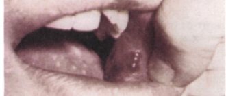
Hyperplasia also develops after changing teeth. Excess enamel appears in the form of drops measuring 1 mm. In the severe stage, pearl drops can reach 5 mm.
Initially, the development of hyperplasia is asymptomatic. The reason why the patient goes to the doctor is to complain about the unaesthetic appearance of his teeth.
Places where the disease spreads:
- at the root of the tooth;
- on the neck of the tooth;
- on the crown of the tooth.
The classification of hyperplasia occurs depending on their structure:
- True enamel.
- Enamel dentin.
- Enamel dentin, filled with pulp inside.
- Enamel drops in the form of nodules, the so-called Rodriguez-Ponty drops.
- Growths that form on the dentin of the crown or inside the tooth itself.
During a visual examination of the teeth, excess enamel may not be detected. When using a drill, you can only accidentally stumble upon a hard area in the dentin.
How does enamel dysplasia manifest?
Your baby just got a few teeth out and you notice a white spot on a brand new incisor or canine? This is a reason to urgently run to the dentist. Your child may be developing enamel dysplasia.
This disease manifests itself as follows:
One or more whitish dots form on the enamel They also differ from healthy dental tissue in that they do not have a glossy shine;- the points expand and deepen ーthe dark dentin becomes visible
; - the enamel is destroyed
, the teeth acquire a yellowish tint due to dentin being exposed; - Dentin begins to deteriorate
, inflammation may occur against this background, and teeth rot.
Typically, destruction begins with the incisors, often in the upper jaw. This disease ultimately leads to tooth decay.
ー not protected by a hard layer of enamel, they are erased right down to the gums. At the same time, the dental canals open, which allows pathogenic bacteria to enter and causes inflammation. The consequences of dysplasia of the enamel of baby teeth are very serious for both the baby and his parents - these are regular inflammations in the canal area, their cleaning, the inability to fully bite off food until the teeth change, and, if necessary, removal of roots. the latter can lead to difficulties with the eruption of permanent ones or their improper growth, because The roots of baby teeth hold space for future permanent teeth.
Treatment of hyperplasia
Therapeutic treatment, in which grinding of enamel drops and filling of pinpoint manifestations of the disease are prescribed. In children, treatment of hyperplasia is carried out using a restorative method.
To restore teeth, photopolymers are used, which completely restore the structure and color of the enamel.
Veneers are installed or artificial crowns are made. Of all types of hyperplasia, only pearl drops on the neck of the tooth can be cured, which over time can cause inflammation of the gums. During the treatment process, excess enamel is removed from the tooth using diamond grinding, and a course of phosphorus-containing medications is prescribed.


