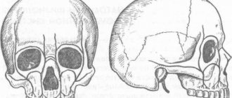
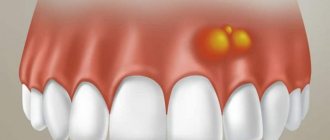
Exostosis of the jaw is a benign formation that manifests itself in the form of an osteochondral growth (osteophyte). The protrusions can be single or multiple and are located on the jaw bone. Maxillary osteophytes most often form on the outer (buccal) surface of the alveolar ridge, and exostosis on the lower jaw - from the inside, on the lingual side. A palatal bone growth (palatal torus) is also found.
Exostoses are painless and do not cause any discomfort at an early stage. But as it increases, the formation begins to cause a lot of discomfort - it complicates eating, affects sound pronunciation, interferes with prosthetics, etc. Treatment of osteophytes is only surgical. Removing exostosis on the gum is a low-traumatic operation that takes about an hour. The operation is performed under anesthesia, so the patient does not experience any discomfort. The method of surgical intervention depends on the location of the osteophyte.
Reasons for appearance
It is not precisely determined why osteophytes appear on the gums. But factors contributing to the disease include:
- Hereditary predisposition;
- frequent inflammation, purulent processes leading to atrophy, deformation of the jaw bone and nearby tissues;
- injuries of the dental system, especially accompanied by fractures of the facial part of the skull with improper reposition of bone fragments;
- complex tooth extraction;
- advanced periodontitis, periodontal disease;
- bite pathologies;
- congenital abnormalities of the jaw.
Often the pathology appears in childhood or adolescence. Also, the appearance of jaw growths may be associated with dysfunction of the endocrine system.
Causes of exostosis
There is still no consensus on the reasons for the appearance of exostoses. A number of authors believe that they are of a tumor nature. But most agree that they are a consequence of disorders of enchondral ossification processes that arise as a result of dysembryogenesis (disorders of embryonic development). Therefore, today the main theory of the formation of bone exostoses is the displacement of the epiphyseal plate during intrauterine development of the fetus. It is a bone growth zone located directly below the epiphysis and is responsible for its lengthening. Therefore, the disease is considered congenital, and its development continues throughout the entire period of growth.
The following can increase the risk of osteochondroma:
- ionizing radiation (in 10% of cases the disease develops in patients who underwent radiation therapy in childhood);
- disturbances in the functioning of the endocrine system, taking hormonal drugs;
- smoking and alcohol abuse of parents.
The disease can also be hereditary and transmitted to the child from one of the parents. Especially often, multiple bone lesions due to exostoses are genetically determined.
The acquired nature of the disease cannot be ruled out. The following can provoke the appearance of neoplasms:
- bone injuries;
- microtraumas caused by excessive physical activity, which is typical for professional sports;
- infectious diseases and chronic inflammatory processes of any localization (for example, a heel spur often develops against the background of plantar or plantar fasciitis);
- pathologies of the periosteum, degenerative-dystrophic diseases of cartilage, ankylosing spondylitis;
- microcirculation disorders in soft tissues;
- muscle dystrophy;
- obesity;
- severe forms of allergic diseases;
- compression of the limbs, including an incorrectly applied plaster cast or tourniquet.
Wearing improperly selected clothing and shoes, in particular frequent hypothermia of exposed areas of the body, increases the risk of developing bone tumors.
Of particular importance in the development of osteochondroma is hormonal imbalance in children during puberty. Often the formation of bone exostoses is observed against the background of increased synthesis of sex hormones in adolescents. This can provoke the growth zones to remain open, which can subsequently cause gigantism. Also, an increase in the size of osteochondral exostoses can continue after the closure of growth plates in women as a result of hormonal changes.
Symptoms
Exostosis of the tooth appears in the form of a convex growth that appears for no apparent reason. Main symptoms:
- Sensation of a foreign body in the mouth;
- discomfort when eating, talking (with large osteophytes);
- pain when pressing on the tumor;
- redness, thinning of the mucous membrane in the pathological area.
A small anomaly can only be detected during a dental examination, since visually it does not manifest itself in any way.
Exostoses of the ribs
The formation of growths on the ribs is more often observed with curvatures of the spine, which also lead to deformation of the chest. Their growth causes fixation of the pathological position of the chest and limitation of the vital volume of the lungs. As a result, respiratory function disorders may occur, which provokes the appearance of signs of hypoxia (oxygen starvation). To a greater extent, the lack of oxygen affects the brain, which can be accompanied by dizziness, headaches and other disorders. The heart, liver, spleen, and other internal organs are also sensitive to changes in the gas composition of the blood.
Why should jaw osteophytes be removed?
A bone growth on the gum is not dangerous until it begins to grow. Increasing in volume, the osteophyte puts pressure on the dentition and bone structures. This leads to tooth displacement, malocclusion, and jaw deformation. Large growths impede the movements of the tongue, complicate diction, and interfere with normal chewing of food. Large growths prevent prosthetics and implantation. Osteophyte of the jaw will not disappear on its own. The only effective method of treatment is surgical removal of the pathological formation.
What is exostosis and why does it occur?
Exostosis in dentistry is called growths - the process of growth of osteochondral or bone tissue, manifested in the form of protrusions on the surface of the palate or lower jaw. They don't cause any pain.
The main problem is that exostosis gradually increases. This leads to discomfort, pressure on teeth and bones increases, and the risk of injury to the oral mucosa increases. With such formations, it is impossible to wear dentures and place implants, and possible consequences include malocclusion and chin displacement.
At the initial stage, exostosis may be invisible, and only a doctor can detect it during a dental examination or on an x-ray. Large growths are easily felt by the tongue.
How to treat?
It is impossible to cure exostoses on the gums at home; surgical intervention cannot be avoided. The operation to correct the alveolar ridge must be performed by a dental surgeon. The procedure has its limitations: contraindications include diabetes, problems with the endocrine system and adrenal glands, and poor blood clotting.
If the growth on the jaw is very small and does not cause any inconvenience, there is no need to rush into performing surgical correction. However, someday you still have to do this. An overgrown exostosis will interfere not only with the tongue, but also with neighboring teeth, which can lead to deformation. And if you need a removable denture, it is the growths on the jaw that will become the main obstacle to the installation of implants, as well as to the successful use of them.
To remove exostosis, a dental surgeon will numb the surrounding tissue growths (local anesthesia is usually used) and make a small incision on the gum. The formation is cut down and smoothed, after which sutures are placed on the jaw mucosa. The duration of the procedure will depend on the size of the exostosis and its location.
What to do after surgery
The first days after surgery, you need to carefully monitor the condition of the stitches and do everything to prevent them from coming apart. It is necessary to temporarily avoid solid and tough foods. Very hot or cold drinks, alcohol and cigarettes will slow down the healing process. It is recommended to reduce physical activity and avoid stress.
Minor swelling and pain are possible; painkillers and decongestants can be used during the recovery period. Particular attention should be paid to oral hygiene: it is better to consult with your dentist about the best means to rinse your mouth to prevent bacterial infection.
If the operation to remove exostosis of the jaw was carried out efficiently and the patient followed all the recommendations, there should be no complications. In case of prolonged pain, fever and swelling that does not subside, you should immediately consult a doctor. In addition, you should not prescribe treatment for yourself - this can only worsen the rehabilitation process.
GUM TREATMENT (SURGICAL METHODS)
Indications
- Rapid growth of osteophyte;
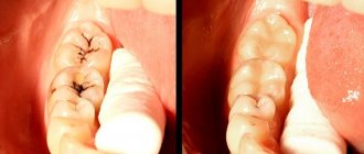
- exostosis after wisdom tooth removal;
- discomfort, pain;
- the appearance of cosmetic defects on the lower or upper jaw (they look like white balls on the jaw, noticeable when smiling or talking);
- the need for implantation, removable or fixed prosthetics;
- the risk of tumor transformation from benign to malignant.
If you need to install prostheses or implants, exostoses will become an obstacle to the procedure. Dentures will injure the bone growth on the gum, and implants will not be able to take root normally in the bone due to the pressure of osteophytes.
Diagnostics
Exostosis of the bone on the jaw does not in all cases manifest itself externally already at the beginning of development. Quite often, pathology is diagnosed during a comprehensive examination aimed at identifying other dental diseases. The clinical picture is formed based on the results of the examination, questioning of the patient, as well as radiographic photography, which clearly demonstrates the structural features of the maxillary region.
Stages and methods of treatment
Removal of growths is realized only using surgical protocols. Depending on the location of the tumor, one of two techniques is used:
- When the torus is located on the surface of the palate, a linear excision is performed, followed by detachment of the mucous flap and removal of the protruding bone area. For complex cases that do not allow separating the entire block, defragmentation is performed. After completing the wound inspection, the doctor tightens the wound and applies suture material.
- A similar technique is used to remove alveolar osteophytes, but the shape of the incision will be different and have a non-standard trapezoidal shape.
The instrument used in the process of removing exostosis is a drill, laser or piezo knife. During the treatment, the adjacent area of the periosteum is also scraped out - as a preventive measure to prevent relapse. In situations where tissue structure deficiency is diagnosed, it is recommended to form a cavity for subsequent placement of plastic material.
In the first hours after surgery, the patient should refrain from eating. During the week allotted for rehabilitation, you should follow medical instructions and recommendations regarding hygienic oral care, daily diet, bad habits and physical activity.
Complications
Adverse manifestations, as a rule, are the result of unscrupulous execution of the instructions of the attending physician. Possible complications include premature rupture of the suture material caused by excessive chewing load, as well as damage to the wound by hard food. In addition, if hygiene standards are not observed, there is a risk of developing an inflammatory process, causing swelling of the mucous tissue and the formation of pus. In such situations, you should immediately contact the clinic to relieve negative symptoms and avoid more serious consequences.
Prevention
Simple preventive measures will help reduce the risk of jaw exostosis or detect the problem as early as possible. Standard recommendations include careful oral hygiene, regular dental examinations (every six months), timely treatment of teeth and gums, and caution during physical activity that can lead to injury. Systematic self-diagnosis is recommended - sometimes you need to carefully examine the oral cavity for the presence of neoplasms, carefully palpating the surface of the jaws and the palatal area.
Indications and contraindications
If the formation is large, leads to inflammation of the mucous membranes of the oral cavity, jaw deformation, or interferes with talking or chewing food, then it is removed. The operation is also prescribed before prosthetics, since even small bumps become an obstacle to the installation of implants.
The patient himself can contact the surgeon if the bone spike spoils the aesthetics of the face. In this case, it is located on the buccal side and the protruding part is visible externally. An aesthetic defect gives a person more psychological discomfort than physical discomfort.
Before the procedure, the surgeon excludes contraindications. The list of restrictions includes infections in the mouth, acute respiratory infections, diabetes mellitus in the stage of decompensation, poor blood clotting, exacerbation of chronic diseases, mental disorders, childhood, pregnancy and a number of other contraindications.
For children, surgery is delayed until they are 18 years old. This is due to the fact that the bones are still growing and the pathology can resolve on its own.
Symptoms of exostosis of the jaw
At the initial stages of development, the pathology usually goes unnoticed, as it manifests itself asymptomatically. It is often discovered when examining a patient for other dental diseases. The formation is clearly visible on an x-ray.
As the growths increase, characteristic symptoms appear. Their intensity depends on the clinical picture: location, shape, size.
By what signs can pathology be detected?
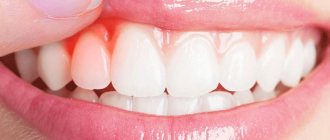
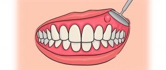
- the appearance of a tubercle or bump on the gums;
- sensation of a foreign object in the mouth, especially if the protrusion is located on the side of the tongue, at the bottom of the mouth;
- pain syndrome of varying intensity (occurs when injured by an acute growth of soft tissue);
- changes in the color of the mucous membranes in the area where the protrusion is located;
- jaw dysfunction if an osteophyte has formed near the jaw joint.
Unlike diseases of an infectious-inflammatory nature, it does not cause an increase in temperature, burning sensation, purulent abscesses, fistulas, and does not affect the tightness of the gums.
Surgical removal of exostosis on the gums
The operation is performed by a dental surgeon. In many cases, local anesthesia is sufficient for pain relief. It is possible to use deep sedation or anesthesia, especially if there are many osteophytes and they are large.
Amputation of bone growths is carried out in stages:
- local or general anesthesia:
- antiseptic treatment of the oral cavity;
- gum incision to access the correction area;
- amputation of a lump with a chisel or laser;
- grinding with a drill until the bone is smooth;
- repeated antisepsis;
- suturing and bandaging.
The operation time depends on the clinical case. It can range from 40 minutes to 2 hours. When using local anesthesia, the patient can go home immediately after the bleeding stops.
After operation
For speedy healing after surgery, the patient should follow the following doctor’s recommendations:
- Don't overheat . Until the wound heals, avoid playing sports, visiting bathhouses, sunbathing on the beach and taking hot baths. An increase in temperature promotes bleeding from the wound.
- Gentle nutrition . During the healing period, avoid rough, dry, spicy and sour foods. You should also not eat food that is too hot or too cold. The consistency of the food should be liquid or puree.
- Avoid smoking . Nicotine and combustion products of tobacco impair wound healing.
Continue brushing your teeth, but avoid the surgical area with your toothbrush. After each meal, rinse your mouth with antiseptic solutions prescribed by your doctor. To relieve pain in the first days after surgery, take painkillers.
Complications
Cartilaginous exostosis in children has a dangerous negative impact on the condition of the growth plates, which can result in shortening of the lower leg, thigh, forearm, shoulder, etc. This can also cause curvature of the arms and legs, increase the risk of fractures and cause disability.
Patients with severe forms of the disease feel inferior, which negatively affects their mental and emotional state.
The most dangerous complication of osteochondroma is its malignancy, i.e. transformation into a malignant tumor - chondrosarcoma. The highest risk of malignancy is with multiple exostoses. In this case, growths localized in the pelvic bones are more likely to undergo malignancy. Less commonly, exostoses of the ribs, scapula, and spine turn into malignant neoplasms. Single osteochondromas transform into a malignant tumor in no more than 1% of cases.
Atypical growth can be observed in any part of the exostosis: the cartilaginous cap, at the base or in the middle part.
Rehabilitation
After removal of the exostosis, the patient remains in the hospital for 3 days, the sutures are removed on days 7-10. Performing marginal resection does not require any serious restrictions even in the early postoperative period. The patient can completely return to his usual lifestyle after the stitches are removed, and forget about exostosis immediately after the operation.
When performing a corrective osteotomy, complete bone fusion occurs after 6 weeks, but full recovery may require up to 3 months. At this time, it is important to strictly follow all the doctor’s recommendations and not to overload the operated part of the body. All patients who have undergone osteotomy are prescribed drug therapy, exercise therapy, and often physical therapy. The duration of rehabilitation in such cases depends on the type of surgical intervention performed and the individual characteristics of the patient.
Thus, osteochondroma is a very common type of tumor. At the same time, statistics on the frequency of its occurrence are not entirely objective, since exostoses often do not appear throughout a person’s life, and therefore are not diagnosed. However, if the formed formation bothers the patient, causes him pain, or reduces the quality of life, you should not hesitate to contact an orthopedist. A specialist will be able to assess the degree of danger of the tumor and, if necessary, remove it using the most gentle method possible.
