CLASSIFICATION
Odontogenic infections (infections of the oral cavity), depending on the anatomical location, are divided into truly odontogenic
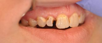
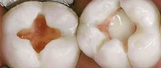
associated with damage to tooth tissue (caries, pulpitis);
periodontal
, associated with damage to the periodontium (periodontitis) and gums (gingivitis, pericoronitis), surrounding tissues (periosteum, bone, soft tissues of the face and neck, maxillary sinus, lymph nodes);
non-odontogenic
, associated with damage to the mucous membranes (stomatitis) and inflammation of the major salivary glands.
These types of infections can cause serious life-threatening complications from the cranial cavity, retropharyngeal, mediastinal and other localizations, as well as hematogenously disseminated lesions of the heart valve apparatus, sepsis.
Purulent infection of the face and neck
may be of non-odontogenic origin and includes folliculitis, furuncle, carbuncle, lymphadenitis, erysipelas, hematogenous osteomyelitis of the jaws.
Specific infections (actinomycosis, tuberculosis, syphilis, HIV) can also be observed in the maxillofacial area.
MAIN PATIENTS
Infections of the oral cavity are associated with the microflora that is constantly present here. Usually this is a mixed flora, including more than 3-5 microorganisms.
With truly odontogenic
infections, along with facultative bacteria, primarily viridans streptococci, in particular (
S.mutans, S.milleri
), anaerobic flora is released:
Peptostreptococcus
spp.,
Fusobacterium
spp.,
Actinomyces
spp.
In case of periodontal infection, five main pathogens are most often isolated: P.gingivalis, P.intermedia, E.corrodens, F.nucleatum, A.actinomycetemcomitans
, less commonly
Capnocytophaga
spp.
Depending on the location and severity of the infection, the patient’s age and concomitant pathology, changes in the microbial spectrum of pathogens are possible. Thus, severe purulent lesions are associated with facultative gram-negative flora ( Enterobacteriaceae
spp.) and
S.aureus
.
In elderly patients and hospitalized patients, Enterobacteriaceae
spp also predominate.
In the conditions of domestic bacteriological laboratories, it is quite difficult to isolate a specific pathogen of a certain odontogenic infection. However, it seems possible to localize pathogens that play a major role in the development of oral infections in supragingival and subgingival plaque.
PRINCIPLES OF TREATMENT OF INFECTIONS OF THE ORAL CAVITY AND MAXILLOFACIAL AREA
Treatment of odontogenic infection
often limited to local therapy, including standard dental procedures.
Systemic antibacterial therapy is carried out only when odontogenic infection spreads beyond the periodontal period (under the periosteum, into the bones, soft tissues of the face and neck), in the presence of elevated body temperature, regional lymphadenitis, and intoxication.
When choosing antimicrobial therapy in a hospital setting, in cases of severe purulent infection, it is necessary to take into account the possibility of the presence of resistant strains among anaerobes such as Prevotella
spp.,
F. nucleatum
to penicillin, which determines the prescription of broad-spectrum drugs, amoxicillin/clavulanate in monotherapy or a combination of fluoroquinolone with metronidazole.
Due to increasing levels of resistance by Streptococcus
spp.
to tetracycline and erythromycin, these drugs can only be used as alternatives. Since metronidazole has moderate activity against anaerobic cocci ( Peptostreptococcus
spp.), simultaneous administration of β-lactam antibiotics is necessary to increase effectiveness.
Preventive systemic use of AMPs when performing dental procedures in the oral cavity or periodontium does not guarantee a reduction in the incidence of infectious complications in somatically healthy patients. Convincing data on the sufficient effectiveness of topical antibiotics for oral infections have not been obtained. At the same time, oral bacteria are a reservoir of determinants of resistance to AMPs. Excessive and unjustified use of antibiotics contributes to their appearance and the development of resistance of pathogenic microflora.
HIV infection in dentistry. Issues of prevention.
Due to the continuing increase in the prevalence of HIV infection, the likelihood of a dentist encountering HIV-infected patients is constantly increasing. Many of these patients may have varying oral manifestations of the disease. Dental health workers cannot refuse to perform their professional duties to AIDS and HIV-infected patients. This will require dentists to know not only the symptoms of oral and periodontal disease, but also the basic principles of HIV prevention.
During dental procedures, damage to the oral mucosa and bleeding are inevitable. In this case, infection of medical instruments, casts, prostheses occurs, and aerosols containing blood and saliva may be dispersed when using high-speed dental units. At a dentist's appointment there may be patients who do not know or are hiding their disease, patients in the incubation stage, who can become a source of infection for other patients and for the staff of the medical institution.
Medical personnel in dental offices and departments belong to the occupational risk group. To prevent infection of personnel during dental procedures, the dentist must know the routes and factors involved in the transmission of HIV, and the provision of dental care must be carried out in compliance with the necessary precautions and with strict adherence to the rules of disinfection and sterilization of medical and dental instruments.
The most common malignant disease of the oral cavity is:
- Kaposi's sarcoma;
- Angular cheilitis caused by the fungus Candida albicans;
- Erythematous candidiasis;
- Pseudomembranous candidiasis;
- HIV gingivitis (linear gingival erythema);
- Necrotizing gingivitis and periodontitis;
- Herpetic ulcers due to HIV infection;
- "Hairy" leukoplakia;
- The wart is viral;
- Aphthous ulcerations;
- B-cell (non-Hodgkin) lymphoma.
HIV infection is a disease caused by the human immunodeficiency virus - an infectious chronic disease characterized by a specific lesion of the immune system, leading to its slow destruction until the formation of acquired immunodeficiency syndrome (AIDS), accompanied by the development of opportunistic infections and secondary malignant neoplasms. AIDS is a condition that develops against the background of HIV infection and is characterized by the appearance of one or more diseases classified as AIDS-indicative.
The source of HIV infection is people infected with HIV at any stage of the disease, including the incubation period. • Mechanism and factors of transmission. • HIV infection can be transmitted through both natural and artificial transmission mechanisms.
The natural mechanism of HIV transmission includes:
- • Contact, which occurs primarily during sexual intercourse (both homo- and heterosexual) and when the mucous or wound surface comes into contact with blood.
- • Vertical (infection of a child from an HIV-infected mother: during pregnancy, childbirth and breastfeeding.)
The artificial (artificial) transmission mechanism includes:
- • Artificial for non-medical invasive procedures , including:
- intravenous drug administration (use of syringes, needles, other injection equipment and materials),
- tattooing,
— when performing cosmetic, manicure and pedicure procedures using non-sterile instruments.
- • Artificial for invasive interventions in health care facilities:
- during transfusion of blood, its components,
- organ and tissue transplantation,
- use of donor sperm, donor breast milk from an HIV-infected donor,
— through medical instruments for parenteral interventions, medical products contaminated with HIV and not processed in accordance with the requirements of regulatory documents.
It can be said with absolute certainty that HIV is not transmitted by mosquitoes, mosquitoes, fleas, bees and wasps. HIV is not transmitted through casual contact. Not a single case of infection through saliva and tear fluid containing blood has been described. Since HIV is not transmitted through saliva, you cannot become infected by sharing glasses, forks, sandwiches or fruit. According to leading experts, contact with intact skin of infected biological fluids (for example, blood) is not enough to transmit the virus.
The main factors of pathogen transmission are human biological fluids:
- • blood,
- • blood components,
- • sperm,
- • vaginal discharge,
- • breast milk.
The main population groups at risk of contracting HIV infection are:
- • injection drug users (IDUs),
- • commercial sex workers (CSWs),
- • men who have sex with men (MSM).
The group at increased risk of contracting HIV includes: clients of sex workers, sexual partners of IDUs, medical workers, including dentists, prisoners, street children, people with a large number of sexual partners, migrating segments of the population (truck drivers, seasonal workers, including including foreign citizens working on a rotational basis and others)
The incubation period for HIV infection is the period from the moment of infection until the body’s response to the introduction of the virus (the appearance of clinical symptoms or the production of antibodies) is usually 2-3 weeks, but can last up to 3-8 months, sometimes up to 12 months. During this period, antibodies to HIV are not detected in the infected person, and therefore the risk of transmission of infection from him in nosocomial foci, including through transfusion of blood and its components, increases.
Stages of HIV infection:
- • acute phase (from 1-5 days to 6 months)
- • latent infection or subclinical stage (from 2 to 10 years)
- • stage of manifest manifestations (prev. AIDS and AIDS).
Acute HIV infection.
In 30-50% of infected people, symptoms of acute HIV infection appear, which is accompanied by various manifestations: fever, lymphadenopathy, erythematous maculopapular rash on the face, trunk, sometimes on the extremities, myalgia or arthralgia, diarrhea, headache, nausea and vomiting, enlarged liver and spleen , neurological symptoms. These symptoms vary in severity. In rare cases, severe secondary diseases may develop already at this stage, leading to the death of patients. During this period, the frequency of referrals of infected people to healthcare facilities increases; the risk of transmission of infection is high, due to the large amount of virus in the blood.
Subclinical stage.
The duration of the subclinical stage averages 5-7 years (from 2 to 10 years, sometimes more), there are no clinical manifestations other than lymphadenopathy. At this stage, in the absence of manifestations, the infected person remains a source of infection for a long time. During the subclinical period, HIV continues to multiply and the number of CD 4 lymphocytes in the blood decreases.
Stage of secondary diseases.
Against the background of increasing immunodeficiency, secondary diseases (infectious and oncological) appear. Initially, these are predominantly lesions of the skin and mucous membranes, then organ and generalized lesions, leading to the death of the patient. Namely: pulmonary tuberculosis extrapulmonary tuberculosis unmotivated weight loss (more than 10% in 6 months) Pneumocystis pneumonia recurrent severe radiologically confirmed pneumonia (2 or more episodes per year) CMV retinitis + colitis infection caused by the herpes simplex virus (chronic or persistent for 1 month or more) HIV-associated cardiopathy, HIV-associated nephropathy, encephalopathy, Kaposi's sarcoma and HIV-associated tumors. toxoplasmosis cryptosporidiosis cryptococcal meningitis progressive multifocal leukoencephalopathy. disseminated fungal infections, non-tuberculous mycobacterial infections or disseminated atypical mycobacteriosis
Laboratory diagnosis of HIV infection is based on the detection of antibodies to HIV and viral antigens. • The standard method for laboratory diagnosis of HIV infection is the determination of antibodies/antigens to HIV using ELISA. To confirm the results, confirmatory tests are used (immune, linear blot).
Diagnostic algorithm for testing for the presence of antibodies to HIV:
- • Stage 1 (screening laboratory).
- • Stage 2 (reference laboratory).
- • Stage 3 confirmatory test (immunoblot)
Stage 1 (screening laboratory).
If a positive result is obtained in the ELISA, the analysis is carried out sequentially 2 more times (with the same serum and in the same test system). If two positive results are obtained from three ELISA tests, the serum is considered primary positive and is sent to the reference laboratory (HIV Diagnostic Laboratory of the Center for Prevention and Control of AIDS) for further research.
Stage 2 (reference laboratory).
The initially positive serum is retested by ELISA in a second test system from another manufacturer, which differs from the first in the composition of antigens, antibodies or test format chosen for confirmation. If a negative result is obtained, the serum is retested in a third test system from another manufacturer, which differs from the first and second in the composition of antigens, antibodies or test format.
If a negative result is obtained (in the second and third test systems), a conclusion is issued about the absence of antibodies to HIV. If a positive result is obtained (in the second and/or third test system), stage 3 is carried out
Stage 3 confirmatory test (immunoblot) (search for antibodies to individual antigens of the core, shell, and enzymes of the virus).
Samples in which antibodies to 2 of 3 HIV glycoproteins (env, gag, pol) are detected are considered positive in the immunoblot. Sera are considered negative (negative) in which no antibodies are detected to any of the HIV antigens (proteins) or there is a weak reaction with protein p 18. Sera are considered undetermined (doubtful) in which antibodies to one HIV glycoprotein and/or any proteins are detected. HIV. If a negative and questionable result is obtained in an immune or line blot, it is recommended to examine the serum after 3, 6, 12 months.
One of the most important problems in HIV testing is the so-called diagnostic window period. This is the period that elapses from the moment of HIV infection until a detectable level of antibodies appears. As a rule, this is the incubation period (from the moment of infection to 6 months). • Taking into account this phenomenon, difficulties arise when examining donated blood from people who are in the mentioned period of HIV infection. Therefore, in most countries of the world, a system of using blood only after it has been stored for 3-6 months has been introduced in order to carry out mandatory re-examination for HIV infection of donors of these doses of blood and its components.
Testing for HIV infection is carried out voluntarily, except in cases where such testing is mandatory. The following are subject to mandatory medical examination for HIV infection: Donors of blood, blood plasma, and other biological fluids, tissues and organs each time donation material is taken; Doctors, paramedical and junior medical staff of centers for the prevention and control of AIDS Medical workers in surgical hospitals (departments) Persons undergoing military service and entering military educational institutions Foreign citizens and stateless persons when applying for a citizenship permit or permit residence.
All instruments and products that come into contact with the wound surface, blood or injectable drugs, as well as certain types of medical instruments that during operation come into contact with the mucous membrane and can cause damage to it are subjected to sterilization: dental instruments: tweezers, probes, spatulas, excavators, pluggers , smoothers, crown removers, skellers, dental mirrors, burs (including diamond-coated) for all types of tips, endodontic instruments, pins, dental discs, cutters, separating metal plates, matrix holders, trays for taking impressions, instruments for removing dental plaque , periodontal surgical instruments (curettes, hooks of various modifications, etc.), instruments for filling tooth canals (pluggers, spreaders), carpule syringes, various types of forceps and nippers for the orthodontic office, vacuum cleaners; ultrasonic handpieces and attachments for them, handpieces, removable micromotor sleeves for mechanical handpieces, cannulas for a dental plaque removal device; surgical instruments: dental forceps, curettage spoons, elevators, chisels, instrument sets for implantology, scalpels, forceps, scissors, clamps, surgical smoothers, suture needles; trays for sterile medical products, instruments for working with sterile material, including tweezers and containers for their storage.
Workplaces are provided with extracts from instructional and methodological documents, first aid kits for emergency prevention in emergency situations.
All medical instruments (as well as dishes, linen, devices, etc.) contaminated with blood, biological fluids, and also in contact with mucous membranes, immediately after use are subject to disinfection in accordance with the order of the Ministry of Health of the Republic of Belarus dated April 2, 1993 No. 66 “On measures to reduce the incidence of viral hepatitis in the Republic of Belarus” and other regulatory documents. Disinfection regimes are similar to those used for the prevention of infection with hepatitis B, C, D.
When carrying out manipulations associated with violating the integrity of the skin, mucous membranes, and also not excluding the splashing of biological fluids during autopsy of corpses, conducting laboratory tests, processing instruments, linen, cleaning, etc., medical workers and technical personnel must use personal protective equipment (surgical gown, mask, goggles or screen, waterproof apron, sleeves, gloves) to avoid contact of the patient’s blood, tissues, biological fluids with the skin and mucous membranes of the personnel. The approach to the use of protective clothing should be differentiated, taking into account the degree of risk of HIV infection.
Medical workers with injuries (wounds) on their hands, exudative skin lesions, and weeping dermatitis are excluded from medical care for patients and contact with care items for the duration of their illness.
Measures for wounds, contact with blood, and other biological materials of patients. Any damage to the skin, mucous membranes, or contamination of them with biological materials from patients during the provision of medical care should be qualified as possible contact with material containing HIV or another agent of an infectious disease.
If contact with blood or other liquids occurs with a violation of the integrity of the skin (injection, cut) , the victim must:
- remove gloves with the working surface facing inward;
- squeeze blood out of the wound;
- treat the damaged area with one of the disinfectants (70% alcohol, 5% tincture of iodine for cuts, 3% hydrogen peroxide solution for injections, etc.);
- wash your hands under running water and soap, and then wipe with 70% alcohol;
Orthopedic dentist, 1st category
Verevkin Dmitry Anatolievich
ODONTOGENIC AND PERIODONTAL INFECTION
PULPITIS
Main pathogens
Viridans streptococci ( S. milleri
), non-spore-forming anaerobes:
Peptococcus
spp.,
Peptostreptococcus
spp.,
Actinomyces
spp.
Choice of antimicrobials
Antimicrobial therapy is indicated only in case of insufficient effectiveness of dental procedures or spread of infection into surrounding tissues (periodontal, periosteal, etc.).
Drugs of choice
: phenoxymethylpenicillin or penicillin (depending on the severity of the disease).
Alternative drugs
: aminopenicillins (amoxicillin, ampicillin), inhibitor-protected penicillins, cefaclor, clindamycin, erythromycin + metronidazole.
Duration of use
: depending on the severity of the current (at least 5 days).
PERIODONTITIS
Main pathogens
Microflora is rarely detected in the periodontal structure and is usually S.sanguis, S.oralis, Actinomyces
spp.
In periodontitis in adults, gram-negative anaerobes and spirochetes predominate. P.gingivalis, B.forsythus, A.actinomycetemcomitans and T.denticola
are highlighted most often.
In juveniles, there is rapid involvement of bone tissue in the process, with A. actinomycetemcomitans
and
Capnocytophaga
spp.
P.gingivalis
is rarely isolated.
In patients with leukemia and neutropenia after chemotherapy along with A. actinomycetemcomitans
C.micros
is isolated , and in prepubertal age -
Fusobacterium
spp.
Choice of antimicrobials
Drugs of choice
: doxycycline; amoxicillin/clavulanate.
Alternative drugs
: spiramycin + metronidazole, cefuroxime axetil, cefaclor + metronidazole.
Duration of therapy
: 5-7 days.
For patients with leukemia or neutropenia after chemotherapy, cefoperazone/sulbactam + aminoglycosides are used; piperacillin/tazobactam or ticarcillin/clavulanate + aminoglycosides; imipenem, meropenem.
Duration of therapy
: depending on the severity of the course, but not less than 10-14 days.
PERIOSTITIS AND OSTEOMYELITIS OF THE JAWS
Main pathogens
With the development of odontogenic periostitis and osteomyelitis, S. aureus
, as well as
Streptococcus
spp., and, as a rule, anaerobic flora prevails:
P.niger, Peptostreptococcus
spp.,
Bacteroides
spp.
Specific pathogens are less commonly identified: A.israelii, T.pallidum
.
Traumatic osteomyelitis is often caused by the presence of S.aureus
, as well as
Enterobacteriaceae
spp.,
P.aeruginosa
.
Choice of antimicrobials
Drugs of choice
: oxacillin, cefazolin, inhibitor-protected penicillins.
Alternative drugs
: lincosamides, cefuroxime.
When P.aeruginosa
, antipseudomonas drugs (ceftazidime, fluoroquinolones) are used.
Duration of therapy
: at least 4 weeks.
ODONTOGENIC MAXILLARY SINUSITIS
Main pathogens
The causative agents of odontogenic maxillary sinusitis are: non-spore-forming anaerobes - Peptostreptococcus
spp.,
Bacteroides
spp., as well as
H.influenzae, S.pneumoniae
, less often
S.intermedius, M.catarrhalis, S.pyogenes
.
Isolation of S.aureus
from the sinus is characteristic of nosocomial sinusitis.
Choice of antimicrobials
Drugs of choice
: amoxicillin/clavulanate. For nosocomial infection - vancomycin.
Alternative drugs
: cefuroxime axetil, co-trimoxazole, ciprofloxacin, chloramphenicol.
Duration of therapy
: 10 days.
PURULENT INFECTION OF SOFT TISSUE OF THE FACE AND NECK
Main pathogens
Purulent odontogenic infection of the soft tissues of the face and neck, tissue of deep fascial spaces is associated with the release of polymicrobial flora: F. nucleatum
, pigmented
Bacteroides, Peptostreptococcus
spp.,
Actinomyces
spp.,
Streptococcus
spp.
ABSCESSES, PHLEGMONS OF THE FACE AND NECK
Main pathogens
With an abscess in the orbital area in adults, a mixed flora is released: Peptostreptococcus
spp.,
Bacteroides
spp.,
Enterobacteriaceae
spp.,
Veillonella
spp.,
Streptococcus
spp.,
Staphylococcus
spp.,
Eikenella
Streptococcus
spp.,
Staphylococcus
. predominate in children .
The causative agents of abscesses and phlegmon of non-odontogenic origin, most often caused by minor skin lesions, are S.aureus, S.pyogenes
.
With putrefactive-necrotic phlegmon of the floor of the mouth, a polymicrobial flora is released, including F.nucleatum, Bacteroides
spp.,
Peptostreptococcus
spp.,
Streptococcus
spp.,
Actinomyces
spp.
S.aureus
can be isolated in patients with severe disease (more often in patients suffering from diabetes and alcoholism).
Choice of antimicrobials
Drugs of choice:
inhibitor-protected penicillins (amoxicillin/clavulanate, ampicillin/sulbactam), cefoperazone/sulbactam.
When P.aeruginosa
- ceftazidime + aminoglycosides.
Alternative drugs:
penicillin or oxacillin + metronidazole, lincosamides + aminoglycosides of the II-III generation, carbapenems, vancomycin.
Duration of therapy:
at least 10-14 days.
BUCCAL CELLULITE
Main pathogens
Usually observed in children under 3-5 years of age. The main pathogen is H. influenzae
type B and
S. pneumoniae
.
In children under 2 years of age, H. influenzae
is the main pathogen, and bacteremia is usually observed.
Choice of antimicrobials
Drugs of choice
: amoxicillin/clavulanate, ampicillin/sulbactam, third generation cephalosporins (cefotaxime, ceftriaxone) IV, in high doses.
Alternative drugs
: chloramphenicol, co-trimoxazole.
Duration of therapy
: depending on the severity of the current, but not less than 7-10 days.
LYMPHADENITIS OF THE FACE AND NECK
Main pathogens
Regional lymphadenitis in the face and neck is observed with infection in the oral cavity and face. Localization of lymphadenitis in the submandibular region, along the anterior and posterior surfaces of the neck in children aged 1-4 years, is usually associated with a viral infection.
Abscess formation of lymph nodes is usually caused by a bacterial infection. With unilateral lymphadenitis on the lateral surface of the neck in children over 4 years of age, GABHS and S.aureus
.
Anaerobic pathogens, such as Bacteroides
spp.,
Peptococcus
spp.,
Peptostreptococcus
spp.,
F.nucleatum, P.acnes
, can cause the development of odontogenic lymphadenitis or inflammatory diseases of the oral mucosa (gingivitis, stomatitis), cellulite.
Choice of antimicrobials
Drugs of choice: AMPs corresponding to the etiology of the primary source of infection.
Processing of dental instruments
Sanitary legislation puts forward strict requirements for the processing of dental instruments, which must be met by all dental offices without exception, regardless of their form of ownership. Such high demands are completely justified, because dental instruments can injure the oral mucosa, which is associated with a high risk of infection.
For the smooth operation of the dental office, it is necessary to have a sufficient number of sterile instruments, at least ten sets for each doctor per shift. This approach will allow us to serve a large number of patients and at the same time have time to systematically process the used instruments.
According to the requirements of sanitary legislation, dental instruments must be processed in three stages:
- Disinfection;
- Pre-sterilization treatment;
- Sterilization.
Disinfection of dental instruments
So, the first stage of processing instruments is disinfection, which is designed to minimize the level of contamination of instruments by microorganisms. Disinfection is carried out using disinfectants. The choice of disinfectant must be approached responsibly. The disinfectant, firstly, must effectively destroy microorganisms and, secondly, not spoil the instruments. These properties are fully met by disinfectants for dentistry “Septolite Tetra” and “Septolite Dental”.
Instruments are disinfected after each client. To do this, used instruments, opened and disassembled, are immersed in a container with a disinfectant. The liquid level should completely cover the instruments. Disinfection must be carried out according to the regimen used for viral infections.
At the end of the holding time, pre-sterilization cleaning begins, which is carried out to clean the instruments from protein contaminants. The products “Septolite Tetra” and “Septolite Dental” allow you to combine disinfection and PSO in one stage. Therefore, after disinfection is completed, the instruments are thoroughly cleaned using brushes directly in the container with the disinfectant.
Disinfection of impressions and prosthetic blanks is carried out before they are sent to the dental laboratory and before installation on the patient. After disinfection, the impressions and dentures are washed with distilled water. This completes the processing of these dental products.
Sterilization of instruments
All products that come into contact with blood and the wound surface, which can lead to injury to the mucous membrane, must be sterilized. Dental instruments are processed using steam or air sterilization. To do this, disinfected instruments are placed in packaging (paper bags or containers) and sent to a sterilizer. Sterilization in an autoclave is carried out for 20 minutes at a temperature of 132 °C, in a dry-heat oven for 60 minutes at a temperature of 180 °C.
Sterile instruments are stored packaged in Kraft bags, and unpacked in UV sterilizers. Please note that the assistant should place sterile instruments on the table immediately before starting the manipulation.
According to sanitary legislation, chemical sterilization using disinfectants is allowed only in cases where other sterilization methods cannot be used. This applies to the sterilization of instruments made from heat-labile materials. Chemical sterilization is carried out strictly in compliance with antiseptic rules. After chemical sterilization is completed, the instruments are washed with distilled water.
NON-ODONTOGENIC AND SPECIFIC INFECTION
NECROTIC STOMATITIS (VINCINS ULCER-NECROTIC GINGIVOSTOMATITIS)
Main pathogens
Fusobacterium is concentrated in the gingival sulcus
, pigmented
Bacteroides
, anaerobic spirochetes. With necrotizing stomatitis, there is a tendency for infection to quickly spread into surrounding tissues.
The causative agents are F. nucleatum, T. vinsentii, P. melaninogenica, P. gingivalis and P. intermedia
.
In patients with AIDS, C. rectus
.
Choice of antimicrobials
Drugs of choice:
phenoxymethylpenicillin, penicillin.
Alternative drugs:
macrolide + metronidazole.
Duration of therapy:
depending on the severity of the current.
ACTINOMYCOSIS
Main pathogens
The main causative agent of actinomycosis is A.israelii
, association with gram-negative bacteria
A. actinomycetemcomitans
and
H. aphrophilus
, which are resistant to penicillin but sensitive to tetracyclines, is also possible.
Choice of antimicrobials
Drugs of choice
: penicillin at a dose of 18-24 million units/day, with positive dynamics - transition to step-down therapy (phenoxymethylpenicillin 2 g/day or amoxicillin 3-4 g/day).
Alternative drugs
: doxycycline 0.2 g/day, oral drugs - tetracycline 3 g/day, erythromycin 2 g/day.
Duration of therapy
: penicillin 3-6 weeks IV, drugs for oral administration - 6-12 months.
