04.01.2020
The operation to remove the nerve of a tooth is familiar to almost everyone who treated teeth in Soviet times or in the 90s. Not surprisingly, most people associate this procedure with arsenic being placed inside the tooth, followed by pain for several days. The memories are terrifying... But is it worth it to be afraid of this procedure now, when the modern industry has made a significant leap forward and modern dental services have appeared in all cities.
The first thing worth saying is that arsenic is a thing of the past. In addition, dentists use modern equipment and innovative materials, including for removing the nerve of a tooth. As a result, the process takes much less time, and the procedure itself is absolutely painless.
Removing the nerve allows you to protect other teeth and maxillofacial tissue from further development of the inflammatory process and the appearance of infectious diseases of the oral cavity.
First, we suggest you figure out what the nerve of a tooth is and when can a dentist decide to remove it?
What is a nerve and how many are there in a tooth?
A tooth consists of several parts. Enamel is the top, most durable layer. It protects the underlying tissue - fairly dense but porous dentin. The infection slowly destroys it and spreads beyond the crown of the tooth, into the root canals. The first thing on the way is pulp. This is a neurovascular bundle consisting of a different number (from 1 to 3) of nerve endings and blood vessels. They are the ones who nourish the tooth, maintain its strong structure and vital functions. In addition, the pulp protects the connective tissues of the periodontium from infection and immediately reports it through pain and temperature sensitivity. When the nerve is removed, the tooth is left without nutrition and protection, and therefore becomes more fragile and susceptible to chipping. It is considered dead, although it retains its functionality.
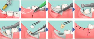
Surgical method
The dentist’s task is to completely clean out the affected nerve from the canals when treating pulpitis.
In this case, treatment of pulpitis involves excision or complete removal of the affected pulp. Removing only the crown part will not work for teeth with one root due to the structure of the nerve. In addition, there are certain restrictions on the age of patients and the presence of allergic reactions.
If the pulp was partially removed, the doctor will also use medications to preserve the remaining part of the nerve.
With complete removal - under anesthesia or using a devitalizing paste - the nerve is completely removed from the tooth, including the root canals.
- Treatment of pulpitis in stages means that it is important for the doctor to remove the tissue completely to avoid recurrence of the infection.
- Afterwards, the canals and the tooth cavity are filled and everything is closed with a permanent filling.
After filling the canals, an x-ray must be taken to check the absence of voids; the cavities are etched and dried to ensure high-quality adhesion of the material to the tooth tissues and to prevent the filling from falling behind after hardening.
How to remove a nerve from a tooth
A couple of decades ago, the only way to remove a nerve from a tooth was with arsenic.
After treating the caries, it was applied to the exposed area of the pulp and a temporary filling was placed. You had to walk with arsenic paste on your tooth for several days for the nerve to die. The danger was that arsenic is a poison that affects the health of the entire body. And with prolonged contact with the tooth, it could be completely destroyed. Modern methods of treating pulpitis and removing the nerve are harmless.
The doctor makes an accurate diagnosis and determines the area of inflammation in the tooth. Then he injects an anesthetic to make the procedure painless. The specialist thoroughly cleans the root canals and removes affected tissue to prevent re-inflammation. If necessary, he applies arsenic-free medicine under a temporary filling, and then replaces it with a permanent one and takes a control photograph. Most often, tooth nerve removal takes place in one session, with a permanent filling immediately installed. It is impossible to carry out treatment without complete removal of the pulp (in adults), since its damage is an irreversible process.
Biological method using antibiotics
This requires high professionalism of the doctor to adequately assess the condition of the pulp. If the pathological process is at an initial stage and has not progressed to tissue destruction, then the doctor can use antibiotics to stop the inflammatory process.
- The tooth is prepared and the pulp chamber is opened;
- the doctor lays down the medicine;
- a microbandage with calcium hydroxide is installed;
- The tooth is filled with a temporary filling.
After the established period, the patient comes for a follow-up appointment. If the condition of the pulp has returned to normal, the filling is replaced with a permanent one.
Tooth hurts after nerve removal
After treatment and the end of anesthesia, the pulpless tooth may hurt. The reason may be temporary, since removal of the nerve is a serious intervention in the structure of the tooth. Usually, after a few days, all unpleasant symptoms disappear. If after two weeks the tooth continues to ache, and the pain even intensifies, consult your doctor immediately. Perhaps it’s all about the neighboring tooth, the pain from which radiates to the recently cured one.
Why does deep caries develop?
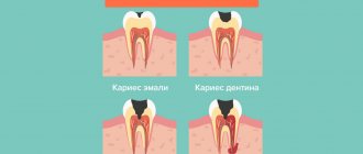
The process of tooth decay under the influence of microorganisms and other factors is caries. Its onset and development is caused by the activity of pathogenic microflora of the oral cavity, in particular the bacteria Streptococcus mutans. They feed on sugars from food, cause fermentation and produce acid. It corrodes tooth tissue, which leads first to demineralization of the enamel, then to deformation of its structure and the formation of carious cavities. Gradually, the process spreads deeper and affects dentin - the bone tissue under the tooth enamel.
Caries most often does not develop at lightning speed and goes through several stages. First, a barely noticeable chalky spot forms on the enamel - an area that is different in color from the rest of the tooth. This is the area of demineralization where the carious process began. Enamel loses the mineral compounds that make up its structure, becomes more fragile and less resistant to harmful influences. This is the initial stage of caries - the spot stage. People often miss it due to the lack of obvious symptoms. Then tooth decay continues, and the disease progresses to the stage of superficial caries. The tooth begins to react to hot/cold, sour/sweet, and a clear defect is noticeable in the enamel.
Without treatment, superficial caries progresses to intermediate caries when dentin begins to deteriorate. Symptoms become more pronounced, pain occurs not only as a reaction to an irritant, but also on its own. According to statistics, most patients seek help from a dentist at the stage of average caries.
But there are also people whom even acute toothache will not force them to go to the dentist. And then the most complex and advanced form can develop - deep caries. At this stage, the carious cavity is in close proximity to the dental pulp; at any moment, inflammation can spread to the dental nerve and lead to pulpitis, and then to periodontitis.
What complications may occur after treatment?
Among the most common undesirable consequences that can be encountered after depulpation are the following:
The pulp was not completely removed or the canals were not completely sealed. This dental error leads to inflammation and requires repeated endodontic treatment. If it is not carried out, a cyst may form or periodontitis or gumboil may occur.- A fragment of a dental instrument remained in the dental canal. This problem is always caused by low qualifications or inexperience of the dentist. You need to take a picture and refill the canals. Otherwise, the tooth will continue to hurt. However, if the complication does not manifest itself in any way (the patient found out about it by chance while taking an X-ray about a neighboring tooth), re-treatment may not be necessary.
- Resealing. The essence of this complication is that the filling composition used by the doctor extends far beyond the top of the tooth root. If the amount of excess is small, pain occurs when pressing on the tooth; if it is significant, severe discomfort sensations persist on an ongoing basis. In any case, surgical treatment is necessary to remove all excess.
- Change in crown color. The tooth darkens after the nerve is removed if low-quality filling material is used. The second reason is that a dead unit becomes covered with microcracks over the years. Coloring pigment accumulates in them. This causes a change in the natural shade.
- Puncture (perforation) of the tooth wall or root. Always caused by mechanical damage. The problem requires a surgical approach.
It is important to understand that these complications are rare. If you have chosen an experienced doctor and a good dental clinic, you can rest assured about the health of the tooth being treated.
Should I remove the nerve in the tooth or not? That is the question!
Agree, it is quite a common situation when, after treating caries with a therapist or treating a tooth with a crown by an orthopedist, the latter may begin to bother you. One of the controversial questions that patients have to face at this moment is when to remove the nerve in a tooth , and under what circumstances is there a chance to keep the tooth alive? In reality, this question is not always clear-cut for dentists either. In this article we will try to figure out what the difficulty is here.
Firstly, you need to immediately decide that a “living” tooth is always and definitely better than a “dead” one. Because under the “nerve” there actually lies an entire organ, consisting not only of the nerve, but also of blood vessels and other tissues and cells needed by the tooth. And all this is called pulp. It provides the tooth with both nutrition and protection from any external influences. And therefore, without it, it is difficult for our teeth to count on a long and happy life.
But often as a result of caries, injury or dentist intervention (for example, when grinding living teeth for crowns), an inflammatory process can occur - pulpitis . And here it is very important how reversible this process is. This is what is important for the doctor to determine when rendering a verdict on the pulp - to execute it (i.e., depulpate the tooth) or to pardon it (leave the tooth “alive”).
And here we can say that 100% reliable methods for diagnosing the reversibility of the inflammatory process in the pulp do not yet exist. If we had the opportunity to look inside the tooth with a microscope, then determining the vitality of the pulp would not be difficult. But it is impossible to do this on a living patient; such an opportunity is currently available only to pathologists. As a rather cynical saying goes, this is why these doctors are least likely to make a mistake in diagnosis. So what remains for practicing dentists? How can they avoid making mistakes? Which path should you take? You can only rely on much less informative indirect signs and use your experience and intuition.
Let us briefly examine what is still available to us when making a decision.
Patient complaints.
Of course, one of the most subjective signs, however, sometimes very informative. Therefore, it is often enough to listen to your feelings to understand how bad things are with your tooth. Depulpation certainly cannot be avoided if:
- You feel a fairly strong aching pain that occurs without any apparent reason and does not go away for a long time. At the same time, it can intensify when changing the position of the body (mainly when trying to bend over or lie down), and also noticeably intensifies at night. Usually in such a situation, many people run to the pharmacy for a pack of painkillers, although the most correct thing in this situation is to call your dentist with a request for an urgent appointment...
- You do not feel aching pain all the time, but only from some irritant (most often from cold ). The important point is that this pain does not go away immediately after removing the irritant for some time (from several seconds or more).
If, upon contact with cold, you feel only a short, instantly passing pain, then, most likely, everything for the pulp, maybe not right away, will end well. Any other sensations of discomfort, for example, painful biting or sensitivity when touched by a brush when brushing your teeth, are usually associated with completely different things and do not talk about the condition of the pulp.
Cold test.
This point smoothly follows from the first and is based on the inadequate reaction of the inflamed pulp to irritation (primarily to cold). This can be used for diagnostic purposes and, so to speak, to conduct reconnaissance in force. The technique is simple, but with a touch of slight sadism. The doctor cools the cotton ball with a special spray and touches the “suspected” teeth with it one by one.
The resulting response is compared with that of obviously healthy teeth that are 100% above suspicion (i.e., they are on the other side, on the other jaw and do not have carious holes or any traces of previous treatment). If suddenly someone wants to “play dentist” and can’t wait to find a bad tooth themselves, then at home you can do this test yourself by applying, for example, an ice cube from the freezer to different teeth one by one.
In general, self-diagnosis is pointless, but in this case it can still be useful to understand your own feelings, and then more confidently poke the doctor at the problem tooth.
It should be noted that this method is good primarily only in order to understand which tooth is actually bothering you. After all, pulp pain is quite poorly localized, and the patient often cannot say which tooth is bothering him. It even happens that the pain from the causative tooth radiates to the opposite jaw. This happens especially often with pulpitis of 8 teeth (wisdom teeth). Therefore, if in such a situation you trust only the feelings of the patient himself, you can break a lot of wood and depulpate innocent teeth for no reason. It is almost impossible to judge the severity of the inflammatory process and its reversibility using a cold test. For each patient, “oh-yoyoy” and “ah-yayayy” can mean completely different things. Although, as in the first point, by the duration and severity of the pain reaction, the doctor can indirectly judge the hopelessness of attempts to keep the pulp alive.
X-ray.
Another highly informative technique for most cases, which is completely useless for determining the neglect and reversibility of the pulpitic process. X-rays can also only indirectly help identify a problematic tooth if there is any doubt about the “culprit” of the pain. The pulp itself, like soft tissue, does not show any changes on x-ray if it is inflamed. What signs can help a doctor suspect something is wrong?
— location of the previously installed filling close to the pulp chamber
In this image: the red line marks the border of the filling, the blue line marks the border of the pulp. As you can see, they are located quite close to each other.
- hidden, invisible during visual inspection, deep carious cavity in the interdental space
In this picture: the black arrow shows a hidden carious cavity that formed on the back side of the lower 7th tooth under the gum level and led to inflammation of the pulp. The reason in this case was the 8 (wisdom tooth) that was not removed in time.
-presence of denticles in the pulp chamber or root canals
In this image: the yellow line marks the border of the filling, which is close to the pulp (its borders are marked in red). A denticle has formed in the thickness of the pulp due to chronic irritation (marked in blue).
-small reactive expansion of the periodontal fissure around the apex of the tooth root
Electroodontometry (EDO).
This is an old, if not ancient, way to check the condition of the pulp using essentially the same method as with the cold test. Those. irritate her. Only in this case it is no longer a low temperature, but an electric current. This time, nothing sadistic, as everyone usually thinks when explaining the essence of the technique. To carry it out, it is not at all necessary to pass current through the entire patient in some kind of electric chair. It is enough to connect a special small device to the tooth, which generates rather weak microcurrents.
By the value to which the tooth “responds” with slight tingling and pinching, one can judge (again, very approximately, unfortunately) the degree of pulp viability. The advantage of this method compared to the cold test is that instead of “ouch” and “ouch”, the doctor deals with impartial readings from the device. Another thing is that 100% of these indications can only be used to judge whether the pulp is alive at all, or whether it died a hero’s death. Everything else, again, is very conditional.
In the end, what do we have? There are 2 main problems with teeth suffering from pulpitis (or very similar pain : difficulties in some clinical cases in determining the source of pain, and difficulties in making a decision about preserving/removing the pulp. As we have seen, it is not easy for even an experienced and knowledgeable doctor to objectively understand these issues in all cases. If with the first question mistakes can almost certainly be avoided, then with the second... while there are no methods for objectively assessing the condition of the pulp, each doctor will make decisions at his own discretion. Someone, playing it safe, depulps all the teeth in a row indiscriminately. Someone is trying to keep them alive. The more data a doctor can collect and interpret correctly, the risk of making an erroneous decision will be minimized. In general, this is an iron law of medicine: the better and more thorough the diagnosis, the more correct and effective the treatment.
What happens if a mistake is made? I'll tell you one example from my practice. A young girl went to the doctor complaining of aching pain in her teeth “somewhere on the right side.” Without any special diagnostics, treatment began... First, one tooth with a deep filling was depulped, which seemed the most suspicious to the doctor. The pain remains. Then - the next one, the pain did not go away. Then both right normally erupted wisdom teeth were removed. The pain even intensified for a while... Instead of stopping and thinking at that moment, the doctor depulped another 3 (!!!) completely intact (i.e. untouched) teeth on this side, but on the other jaw. When the pain did not go away even after that, the girl, suspecting something was wrong, went to look for reasons in other places. Ultimately, she was diagnosed with trigeminal neuralgia, which, although it is not always well and quickly treated, in her case it was released quite quickly due to the treatment prescribed with the help of a neurologist. The result of the diagnosis using the “finger in the sky” method was 5 completely needlessly killed teeth.
Therefore, it is so important to collect as completely as possible all possible subjective and objective information if the situation is confusing. In this case, the probability of making a wrong decision will tend to zero. In addition, you should strive to preserve the pulp in the tooth, and do not rush into radical decisions if in doubt. At the same time, in borderline cases, you should always monitor the result of treatment, even if the pain has subsided. A cold test, x-rays, and odontometry will be able to promptly identify asymptomatic (“silent”) pulp death and the need for its removal. Otherwise, a cyst may grow completely unnoticed near the root of the tooth. And then there is a risk of losing the entire tooth.
How to treat deep caries with pulp damage?
Caries complicated by inflammation of the pulp is treated in several stages. In case of inflammation at an early stage, the dentist may try to preserve the pulp completely or partially, in which case antibiotic therapy is carried out. But most often, in case of pulpitis, the nerve is removed, and the root canals are cleaned and treated.
Further treatment tactics depend on the individual characteristics of the disease. Root canals are often filled, after which a filling is placed in the crown of the tooth. If your own tooth is severely damaged, a stump inlay or an artificial crown is installed.
Treating deep caries while preserving or removing the nerve is difficult, expensive and time-consuming. Therefore, we do not recommend starting the carious process to such an extent that the nerve needs to be removed. The best option is to consult a dentist at the first signs of the disease, and if there are no complaints, visit a professional examination twice a year.