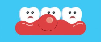
Quite often, parents may notice a soft bump that appears on their child’s gum when pressed. What could be the reasons for this phenomenon?
- inflammation of the roots (both baby teeth and molars, permanent ones);
- the beginning of tooth eruption.
Why does an abscess occur?
There can be many reasons for the formation of a lump (fistula, swelling), but the most common of them is undetected or ignored caries. Not all children maintain oral hygiene and eat properly, and not all parents carefully control this, which causes caries to appear on one or several teeth at once.
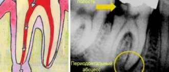
If caries is not treated, it will develop into pulpitis, extending beyond the tooth area and affecting the upper part of the root. As a rule, a lump appears just near the tooth whose root is inflamed. Typically, a lump can be noticed near a tooth with caries or with a filling that was placed long ago or poorly; Another common reason for its appearance is an injury received from a strong blow or an accidental fall, which can trigger the onset of the inflammatory process.
Stages of development of an abscess:
- As a result of caries, the infection penetrates into the pulp, affecting the root tip.
- Pus begins to form near the top of the root.
- Purulent formations fall under the mucous membrane of the gums.
- A cyst appears, looking like a small lump from the outside.
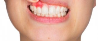
The most important symptom of an abscess is a soft and painful swelling, even with slight pressure.
Sometimes, due to an excess of pus, the lump may burst under pressure, and then a fistula appears - a small hole in the gum, which is connected with the source of inflammation located in the upper region of the root. A feature of the fistula is the constant release of purulent formations.
If inflammation decreases for some reason, the fistula may close on its own. However, when the child’s immunity decreases and the inflammatory process begins again, accompanied by pus formation, the appearance of a fistula will not be long in coming.
Tooth cyst (radicular cyst) - symptoms and treatment
Types of treatment for radicular cysts can be divided into conservative, surgical and conservative-surgical.
Conservative treatment
The conservative method is aimed at removing the necrotic neurovascular bundle of the tooth (pulp) and eliminating pathological changes in the bone behind the root apex.
The decayed pulp is removed with special endodontic instruments. Then the root canal is expanded and antiseptic treatment is carried out. Medicinal pastes are introduced into the root canal, which eliminate the microbial flora and restore the affected bone tissue. These pastes are administered for a period from several days to 1–1.5 months. Usually this process is well tolerated, but sometimes the pastes cause an exacerbation of the process with severe pain. To prevent the paste from getting into the mouth, the tooth cavity is hermetically sealed with a temporary filling.
If the inflammation of the bone tissue is eliminated, the root canals are filled with permanent material, and the defect in the hard tissues of the tooth is closed with a filling.
The fact is that there are no microorganisms in the cyst cavity itself. This is a response to a high concentration of microorganisms in infected root canals. After proper treatment of the canals, the number of microbes decreases and the body, with the help of cellular immunity, copes with a small number of microorganisms in the area of the root apex. The “barrier” in the form of a thin connective tissue membrane of the cyst becomes unnecessary, and macrophages completely destroy the membrane. With a small size of the cyst, rational treatment and the body’s ability to resist the disease, conservative treatment gives a good result - the shell of the radicular cyst is completely resorbed, the alveolar fascicle bone is restored.
After final treatment of the canals, it is recommended to do a CT scan after 6 and 12 months. If the cyst is completely resolved, there is no need to fear relapses, since the cause (bacterial infection of the canals) has been completely eliminated.
After root canal treatment, the tooth does not receive adequate nutrition, so it may deteriorate over time. To prevent this, it is recommended to cover pulpless teeth with artificial crowns.
If the conservative method is ineffective and the focus of destruction remains, surgical removal of the cyst is performed. This combination of methods is called conservative-surgical treatment.
Surgery
The surgical method involves removing a cyst from a previously pulpless tooth, and often the apex of the tooth root is cut off. The operation is carried out strictly according to indications: in case of a broken instrument, poor-quality treatment of the measles canal, or if the root protrudes into the cavity of the cyst. The extent of surgical intervention is determined during diagnosis.
To improve the prognosis after surgical treatment, bone defects are more often replaced with osteoplastic materials [15][16]. Such materials restore lost bone tissue and stimulate the formation of new one.
One of the main directions in modern reconstructive surgery is the use of biotechnologies, which allow accelerating the processes of regeneration of damaged tissues [17]. We are talking about platelet-derived growth factors obtained from the patient’s own blood. First, venous blood is taken from the patient. Then, using a centrifuge, the blood is separated into fractions. The platelet-rich fraction is used to restore bone tissue defects. This method is available almost everywhere.
There are two main surgical methods for treating radicular cysts: cystectomy and cystotomy.
1. Cystectomy is an operation in which the fibrous membrane of the cyst is completely removed. Usually performed in an outpatient setting under local anesthesia. If the cyst is large and affects the nasal cavity, maxillary sinus or mandibular canal, the operation is performed in a maxillofacial surgery hospital.
Surgical technique:
- Under local or general anesthesia, a section of the jaw bone in the projection of the cyst is skeletonized (exposed).
- Access to the cyst cavity is created, all affected bone tissue and the cyst shell are removed.
- According to indications, the root apex or part of it is cut off and retrograde filling of the root canal is performed (filling through the root apex) and filling of the apical openings of the canals with special cement intended for these purposes.
- The surgical wound is treated with antiseptic solutions, filled with osteoplastic material and sutured.
- Postoperative material is sent for pathohistological examination.
A dental microscope is used for treatment. It allows you to minimize the volume of removal of the root apex and effectively close the root canals.
You must visit the doctor the next day after the operation, then if the course is calm after 10–14 days, to remove the stitches. The patient should be under the supervision of the attending physician until the postoperative wound is completely healed, then return every six months to monitor the restoration of bone tissue and exclude relapse. As practice shows, relapses are rare; they are usually detected in the first year after surgery.
Types of material for filling a bone tissue defect after surgery:
- Autogenous material is obtained from another area of the patient’s bone tissue (branch of the jaw, chin, etc.). Due to its complete biological compatibility, such material is rarely rejected and quickly restores the defect. However, when the patient’s tissue is taken, the operation time increases, the intervention becomes more traumatic, which negatively affects the patient’s recovery.
- Xenogeneic material is obtained after special processing of mammalian bone tissue. The most commonly used bones are ox and pork. Processing removes substances that can cause immune and allergic reactions.
- The synthetic material contains calcium salts. For better compatibility it is often used in combination with autogenous material.
2. Cystotomy is an operation during which the cyst shell is not removed, but only its anterior wall is excised, which allows the contents of the cyst to be evacuated. With cystotomy, the scope of surgical intervention is smaller, but recovery after such an operation lasts longer (about a month). The resulting bone defect is filled with turunda with iodoform, then dressings are made with a certain frequency, replacing the turunda. This method is used less and less due to protracted recovery, the introduction of new techniques and the improvement of the material and technical base.
Oronasal (between the mouth and nose) cystectomy and oronasal cystotomy are performed if the cyst penetrates into the maxillary sinus or pushes it aside.
Surgical methods using laser technology are increasingly used in modern dentistry. Laser techniques are less traumatic. In addition, the laser has a bactericidal property, which reduces the likelihood of developing purulent-inflammatory complications [18][19][20].
Removal of a tooth. A tooth is removed only if saving it is not practical. Indications for removal:
- complete destruction of the crown part of the tooth;
- destruction of the hard tissues of the tooth below the level of the gingival margin by more than 2 mm, when it is not possible to advance the root orthodontically or when the root of the tooth is too short, which will complicate further prosthetics;
- lack of lumen in the root canal (due to structural features or previous treatment).
In all other cases, tooth-preserving operations described above are performed.
Complications after treatment
Complications after cyst removal occur infrequently. These include perforation of the maxillary sinus, damage to the inferior alveolar nerve, as well as the formation of a residual cyst, which is formed during incomplete excision of the radicular cyst.
How to treat an abscess on the gum?
If an abscess or fistula appears, then you should not expect that the situation will resolve on its own: you need to make an appointment with a dentist. Treatment of an abscess on a baby tooth and a molar one will be different.
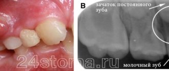
The appearance of an abscess on a baby tooth indicates periodontal inflammation. Such teeth must be removed, since the inflammatory process in the upper part of the root, accompanied by the formation of pus, may well spoil the molar, which will soon erupt in place of the milk tooth. This happens because the roots of temporary teeth are located next to the rudiments of molars. Bad bacteria and infection can also enter the lymph nodes under the jaw, causing them to become inflamed. The occurrence of a fistula means that pus will constantly seep into the mouth, which can cause the development of tonsillitis, since the tonsils will become infected. Various colds in a child are a direct consequence of an abscess on a tooth. Also, do not forget that toxins formed in the area of inflammation will definitely enter the bloodstream: this can result in allergic reactions, asthma and other serious general somatic diseases.
If we are talking about an abscess on a molar, then ordinary treatment is required for adults (provided that the tooth is not fundamentally damaged and can still be saved from removal).
Having discovered an abscess in a child, you should not try to cure it on your own. You can easily find a lot of advice on the Internet on how to relieve inflammation, but no amount of rinsing or even taking powerful antibiotics can eliminate the inflammatory focus located at the root of the tooth. Moreover, it is useless to hope that the tooth ache will stop. A bursting lump (this often happens) indicates only a short respite, during which, meanwhile, bacteria will still continue to enter the bloodstream and spread throughout the body. This situation can turn into a serious problem if the child has diseases of the respiratory system, heart and other internal organs.
Often, dentists suggest leaving teeth near which the inflammatory process is occurring, persuading the parents of a young patient to carry out the silvering procedure. The arguments for refusing removal can be very compelling: possible problems with diction and pain during the removal operation. However, such dentists neglect the information contained in every textbook on dentistry, which clearly states that a baby tooth with a purulent focus must be removed. Silvering will only complicate the situation, and an unresolved inflammatory process can lead to asthma, tonsillitis and even diabetes: in comparison with these diseases, temporary problems with diction will seem like sheer nonsense.
But the formation of an abscess can be prevented, and this is not difficult to do - just teach the child to maintain oral hygiene. At an early age, a child cannot be adequately responsible for his actions, including those related to the care of the oral cavity, teeth and gums, so responsibility for the procedure lies entirely with his parents, who must control how and when the child brushes his gums and teeth . It is the parents who should buy the most effective baby toothpaste that guarantees maximum protection against caries.
Causes of Teething Cysts in Children
During teething, a hematoma is formed; it occurs due to the fact that the tooth cannot break through the mucous membrane and thereby injures the gums and ruptures blood vessels. The longer the formation is in the oral cavity, the larger its size; in addition, the amount of swelling will depend on the group affiliation of the tooth.
The main causes of the tumor:
- pulpitis;
- caries;
- periodontitis;
- improper tooth treatment or lack thereof;
- eruption injuries.
The baby’s body tries to limit the source of infection, which is why a dense protective capsule is formed, which eventually develops into a cyst. The occurrence of such formations is quite common. An infection caused by carelessness, an unsuccessful attempt to chew hard food, a fall that led to a tooth injury - all this can serve as a starting point for gum inflammation and further development of a cyst.
Another fairly common mistake parents make, which can lead to the appearance of a tumor during teething, is negligence in caring for baby teeth.
It is generally believed that you should take serious care of your dental health after changing them. Since there is no point in treating non-permanent baby teeth - they will soon fall out. In fact, not taking things seriously leads to many problems. Any gum diseases and oral infections should be eliminated in a timely manner, regardless of how many teeth the baby has and whether they are permanent or not.
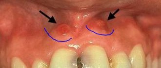
A lump on an area of the gum where there is no tooth yet: what to do in this case?
A couple of weeks before the tooth erupts, a cyst may appear on the gum, looking like a lump filled with a clear or bluish liquid. Such bumps are by no means common: dentists do not consider such formations to be a pathology requiring treatment. In addition, such formations on the gums in no way indicate an inflammatory process.
According to statistics, a small percentage of children are affected by the appearance of such a cyst. The child may not even suspect that he has a lump in his mouth, because if he touches it, there is no pain. However, a dental examination is still necessary, because the inflammatory process may still begin, in which case intervention will be required. The presence of inflammation is indicated by such signs as increased body temperature, pain when touching the lump, and swelling of the mucous membrane.
Most parents do not like the fact that their child has a lump in his mouth: in this case, you can ask the dentist to make an incision under anesthesia, as a result of which the fluid inside the cyst will come out. As a rule, when part of the cyst is removed, the crown of the tooth about to erupt is visible.
Treatment of inflammation
Removal of a lump with pus on a child’s gum should be started immediately. Only a dentist can help the baby.
IMPORTANT! Self-medication, as well as piercing and squeezing are strictly prohibited! Such an intervention will lead to serious complications.
When an abscess forms over a baby's baby tooth, the doctor most often removes it. Such measures are necessary to exclude the possibility of destruction of the rudiments of the root. If a child has a lump on his gum next to a permanent tooth, treatment begins with a thorough examination. Sometimes the dentist will send you for an x-ray to assess the extent of the damage. Removal of a permanent tooth is carried out in extreme cases.
How to help a child at home with suppuration?
If suppuration and the formation of a lump on the gum appear, it is recommended to contact a dentist as soon as possible, but, unfortunately, for various reasons this is not always possible.
If an adult can endure pain (although, of course, situations are different), then it is much more difficult for a child to endure painful sensations, and it is not easy for parents to watch their child suffer. There are ways to alleviate the baby's condition. Naturally, they will not replace full-fledged treatment, so you can resort to them only in situations where going to the doctor is impossible right now.
These include:
- take as much warm liquid as possible, which reduces intoxication of the body;
- eat liquid food (moderately warm, but not hot!), as it injures already damaged gums;
- If the pain becomes severe, you can use painkillers. For example, Nurofen or Paracetamol - the exact dosage depends on the age of the child, so before use you should carefully read the instructions for the drug;
- to reduce swelling, you can resort to cold - any frozen product from the freezer compartment of the refrigerator is wrapped in a soft cloth and applied to the cheek;
- use rinsing solutions that temporarily relieve pain - chamomile decoction or the drug chlorhexidine, which reduces irritation.
Preventive measures to prevent the formation of lumps on the gums
To reduce the likelihood of an abscess to a minimum, a child from childhood should be taught to carefully observe oral hygiene.
The list of the most effective measures includes:
- Brushing your child's teeth twice a day. First, parents should brush their child’s teeth themselves, showing how to do it correctly, gradually giving the child more independence in this matter;
- rinsing your mouth after every meal;
- minimizing sweets in the diet - it is important to ensure that the child keeps caramel and candies in his mouth as little as possible;
- Do not introduce your child to chewing gum (the product is especially harmful if it contains sugar).