53600
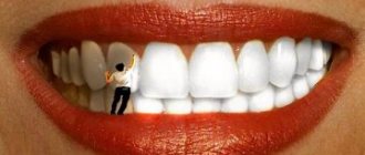
Crooked, unhealthy teeth can ruin the appearance of an otherwise attractive person. Oral health is important not only for our attractiveness, because the main function of teeth is to grind food. The condition of the stomach and intestines depends on this, which directly affects health and life expectancy .
How is the bite formed?
The process of bite formation is long - from birth to adolescence. There are 5 periods.
- Initial (up to 6 months): no teeth, the upper jaw is larger in size than the lower jaw. At this time, an active feeding process is extremely important for the proper development of the jaw apparatus.
- Emerging temporary bite (6 months - 3 years): baby teeth and a bite appear, but it is still temporary. This stage is accompanied by inflammation of the gums, a rise in temperature, often grinding of teeth (bruxism), and emerging teeth may be crooked. It is important to maintain hygiene without taking any orthodontic measures.
- Formed temporary occlusion (3 - 6 years): baby teeth have erupted and can close in different ways, jaw growth continues. This is the time of active use of teeth. Characteristic wear of temporary teeth is considered normal before the upcoming change.
- Changeable dentition (6-12 years): jaw growth, loss of baby teeth and appearance of permanent teeth. Often new teeth, especially the lower ones, erupt unevenly. During this period, it is necessary to decide on a plan for further correction of the bite.
- Permanent dentition (12 - 15 years): replacement of milk teeth with permanent ones, all 28 teeth are visible, no wisdom teeth. The period is most favorable for orthodontic treatment.
In order for the bite to form correctly, there must be careful attention to teething, oral hygiene, and a timely plan to correct emerging problems.
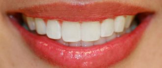
Correct teeth bite
An ideal orthognathic bite implies the presence of the following features:
- The upper dental arch is slightly inclined forward and has the shape of a semi-ellipse, the lower, in the shape of a parabola, is slightly backward.
- When the jaws close, each upper tooth contacts the opposite tooth below.
- There are no obvious gaps between the teeth.
- The upper teeth overlap the lower teeth by about a third of their height.
Despite the fact that there are the above parameters for a perfect smile, the question “What should be the bite of the teeth?” cannot be answered unambiguously. In addition to the orthognathic bite, modern orthodontics allows for several options for the normal structure of the dentition and the location of the jaws.
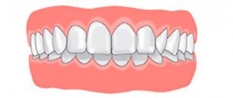
Orthognathic
The upper row of teeth overlaps the lower one by 30% of the height, there are no gaps between the teeth or spaces between the rows.
Biprognathic
The dentition is slightly directed towards the vestibule of the mouth.
Progenic
The lower jaw moves forward slightly.
Straight
When the jaws close, the upper teeth come into contact with the lower cutting edges.
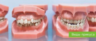
Varieties
| View | Short description |
| Orthognathic (1) | The bite is perfect, there are no gaps when both dentitions are closed, the incisors are straight |
| Straight (2) | The upper teeth are not able to overlap the lower row, but are closed only by the cutting edges. May have a negative character, fraught with abrasion of the front dental elements through an increase in the load on their cutting part |
| Progenic (3) | This bite can be considered as incorrect, since it is characterized by a slight forward movement of the lower jaw |
| Biprognathic (4) | The rows have an inclination towards the lips, which is noticeable when viewing the jaw from the side |
| Opisthognathic | Visible inclined inwards the oral cavity. In this case, the front teeth look almost perfectly straight |
Types of human bite
Attention! All types of correct dental closure can provide an aesthetic appearance of a smile and healthy functioning of the jaws, but based on statistics, there is an average of only 10-20% of the population with ideal teeth alignment.
Perfect closure (opistognathic appearance)
Malocclusion of teeth
Look at your reflection in the mirror. If you notice an excessively protruding upper or lower lip, teeth that “overlap” each other, or gaps between the dentition when the jaws are closed, this is a clear reason to consult a specialist. Descriptions of dental anomalies and photographs of teeth with malocclusion pathologies are presented in the table below.
Distal (prognathic)
In a distal bite, the upper jaw is more developed than the lower jaw.
Mesial (medial)
With a mesial bite, the lower jaw is pushed forward.
Cross
In a crossbite, the rows of teeth intersect in the manner of scissors when the jaws close.
Deep
In a deep bite, the upper teeth significantly overlap the lower teeth.
Open
The presence of pronounced gaps between the dentition when the jaws are closed indicates an open bite.
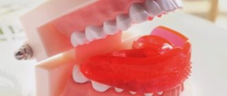
Bite in the absence of teeth
A person without teeth has problems with the functioning of the temporomandibular joints, facial aesthetics deteriorate, and wrinkles appear due to loss of skin tone. To avoid unpleasant consequences, it is necessary to restore the bite using a complete denture made using a special technique.
Bite restoration with complete dentures
- The central ratio of the jaws is determined - the position of the lower jaw in relation to the upper in three planes: vertical, sagittal and transversal. The role of the dentition is played by wax rollers.
- Measurements are taken using a device consisting of an external facebow-ruler and an intraoral plate with a flat frontal part and curved distal parts.
- The plaster model marks the boundaries of the future structure, the line of the middle of the alveolar process, the tubercles of the upper and lower jaw, and the midline.
- Artificial teeth are placed in such a way that when you smile, the part of the prosthesis that imitates the gum is not visible.
Scientific rationale for the modeling sequence of aesthetic restorations
The anatomy of human teeth, their classical features and forms are considered to be well studied for a long time [2, 4]. When using dental materials from previous generations, this knowledge was absolutely satisfactory to dentists. However, the appearance on the market of dental services of ceramics and composites, providing high aesthetic qualities of structures, showed limited information about the fine details of the tooth structure [3, 5]. On the other hand, the insufficiency and lack of demand for medical knowledge in the field of tooth morphology leads to the fact that even classical signs are not always reproduced in the design [7]. The result is more or less pronounced differences in the shape and relief of the restoration from the natural appearance of the tooth.
We studied the aesthetic characteristics of permanent teeth in 350 people using odontoscopy and odontometry.
The need to bite, tear and grind food contributed to the formation of the main groups of teeth: incisors, canines, molars, premolars differ in shape, size, number of roots and are located in the dental arches. The upper arch is usually more rounded, the lower one is slightly compressed in the transverse direction. The anatomical crown, the border of which runs along the neck of the tooth, and the clinical crown, which is located above the gingival edge immediately after the tooth erupts, are identical in height. With age, the anatomical crown shortens as a result of tooth wear. The clinical crown also decreases, but may also lengthen due to gum recession (Fig. 1).
Rice. 1a. Changing the size of the central incisor. Rice. 1b. Changing the size of the central incisor.
Based on general characteristics, teeth were distinguished according to the signs of belonging to the right or left side, which relate to the curvature of the crowns, the ratio of the distal and mesial angles of the crown, and the inclination of the roots.
A sign of crown curvature is a greater convexity of the vestibular part of the crown, located near its mesial edge, and a gentle slope of the distal one. It is more clearly defined when examining the tooth from the occlusal surface and is expressed in 71% of central incisors (Fig. 2).
Rice. 2a. The sign of crown curvature is determined. Rice. 2b. The sign of crown curvature is determined.
In 23% of cases, the convexity is shifted to the distal side, in 6% of teeth the sign of curvature is not detected. The sign of the crown angle characterizes the situation when the mesial angles, composed of the mesial surface and the cutting edge (masticatory surface), are sharper than the distal ones (Fig. 3).
Rice. 3a. There is a pronounced sign of distal deviation of the periodontal dome. Rice. 3b. There is a pronounced sign of distal deviation of the periodontal dome.
This inequality of angles is observed in 85% of central incisors. In molars, the sign of angle is due to more massive mesial cusps. The sign of root inclination means that the root or its apex is curved in the distal direction in relation to the longitudinal axis of the tooth. In the oral cavity, the sign manifests itself as a distal displacement of the apex of the dentogingival dome.
Based on their appearance, teeth were classified into separate geometric shapes (rectangle, triangle, oval), which are characterized by their own characteristics (Fig. 4).
Rice. 4. Basic geometric shapes of permanent teeth.
Rectangular incisors in the absence of abrasion are detected in 55% of cases: the transverse dimensions of the vestibular surface in the gingival, equatorial region and at the cutting edge are close in value, as a result of which the proximal surfaces are almost parallel (Fig. 5).
Rice. 5a. Rectangular cutters. Rice. 5 B. Rectangular cutters.
The angle sign may be weakly expressed. The length of contact with adjacent teeth is significant: in 72% of cases, contact between teeth begins at the apex of the gingival papilla and ends at the cutting edge. With a triangular crown shape, which is characteristic of 39% of intact incisors, the transverse dimensions of the vestibular surface increase from the neck to the cutting edge (Fig. 6).
Rice. 6. Triangular shaped teeth.
The crown angle sign is well expressed in 88% of cases. The length of contact with adjacent teeth is insignificant. The oval shape of the crown occurs in 7% of cases, having similar values of transverse dimensions in the cervical region and near the cutting edge. The largest diameter is at the equator. The lateral surfaces are presented in the form of convex arcs. The corners at the cutting edge are smoothed. Contacts with neighboring teeth are point-like.
The vestibular surfaces of canines, premolars and molars have a more complex geometric shape due to protruding cusps.
According to a survey of young people, in 53% of cases the upper dental arch has an oval shape, in 42% - round, and in these arches there are teeth of any geometric shape: rectangular, triangular, oval. However, the rare rectangular and triangular dental arches (2.6% of cases) are characterized by rectangular and triangular teeth, respectively.
The visual perception of the geometric shape of a tooth can be influenced by the individual characteristics of the dentogingival contour. The dome-shaped gingival margin, characteristic of 56% of incisors, resembles a wedge or triangle in shape and gives the tooth a triangular shape. 39% of incisors have a rounded periodontal edge, 5% of teeth have a flattened, almost straight gingival contour. The shape of the incisal edge also reflects the individual characteristics of the tooth (Fig. 7).
Rice. 7a. The relief of the vestibular surface and scalloped cutting edge are pronounced. Rice. 7b. The relief of the vestibular surface and scalloped cutting edge are pronounced.
Immediately after eruption, the cutting edge is serrated. Then abrasion facets appear. An uneven (convex or concave) surface can be explained by the peculiarity of contact with antagonist teeth.
The relief of the vestibular surface of the tooth is of great aesthetic importance: vertical ridges, bridges, convexities, depressions, and areas. A single ridge is usually characteristic of the middle part of the vestibular surface. If there are two ridges, the most common ones are mesial and distal. The three enamel ridges are usually located mesially, medially and distally (Fig. 8).
Rice. 8a. Vertical ridges on the vestibular surface of the incisors. Rice. 8b. Vertical ridges on the vestibular surface of the incisors. Rice. 8th century Vertical ridges on the vestibular surface of the incisors.
Physiological wear of teeth leads to the formation of a smooth vestibular surface.
Modeling of aesthetic restorations should pursue the goal of reproducing in detail the natural classical, as well as individual characteristics of permanent teeth. However, an analysis of the quality of 145 restorations that were created by reproducing the characteristics of a symmetrical tooth showed differences between the artificial structure and the intact tooth in almost half of the cases [1, 6]. In most cases, this concerns the restoration of rectangular-shaped features where triangular or oval ones are required. In some cases, there is a recreation of oval (rectangular) tooth shapes in a triangular jaw.
The absence of signs of tooth affiliation was found in an average of 30% of restorations. Of these, 77.14% were modeled without taking into account the sign of crown curvature. Many patients had differences in height, width, or asymmetry in the size of individual sections of aesthetic structures (Fig. 9).
Rice. 9a. A veneer differs in size from an intact symmetrical tooth. Rice. 9b. A veneer differs in size from an intact symmetrical tooth. Rice. 9th century A veneer differs in size from an intact symmetrical tooth.
Among the age-related features characterizing the surface relief, there was practically no re-creation of perikymatia on structures in adolescents. The lengthening of the clinical crown of the tooth due to gum recession was not taken into account: the height increased significantly due to the filling material. No gum-mimicking polymers were used.
Individual features of the relief were absent in most restorations (vertical ridges, relief cutting edge).
At the filling stage, insufficient or excessive use of material changed the shape, size, and characteristics of the tooth. Thus, excess filling material on the vestibular surface caused an unnatural convexity of the crown (Fig. 9).
The lack of composite material led to the loss of proper anatomical volume, a decrease in the overall height and proximal slopes, and flattening of the crown. Single structures stood out as a result of asymmetry - differences from a similar tooth on the opposite side. Paired or multiple restorations looked unnatural if a single geometric type of crowns was not maintained, signs of tooth affiliation, and individual characteristics were not observed.
The results of scientific research, including analysis of the aesthetic characteristics of teeth and the quality of existing restorations, made it possible to develop recommendations for step-by-step planning of the sizes and shapes of aesthetic restorations.
The algorithm for planning the size and shape of the structure represents a certain sequence of measuring and describing specific anatomical structures of the tooth (Scheme 1).
Scheme No. 1. Restoration sequence algorithm.
First, it is necessary to conduct a comparative assessment of the size of the clinical and anatomical crown of the tooth. The presence of wear areas in the area of the cutting edge indicates a decrease in the height of the clinical crown. Gingival recession with exposure of the neck and root of the tooth is a sign of an increase in the vertical size of the clinical crown (Fig. 1).
The measurement (odontometry) is made with a micrometer (Fig. 10).
Rice. 10a. Measuring the transverse dimensions of a tooth. Rice. 10b. Measuring the transverse dimensions of a tooth. Rice. 10th century Measuring the transverse dimensions of a tooth.
The height of the clinical crown of the central and lateral incisors is assessed by the distance from the cutting edge to the marginal level of the gums along the vertical midline. Similar dimensions of the canine and premolars are measured along the midline from the apex of the cusp to the marginal level of the gingiva. The height of the molar crown is the distance from the gum level to the top of the most prominent cusp.
The shape of the tooth crown is considered rectangular if the lateral edges of the vestibular surface are parallel, and square if the height and width are equal. Mesiodistal dimensions in the neck of any tooth are measured by the distance between two points of opposite proximal surfaces at the level of the apexes of the interdental papillae. The horizontal parameters of the incisors in the equatorial region are determined at the level of the middle third of the crown height. Similar values of the central and lateral incisors in the area of the cutting edge are assessed by the distance between the protruding points of the mesial and distal edges of the crown. The mesiodistal dimensions of the canines and premolars are measured between the lateral sections of the crown located at the greatest distance. The transverse parameters of molars in the equatorial region are assessed as the distance between the most convex sections of the proximal surfaces.
Visual assessment and measurement results make it possible to describe the geometric shape of the tooth crown based on the relative position of the lateral surfaces.
The shape of the tooth crown is considered rectangular when the lateral edges of the vestibular surface are parallel (square when the height and width are equal). The triangular shape of the tooth is characterized by its maximum horizontal size at the cutting edge. A crown is considered oval in shape when the lateral surfaces have a rounded outline with the largest horizontal dimension in the area of the middle third of the tooth.
Next, the severity of the signs of belonging to the side of the teeth is assessed. The sign of the crown angle is recorded in the case of a predominance of the distal angle of the vestibular surface over the mesial one (Fig. 11).
Rice. 11. No signs of crown angle.
The sign of crown curvature is considered positive if the convexity of the vestibular surface is located closer to the mesial edge. In some cases, there is no sign of crown curvature (Fig. 12).
Rice. 12. Smooth vestibular surface of the central incisors.
A sign of tooth root deviation is noted in the map when there is a distal displacement of the apex of the dentogingival contour. The description of the individual characteristics of the tooth includes the surface topography, the shape of the gingival contour of the tooth, the shape of the cutting edge, and the length of contact with neighboring teeth. The type of relief of the vestibular surface of the incisors is determined by the presence or absence of vertical enamel ridges (Fig. 8).
The shape of the dentogingival contour is assessed by the upper border of the tooth crown, which starts from the top of one interdental papilla, then goes along the edge of the gums and ends at the top of the other interdental papilla (Fig. 13).
Rice. 13. Contours of the gingival dome.
The dentogingival contour can be round, dome-shaped or flat. It is necessary to plan the length of proximal contacts between teeth in such a way that there is enough space for the interdental papilla. The planning stage is completed by choosing the shape of the cutting edge of the teeth (Fig. 14).
Rice. 14. Planning the area of the cutting edge of the central incisors.
The anatomical features of chewing teeth require a careful assessment of the relationship of the cusps on the occlusal surface, and their shape can change significantly due to abrasion. Modeling restorations is a complex and demanding process.
The first stage is the formation of the base of the restoration, which includes the contours of the geometric shape of the dentin and mamelons at the cutting edge, with a clear designation of the lateral and lower boundaries of the dentinal layer (Fig. 15).
Rice. 15. Opaque base of restoration (diagram).
The second stage involves modeling the signs of teeth belonging to the side (signs of curvature, crown angle, deviation of the periodontal dome). The third stage is the reproduction of the individual characteristics of the tooth, including macro- and microrelief, the shape of the cutting edge and the gingival dome. The sequence of restoration of morphological elements corresponds to the sequence of odontoscopic examination and restoration planning. There is a gradual transition from recreating large details (the geometric shape of the vestibular surface) to reproducing medium-sized ones (signs of the angle and curvature of the crown), and then to modeling smaller elements (enamel ridges, teeth in the area of the cutting edge).
Using imaginary lines (two vertical and two horizontal), it is necessary to divide the vestibular surface of the tooth into segments, which will make it possible to more clearly determine the topographic position of a particular morphological element (Fig. 16).
Rice. 16. Conventional division of the vestibular surfaces of teeth into segments.
For example, in the upper tier the dentogingival contour is modeled, as well as a sign of tooth root deviation in the form of a displacement of the top of the gingival dome to the distal side. Predominantly in the middle part of the tooth, a sign of crown curvature is created through the formation of the largest convexity in the mesial region. Individual features of the cutting edge, expressed by varying degrees of transparency and mamelons, are located in the lower tier (Fig. 17).
Rice. 17a. Modeling the basis of the restoration. Rice. 17b. Modeling the incisal edge of the restoration.
Contacts between teeth, proximal slopes, as well as vertical enamel ridges are modeled, respectively, in the mesial and distal parts throughout the middle and lower tier. In the mesial region there may be a median enamel ridge (Fig. 18).
Rice. 18. Modeling of the individual relief of the vestibular surface.
Restoration of large morphological details must be carried out using opaque shades of the composite. The main guideline when working with opaque filling material is the border of the transparent tooth enamel. So, if the enamel evenly covers the entire surface, then for the corresponding layer of filling material it is necessary to leave no more than 0.5 mm of free space around the entire perimeter of the tooth. If the transparent strip is located in the area of the cutting edge, then the opaque is not brought to the lower border by 1.0 mm. In the case where a layer of transparent enamel is observed on the proximal edges, 1.0-1.5 mm should be left for the enamel layer. With severe abrasion, the dentinal layer is formed over the entire height of the tooth crown up to the cutting edge.
During the work, it is necessary to ensure that the thickness of the opaque layers does not exceed the amount of lost dentin. It is important that the opaque layer is not longer or shorter than on a symmetrical tooth and does not interfere with the type of transparency.
In accordance with the algorithm for planning the anatomical shape and relief, developed by the staff of the Department of Therapeutic Dentistry of BelMAPO based on their own odontometric and odontoscopic observations, 166 structures were made.
The results of visual examination of restorations modeled using the technique of preliminary planning of their morphology showed a significant improvement in quality compared to the previous period (see table).
In most cases, the optimal anatomical shape of the teeth was recreated, which corresponded to group affiliation, age and individual characteristics. The frequency of occurrence of signs of angle, crown curvature, and deviation of the gingival dome on restorations has increased almost 10 times. The vertical and horizontal dimensions of the created structures did not differ significantly from intact symmetrical teeth. Thanks to this, a single geometric type was maintained, characteristic of this group of teeth. Age-related features were simulated, including root exposure and age-related abrasion. In the vast majority of young people, enamel ridges and grooves on the vestibular surface were modeled. The alternation of highlights and shadows, due to the presence of macrorelief, emphasized the naturalness of the created restoration (Fig. 19).
Rice. 19a. Planning of aesthetic restorations. Rice. 19b. Modeling of aesthetic restorations.
Thus, the results obtained made it possible to identify errors made in the manufacture of composite restorations regarding sizes, shapes, and surface topography. Inaccuracies were allowed at the planning stage if signs associated with the group affiliation of the tooth, its age or individual characteristics were not recorded.
The lack of a strict sequence of actions, and often the exclusion of the very stage of planning the anatomical shape, caused a significant violation of the aesthetics of restorations. In turn, the use of the proposed algorithm of actions for assessing and reproducing the characteristics of tooth morphology in conjunction with the skills of odontoscopic examination helps to significantly reduce the number of errors and complications during the restoration of the anatomical shape of the restoration.
LITERATURE
- Danilova D.V. // Sat. Proceedings of the 5th Congress of Dentists of the Republic of Belarus. - Brest, 2004. - pp. 138-140.
- Dmitrienko S.V., Ivanov L.P., Krayushkin A.I., Pozharitskaya M.M. Practical guide to dental modeling. - M., 2001. - 239 p.
- Leonova L. E., Zheleznitskikh M. V., Maksimovskaya L. N. // Klin. dentistry. - 2002, No. 1. - P. 8-11.
- Lomiashvili L.M. // Institute of Dentistry. - 2003, No. 2. - P. 6-31.
- Lutskaya I.K. // Modern. dentistry. - 2003, No. 1. - P. 30-37.
- Lutskaya I.K., Danilova D.V. // Sovrem. dentistry. - 2004, No. 1. - P. 22-25.
- Novak I.V., Danilova D.V. // Sat. works of young scientists. - Mn., 2005. - P. 49-51.
Bite after wisdom tooth removal
Some doctors are convinced that wisdom teeth are an atavism and must be gotten rid of. In ancient times, they were used to chew tough foods, such as raw meat. With the development of civilization, the human diet changed, his jaw began to gradually decrease - and the “eights” turned out to be superfluous. Most often, before starting to correct the bite, wisdom teeth are removed. This frees up space for the correct formation of rows, and the result of teeth alignment will be much better.
What affects the bite of teeth?
The negative impact of defects in the dental system on the condition of the entire organism has been repeatedly proven by scientists. The list of identified health risks is impressive even for ardent optimists.
- Due to improper distribution of the chewing load, tooth enamel wears off faster, teeth begin to react painfully to cold and hot, their necks become exposed, and noticeable gaps form between the teeth.
- Diseases of the temporomandibular joints occur, accompanied by headaches and a characteristic clicking sound when the jaws move.
- The aesthetics of the face suffers: its proportions change, wrinkles form, teeth become crooked, and the smile becomes unattractive.
- It becomes difficult to bite food, chewing functions are disrupted, causing diseases of the gastrointestinal tract.
- Speech defects appear, facial expressions are distorted, and self-esteem decreases.
- Crowding and crooked teeth cause accelerated tooth decay due to frequent chipping and carious lesions.
- The oral mucosa is injured - cheeks, gums, tongue, and the tissues of the oral cavity become inflamed.
- The quality of breathing deteriorates, which leads to diseases of the nasopharynx, trachea, and hearing aid.
Features of correct bite in a child
If we consider this problem in terms of age, then the characteristics of the correct bite at any period of a person’s life will be similar. The only exceptions we can put aside are children, in whom only after changing their baby teeth can we say with 100% probability that they have a correct or incorrect bite.
In fact, an adult bite is no different from a child’s bite from an anatomical point of view.
The correct arrangement of the dentition is comparable at any age, as is the structure of the jaw. But still, it is worth paying attention to the state of the bite of children more often due to the rapid growth of all parts of the chewing apparatus and the gradual replacement of milk teeth with primary ones.
The bite is formed from birth, and the process lasts up to 15 years. Development occurs in stages, from the moment the first primary incisors appear. After changing all the elements, the bite becomes permanent. Its formation is directly influenced by both hereditary and external factors.
In childhood, it is important to pay increased attention to the prevention of the development of correct bite, so that the jaw bones and teeth alignment correspond to the physiological norm.
For proper development of the bite, it is necessary to: keep the child breastfed from birth; Try not to accustom your baby to a pacifier and pacifier. If this is difficult to achieve, you need to use rubber as little as possible; prevent the development of bad habits such as sucking fingers and toys. During sleep, the child's mouth should be closed and the head should not be thrown back. From the beginning of the growth of baby teeth, it is necessary to include solid foods in the diet.
You should consider visiting an orthodontist after teething.
If necessary, the specialist will prescribe a comprehensive examination and radiography to identify the causes of dental pathologies. Then you should get advice from doctors in related fields. Specialists will determine contraindications, the likelihood of complications and assess the condition of the baby’s entire body.
Based on the data obtained, the orthodontist will draw up a treatment plan for approximately 2 years with monthly clinic visits.
There are several risk factors that may indicate that your child needs to correct his bite: deformation of the spinal column, a genetic predisposition to pathologies of the development of the dental system, difficulty breathing through the nose, diseases of the central nervous system and bad habits (grinding teeth, pushing the lower jaw forward or swallowing, clicking jaw joints, etc.). But only a doctor can give an accurate conclusion on the issue of correcting the bite.
Therefore, it is best to contact myofunctional centers, which unite orthopedists, pediatric dentists, orthodontists and osteopaths. The joint work of specialists guarantees accurate diagnosis and successful resolution of the issue.
In modern dentistry, there are various devices for correcting malocclusion in children, designed to achieve the following goals:
- prevention of bad habits;
- expansion of the jaw arch;
- correction of speech defects;
- normalization of the functioning of the masticatory muscles;
- tongue position control.
Treatment of dental malocclusion
How to change the bite of teeth? Pathologies of the dental system can only be eliminated through orthodontic treatment. An exception is the correction of a low bite, when the height of the teeth decreases with age due to their wear. In such cases, the bite is corrected by dental prosthetics with crowns or veneers, as well as by implantation. Other anomalies of teeth and bite can be removed at any age with special orthodontic structures or through surgical intervention.
However, maxillofacial operations are performed only in case of serious deviations from the norm. Usually, to correct dental malocclusion, doctors suggest correction with braces or aligners.
Now that you know about all the health risks that can cause pathologies of the dental system, and how to correct your dental bite, all that remains is to take specific actions. Even if at first glance it seems that your teeth are straight, make an appointment with an orthodontist. Diagnosing the condition of the dentition will help maintain the health of other organs and systems. And timely orthodontic treatment will make your smile more attractive and significantly reduce the number of future visits to the dentist.
A perfect smile with veneers and lumineers
Restoration of the smile area using veneers and lumineers is suitable if you have absolutely healthy, but not very beautiful teeth. Indications for the use of veneers are:
- Chips, microcracks in teeth;
- Darkening of the enamel that cannot be eliminated by bleaching;
- Noticeable stains on the surface of the teeth;
- High-quality fillings, but darkened over time;
- Large interdental spaces;
- Slight crowding of teeth;
- Deviation of the position of the teeth from the axis;
Microprostheses are fixed on the outer surface of the teeth to change the visual perception of the dentition and create a beautiful smile. The color of the veneer is chosen so that it matches your own teeth. In this case, the presence of veneers is invisible. People around you simply notice your beautiful smile. They are not suitable for restoring severely damaged teeth. In such cases, it is better to choose ceramic crowns rather than veneers.
Installation of veneers and lumineers requires grinding of teeth. Since without it, the lining will protrude above the teeth due to its own thickness. Your teeth won't look beautiful and your smile won't look perfect. The amount of grinding depends on the individual characteristics of the patient and his teeth. In some cases, it is indeed possible to completely do without turning, but the doctor will be able to tell how microprosthetics will work in your case only after an examination.
Dental restoration with lumineers and veneers takes time, since microprostheses are made individually. The entire process of creating a Hollywood smile takes place in several stages:
- Preliminary inspection. The task of this stage is to identify indications and contraindications for the installation of veneers;
- Sanitation of the oral cavity. Plaque and stone are removed from the surface of the teeth;
- Grinding of teeth for veneers. This step is not always required;
- Preparation of casts. After the teeth are completely prepared for restoration, the doctor takes impressions to make veneers;
- Manufacturing. Veneers are made in a dental laboratory using CAD/CAM technology. Lumineers must be ordered from the manufacturer;
- Installation. Once the veneers are ready, they are fixed to the teeth with dental adhesive.
You can see the result of dental restoration and decide whether it is necessary even at the preparation stage. To do this, the doctor will make a 3D computer model of your new beautiful smile.