25.11.2019
“Bite” refers to the type of closure of teeth. Normally, the upper teeth should slightly overlap the lower teeth. In this case, the chewing function is performed most fully. Due to impaired jaw growth, the child experiences bite deviations: for example, the upper jaw may be overdeveloped or, conversely, underdeveloped, and the teeth may not be in their proper places.
Unfortunately, parents rarely take any effective measures to correct malocclusion in children, believing that “everything will go away on its own.” By doing this, they make a huge mistake, since a child with malocclusion suffers from digestive and swallowing disorders. Sometimes cosmetic inconveniences lead to the development of an inferiority complex; children withdraw into themselves, are reluctant to communicate with peers, and become capricious and irritable.
Causes of malocclusion in children
Among the many reasons for the formation of malocclusion, we name the following:
- Features of the structure of the jaws, which are most often inherited;
- Disturbances in the growth of jaw bones;
- Having bad habits (this includes thumb sucking);
- Using a pacifier, as well as using a pacifier that has a large hole. Parents should be aware that the hole in the nipple must be made as small as possible so that milk flows out of the bottle only if the child makes the correct sucking movements;
- Diseases of the ear, nose and throat;
- Sometimes incorrect posture also contributes to the formation of malocclusion in children;
- Prolonged stay of the child on artificial feeding;
- Keeping the child in one position while sleeping.
Types and differential diagnosis
Like most diseases, periodontitis can occur in acute or chronic form. Sluggish periodontal inflammation is divided into three types:
Granulating
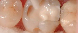
Depending on the type of infection that affects the tooth tissue, and on the specific immune response of the body, various kinds of altered tissues are formed in the periodontal cavity. With chronic periodontitis of this type, granulating tissue appears. It looks like a loose red formation near the top of the tooth root, which quickly grows and thereby destroys the bone tissue of the jaw. Of all three types of chronic inflammation, granulating periodontitis is considered the most active and pronounced. Its characteristic manifestation is the formation of fistulas, which can keep the tooth in a latent form of the disease for a long time. Connective tissue can not only affect the plate of the alveolar process (jaw), but also spread even to the skin. Therefore, advanced cases of periodontitis are much more difficult to treat and can affect large areas of tissue, including adjacent teeth. An X-ray image allows differentiation of granulating periodontitis from other types of periodontal disease. It clearly shows darkening in the area of the apex of the tooth root with circular outlines of tissue or in the form of flames. At the same time, the image does not show sharp boundaries, as in some other classifications of periodontitis. These dark spots indicate that the bone tissue has dissolved as a result of inflammation and has been replaced by connective tissue.
Fibrous
This form of the disease is quite rare and is accompanied by an asymptomatic chronic course. As a result of sluggish periodontitis, connective fibrous tissue forms around the tooth root. It is denser and is visible on an x-ray as a periodontal tooth gap, more widened than usual. The result of the appearance of fibrous tissue can be:
- suffering from acute periodontitis, which was successfully treated;
- poor-quality filling of tooth root canals;
- lack of treatment for granulating periodontal inflammation.
If the disease has entered the stage of remission (quiescence), and the patient does not complain about the causative tooth, then fibrous periodontitis cannot be treated. However, in cases of exacerbation, therapy in the form of refilling the root canals of the tooth is possible.
Granulomatous
If granulating periodontitis does not have clear outlines on an x-ray, then granulomatous periodontitis is characterized by the presence of a capsule with purulent contents. It occurs at the apex of the tooth root and gradually grows, affecting the bone tissue.
There are three degrees of development of periodontal abscess:
- Granuloma. The size of the capsule with this diagnosis reaches 0.5 cm in diameter.
- Cystogranuloma. It is diagnosed when the dense membrane reaches 1 cm.
- Cyst. More than one centimeter in diameter suggests the cystic development of periodontitis.
An increase in granuloma indicates that the inflammatory process does not find a way out and continues to dissolve the bone tissue near the tooth. In this case, a whole chain of pathological processes occurs in the human body. Sometimes particularly advanced cases showed a cyst of up to 4-5 cm in the tissue, which significantly impaired the integrity of the lower jaw, increasing the risk of its fracture.
On the other hand, the accumulation of serous fluid in the tooth area regularly causes additional intoxication to the entire body, which can be accompanied by periods of weakness, fatigue, low-grade fever and other symptoms of periodontitis.
Among all forms of chronic
Granulomatous periodontitis is considered to be the most sluggish. The fibrous tissue of the capsule serves as a protective barrier that prevents pathogenic bacteria from entering the blood. Therefore, the liquid in the capsule can remain in the tooth tissue for years without showing itself. Depending on their location, granulomas can be located either immediately under the root apex or in the tissues of the lateral cavity.
Classification of malocclusion in children
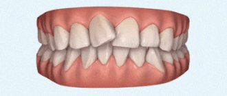
Normal types of occlusion in children are orthognathic, progenic, direct, biprognathic. Orthognathic occlusion is ideal. In this case, the lower and upper teeth are in perfect contact. A progenic bite is characterized by the fact that at the moment of closure of the teeth, the front teeth protrude somewhat forward. In a straight bite, the upper incisors meet the lower cutting edges. Biprognathic occlusion is characterized by the fact that the lower incisors are slightly inclined towards the vestibule of the mouth.
Among the pathological types of bite, the following types are distinguished:
- Prognathia (or distal bite). In this case, the upper jaw sharply protrudes forward.
- Mesial occlusion, or progenia. In this case, the lower jaw protrudes forward.
- With a deep bite, the upper incisors overlap the lower ones by more than 50 percent.
- With a crossbite, the normal relationship of the dental arches is disrupted. Therefore, the teeth begin to move sideways relative to each other.
- An open bite is characterized by non-occlusion of the lower and upper incisors. There are its varieties. So, with an open anterior bite, the incisors do not close, and with an open lateral bite, the molars do not close.
Periodontitis by location
There are two main divisions in the classification of periodontitis based on the location of the disease: apical and marginal. The first option (apical) is also apical, based near the apex of the tooth root. This type is the most common, since periodontal infection in most cases occurs through a descending channel: from caries to inflammation of the pulp, and then through the root canal the infection descends into the periodontal tissue. This process most often becomes chronic, since the protective mechanisms of the periodontium are much more powerful than those of the pulp. Therefore, the infection can enter the deep tissues of the tooth for years without causing signs of disease.
Marginal chronic periodontitis is located on the lateral walls of the tooth root and has the etiology of microtraumas.
Periodontitis, the treatment stages of which are a complex, complex process, requires immediate attention to a dental clinic. It is safe to say that such a diagnosis requires highly qualified specialists with extensive experience in solving such problems. Dentists of the LeaderStom network of clinics are rightfully considered the best in this area of dental therapy. The latest technologies, progressive techniques and extensive practice allow them to cope with the most complex, advanced cases.
Signs of malocclusion
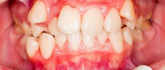
Often, parents do not need to carefully examine the child’s oral cavity to notice that he has an incorrect bite, since its signs are very obvious. In such children, the jaws move incorrectly (forward or backward), the dentition is uneven, and in some areas of the jaw the teeth are very crowded. The so-called hyperdentia, that is, the presence of supernumerary teeth, is often noted.
Another important sign of malocclusion in children is dystopia, that is, their incorrect position. Malocclusion is characterized by the presence of diastemas and three.
Children with malocclusion often complain of difficulty swallowing and chewing. They constantly bite their tongue and cheeks, which causes pain in the mouth, and they often develop stomatitis. Pain in the facial muscles and headaches are often noted.
You can suspect malocclusion in children based on a number of indirect signs. In such children, breathing through the mouth predominates. When swallowing, such children strain their lips and chin excessively. With significant deviations of the bite, facial asymmetry is noticeable. You may also notice a deviation in the timing of the eruption of permanent teeth.
In children with malocclusion, tooth enamel wears off much faster. Increased sensitivity of the tooth occurs, i.e. reaction of the tooth surface, when it can hurt from sour, salty, sweet, as well as cold and hot. Finally, improperly positioned teeth cause the development of ulcerative inflammation of the gums, bleeding and even damage to the jaw joints.
If children have an anterior open bite, biting food becomes significantly more difficult. An open lateral bite, on the contrary, makes chewing difficult. With a deep distal bite, the child's breathing is impaired.
And finally, all types of abnormal bite lead to impaired pronunciation of sounds, which results in diction defects. And since malocclusion is also an aesthetic problem, it is easy to notice that the child becomes withdrawn, sad, and sometimes irritable.
Consequences of malocclusion
If a malocclusion is suspected, children should be examined by a pediatric dentist, otolaryngologist, orthodontist, or speech therapist. In this case, the main role in determining malocclusion belongs, of course, to the orthodontist. To carry out calculations and accurately diagnose, a panoramic image is required, as well as an X-ray examination of the skull. In addition, a photograph of the child's face may be required.
Finally, to determine the exact position of the teeth in the oral cavity, an alginate impression is made, on the basis of which a plaster model of the dentition can be made. With its help you can see the exact location of the teeth. To clarify the diagnosis, computed tomography and myography may be required.
Pathological bite is not just an aesthetic problem. Any deviation from the normal position of the teeth leads to improper load on the teeth. In the process of chewing food, teeth begin to wear down excessively, which can lead to the development of bruxism, when a child grinds his teeth heavily during sleep. Because of this, the tooth begins to be affected by caries.
In addition, abnormal tooth mobility develops due to improper positioning of the teeth. If this condition is not treated, teeth will begin to fall out over time. Due to the pathological development of the temporomandibular joint, frequent headaches and clicking sounds appear when opening the mouth. Since the incorrect position of the teeth strains the joint and muscles, constant muscle pain occurs.
Aesthetic problems manifest themselves in the fact that the child’s facial profile changes. He has an ugly smile because his teeth are crooked. Sometimes the degree of tooth curvature is so pronounced that the child is completely ashamed to smile.
All these consequences together significantly worsen the child’s quality of life. So this pathology must be corrected in time.
Preventing protruding teeth
It is easier to prevent any problem than to treat it. Therefore, at the first signs of dental deformation, it is recommended to consult a doctor.
To achieve a positive result you must:
- 1. Correctly use braces, veneers, Herbst apparatus and other modifications of orthodontic devices. Do not remove them ahead of time and if you suspect a violation or breakdown, immediately contact a specialist.
- 2. Listen to your doctor's advice. The orthodontist sees the big picture, so he or she will know better which treatments to use and for how long. It is important to attend all appointments to ensure timely adjustments to the devices and monitor changes.
- 3. Provide a psychologically healthy climate. Stressful situations, poor sleep, excessive physical and mental stress negatively affect all organs and systems, including bone tissue that needs to be corrected.
- 4. Eat right. A balanced diet and the intake of vitamins and minerals have a positive effect on regeneration processes in bone tissue and help eliminate malocclusions.
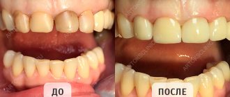
Beautiful teeth are not always a gift from nature. Most often, the painstaking work of an orthodontist is hidden behind a Hollywood smile. It is necessary to understand that the older the patient, the more advanced the problem, the longer it will take to correct the defect. After 25 years, processes in bone tissue slow down. Therefore, if there is such an opportunity, you need to start treatment in childhood or adolescence.
Also, we must not forget about the retention period. It lasts no less than the period of wearing braces. Consolidation of the result is necessary, because otherwise the ligaments will return the teeth to their original place. Thanks to the methods of modern medicine, the process of teeth straightening occurs relatively quickly and painlessly.
Treatment of malocclusion in children
The process of correcting malocclusion in children occurs much faster than in adults. But parents do not need to take advantage of this feature of children’s jaws, but start treatment as early as possible. The optimal period for starting bite correction is 5-6 years. Before correcting the bite, it is necessary to carry out a complete sanitation of the oral cavity and professional teeth cleaning. If necessary, some teeth are removed; the dentist will tell you which ones.
Today, there are many ways to professionally correct your bite. Let's look at some of them.
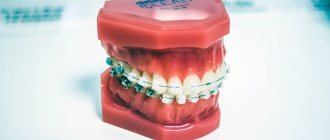
Braces
Braces (bracket systems) are devices that help correct an abnormal bite. Treatment with this method involves wearing a brace system for a long time, which helps the tooth or group of teeth return to their normal position in the socket.
The position of the teeth can be changed because they do not fuse with the jaw bones, but are only tightly fixed in the sockets. When constant pressure is applied to the wall of such a hole (of course, it will not be too great), then the bone tissue in such a place gradually resolves, and this causes the tooth to change position. In place of the connective tissue, new bone tissue gradually forms, which fixes the tooth in its new position.
Braces are a non-removable system. They are fixed to the tooth thanks to the base. The bracket groove contains the main element – the orthodontic arch. It is made of materials that are able to “remember” its position. An orthodontic arch has the shape of an anatomically correct dentition. The force exerted by the arch leads to the gradual alignment of the teeth in the dentition.
Straightening teeth with braces takes a long period of time, takes place over a long period of time and under the supervision of a doctor. Treatment in some cases can take from six months to three years, and sometimes longer. All this time, patients constantly visit the dentist and follow all his instructions. Only then will treatment with braces be effective.
Mouthguards
Mouth guards are special transparent plates made of high-quality silicone that help effectively get rid of uneven teeth. Their design allows the teeth to gradually move in the dentition. The aligners are worn all day and removed at night, as well as when eating and brushing your teeth. They have a mechanical effect on the teeth, closing the gap between them and straightening the jaw.
Like braces, mouthguards are worn for a long time. They can only be purchased at specialized dental clinics. An individual model of mouth guards is made for each person. They are not felt in the mouth and are beneficial if you follow all the doctor’s instructions when wearing them.
Caring for such devices is not difficult. At night, when removing the mouth guard, rinse it with clean boiled water. Boiling water cannot be used, as the silicone may lose its properties.
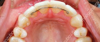
Dental plates
Plates are placed on teeth if minor malocclusions need to be corrected. They are very well adapted to be installed by small children. The period of wearing them in this case is much shorter than for adults.
Such plates can be removable or non-removable. Fixed plates are worn for approximately two years. Removable plates have a simpler design and are made of high-quality plastics. They are very easy to attach using hooks.
The good thing about removable plates is that they can be removed regularly. However, they are effective only for minor violations of the structure of the dentition.
Trainers
This is an elastic splint that easily adapts to any type of jaw. They, unlike braces, eliminate the cause of malocclusion. Due to its physiological effect on the teeth, it easily corrects the bite, and therefore is most suitable for young patients.
Trainers are unnoticeable and can be worn for a short time, say, an hour. They do not cause any physical discomfort.
Causes of protruding teeth
Most often, the two central teeth protrude forward due to the large grouping and strong pressure of the dentition. Also, the reason may be their initially incorrect position. But it is possible that there is a malocclusion after correction. Many people who wear braces experience a similar problem: protruding front teeth.
There are several causes of the pathology:
- 1. Premature cessation of treatment. External improvements do not always mean full recovery. When braces are removed prematurely, the central teeth gradually move forward.
- 2. Failure to comply with doctor's recommendations. Unauthorized cessation of alignment naturally leads to the return of bone tissue to its original place.
- 3. Incorrect installation of braces. It is possible that the arc tension is insufficient or certain anatomical features of the patient are ignored.
- 4. Rare visits to the supervising orthodontist. Throughout the entire correction period, it is important to make changes and also monitor the design of the braces, which can break without a person noticing.
5. The most common reason for protruding front teeth is ignoring the retention period. After the braces are removed, the treatment does not end, but continues. Every competent doctor knows how important it is to consolidate the result. A frivolous attitude and refusal to further consult an orthodontist will inevitably lead to the return of previous defects.