Sialadenosis is a non-inflammatory disease of the salivary glands, leading to enlargement and/or disruption of their function. The lesion is dystrophic in nature and is not associated with the appearance of tumors. Structural changes in gland tissue usually appear against the background of other systemic pathologies. Dentists quite often encounter this phenomenon: 1 case out of 10 pathologies of the salivary glands is represented by sialadenosis.
The disease usually occurs in middle-aged people. Since it often accompanies endocrine, allergic, dysmetabolic and other systemic diseases, the approach to treatment should be carried out not only from the dental side.
Types of sialadenoses
Based on the location of pathological changes, the following types of sialadenoses are distinguished:
- interstitial;
- parenchymal;
- ductal
Pathological areas can be located both in the tissues of the gland itself and in the ducts.
There are several stages based on severity:
- First. The salivary glands are of normal size and are faintly palpable.
- Second. There is a slight enlargement of the glands, which is not noticeable during visual inspection, but is palpable.
- Third. Enlargement of the glands, which is noticeable both during visual inspection and palpation.
According to the nature of origin, neurogenic, allergic, endocrine sialadenoses are distinguished, as well as nutritional ones - associated with dietary habits.
Ask a Question
Sialadenitis
What is sialadenitis?
The salivary glands contain a network of tiny tubules called ducts.
Saliva enters the mouth through them. If the flow of saliva slows or stops for any reason, bacterial growth can occur in the “stagnant” saliva. This infectious disease is called sialadenitis. The most commonly inflamed glands are the parotid glands (in front of the ear) and the submandibular glands (under the chin). Inflammation is often caused by the bacteria Staphylococcus aureus. Sialadenitis occurs most often with a combination of the following factors:
• Old age (over 50 years).
• Weakened state after illness or dehydration.
• Dry mouth (xerostomia).
Saliva production sometimes decreases during illness or after surgery, as well as in old age. The formation of stones (sialolith) or twisting of the duct leads to stagnation in the salivary gland. In some diseases, such as Sjögren's syndrome, saliva production decreases, causing sialadenitis. People undergoing cancer treatment are also susceptible to this infection.
Symptoms
Sialadenitis appears as a swelling, painful lump in the cheek or under the chin. Pus may leak through the gland into the mouth. The spread of infection is indicated by fever, chills and general malaise.
Diagnostics
During an external examination, the dentist will detect swelling in the area of the salivary gland. If there is pus, you will be tested for bacteria. You may be referred for other tests and examination of your salivary glands and ducts.
Expected duration of illness
Sialadenitis is usually cured within one week. But a mild or untreated infection can become chronic (long-term). In this case, treatment lasts from several weeks to several months, with temporary deterioration of the condition.
Prevention
Always drink plenty of fluids. This is especially important after surgery, during illness and in old age.
Treatment
The first step is to ensure your body has enough fluids. You may be given fluids intravenously. The doctor will prescribe antibiotics to kill the bacterial infection.
Once fluid balance is restored, your doctor will recommend eating foods that stimulate saliva secretion, such as sugar-free sour candies and chewing gum.
If the infection cannot be cleared with medication, surgery may be required to open the ducts or remove the stones.
When to see a doctor
According to information on the website of the Russian Dental Association, sialadenitis is treated by a dentist. Additionally, to find out the causes of the disease, you may need to consult other specialists. If you notice redness and painful swelling in front of the ear or under the chin, contact your dentist, especially if you are at risk for developing sialadenitis.
Forecast
With prompt diagnosis and intensive treatment, the outcome of sialadenitis is favorable in most cases.
Why does sialadenosis occur?
Sialosis can develop against the background of underlying diseases or be caused by certain physiological reasons. For example, pathology occurs in women during pregnancy and lactation. Pathological changes are often provoked by autoimmune diseases - systemic scleroderma, rheumatism, psoriasis, etc., as well as metabolic and endocrine disorders.
Eating disorders that lead to sialosis include strict long-term diets and restrictions, and anorexia. In general, the most common causes are diabetes mellitus, menstrual dysfunction and thyroid dysfunction, chronic pancreatitis and other gastrointestinal pathologies.
The allergic nature of the disease may be associated with drug allergies. In some cases, sialosis of the salivary glands develops after surgery or trauma in the area of the dental system.
Symptoms of the disease
Sialosis most often affects the parotid glands, rarely - the submandibular and sublingual glands. Usually we are talking about a bilateral pathological process. The picture of the disease is nonspecific - painful swelling appears in the area of the affected glands, their increase in the 2nd and 3rd stages. The increase persists over time.
One of the manifestations of the disease is dry mouth. This is due to the fact that salivation during illness may be insufficient.
During the examination, the doctor will determine the characteristic swelling of the soft tissues. Palpation does not cause severe pain; sometimes discomfort or mild pain appears. The nearby lymph nodes are not changed, and there are no restrictions in opening the mouth. During a massage of the salivary gland, clear saliva is released without external features.
It is worth noting that specific symptoms may accompany sialosis, occurring with decreased or increased salivation without an increase in the glands themselves. For example, this is observed with stomatitis, duodenal ulcers, parasitosis, neurasthenia, etc.
SURGICAL METHOD FOR TREATING SIALOLITHIASISIS
A. Ya. Sarkisov
Assistant at the Department of Maxillofacial Surgery and Surgical Dentistry, Stavropol State Medical Academy
L. G. Tagieva
Resident, Department of Therapeutic Dentistry, Stavropol State Medical Academy
Stones in the salivary glands and their excretory ducts are not that uncommon. Sialolithiasis affects almost 1.3% of the world's population. Among all diseases of the salivary glands, salivary stone disease is one of the most common: according to various authors, it accounts for from 20.5 to 78%. This obliges practitioners to thoroughly study all aspects of salivary stone disease.
Information about diseases of the salivary glands goes back centuries. The localization of stones in the area of the salivary glands was known back in the time of Hippocrates, as indicated in his writings.
The first description of stones in the oral cavity was given in 1556 by Ambroise Pare, who surgically removed stones from the bottom of the cavity in two patients. The name "saliva stones" comes from Scherer (1737). It is impossible to leave sialolithiasis without treatment, since the salivary glands, although their duct is blocked by a stone, continue to produce a certain amount of secretion, which becomes a favorable environment for infectious agents. In this case, the salivary gland becomes inflamed, which results in pain.
The formation of salivary stones is associated with dyskinesia of the ducts, their inflammation, stagnation and alkalization of saliva (pH 7.1-7.4), an increase in its viscosity, and the entry of foreign bodies into the ducts. These factors contribute to the loss of various salts from saliva (calcium phosphate, calcium carbonate) with their crystallization on an organic basis - a matrix (desquamated epithelial cells, mucin). Modern authors interpret the process of stone formation as follows: inflammation in the excretory ducts and parenchyma of the salivary glands leads to swelling of the walls of the ducts and narrowing of their lumen; this entails stagnation of saliva.
In addition, inflammation and exposure to microorganisms disrupt the physicochemical structure of the wall of the excretory duct, causing rejection of the cellular elements of the duct walls and loss of gel. The rejected cells and gel form lumps that form the core of the future stone. This core is gradually encrusted with lime salts that fall out of saliva as a result of stagnation or changes in its composition. In particular, a significant role in stone formation is played by such a factor as an increase in the content of calcium and phosphorus in the blood plasma, noted in patients with salivary stone disease.
Stones come in different sizes (from grains of sand to 2 cm in diameter), shape (oval or oblong), color (gray, yellowish), consistency (soft, dense). When the duct is obstructed, inflammation occurs or worsens in it - sialodochitis. Purulent sialadenitis develops. Over time, sialadenitis becomes chronic, with periodic exacerbations. In a chronic course, sclerosis (cirrhosis) of the gland develops. The clinical picture depends on the duration of the disease, the size and location of the stone, the degree of destruction and damage to the gland parenchyma, the presence or absence of an exacerbation, the degree of activity and resistance of the patient’s body, and other factors.
Of great importance are the degree of elasticity and the possibility of stretching the tissues that make up the wall of the excretory duct of the salivary gland. The first symptoms of salivary stone disease are the occurrence of pain and enlargement of the salivary gland when eating, that is, during the period of increased salivation. After stopping food intake, “salivary colic” disappears, and the volume of the gland decreases. Apart from minor pain and “some awkwardness” in the area of the duct, the patient does not experience anything. As the stone grows, which increasingly impedes the flow of saliva, the symptoms of the disease become more clear-cut. Aching, shooting pain of significant intensity appears, especially when eating acidic food, and the gland swells. The disease proceeds as a chronic process until either complete obstruction of the salivary duct occurs, or an ascending infection occurs. When the evacuation of saliva stops, the iron sharply increases in volume, which is accompanied by increased pain. Palpation clearly identifies an enlarged, sharply painful salivary gland. Often, an associated ascending infection causes the development of an inflammatory process with all its inherent signs. Often the picture of acute inflammation of the salivary glands may be similar to the manifestation of phlegmon. An exacerbation of the disease in young people compared to the elderly is accompanied by a rise in body temperature to 38.5-39 ° C with pronounced, varying degrees of signs of intoxication.
Clinical case
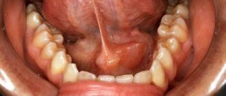
Patient K., 45 years old, came to the clinic with complaints of pain when eating. Dry mouth was sometimes noted. An objective examination revealed the following: in the sublingual area on the right there is a painful seal on palpation.
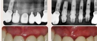
An orthopantomogram in the area of the lower jaw on the right reveals an oval-shaped, homogeneous formation with clear contours measuring about 1×1.7 cm, resembling bone tissue in density (Fig. 1). After a clinical, laboratory and x-ray examination, the patient was diagnosed with calculous sialodochitis of the submandibular salivary gland on the right.
Rice. 1
Treatment
Before the operation to remove the stone from the duct, conduction anesthesia of the lingual nerve is performed (mandibular type); in addition, the mucous membrane of the floor of the mouth is lubricated on the corresponding side with a 10% lidocaine solution. Infiltration anesthesia should be avoided in this case, as it makes it difficult to navigate when searching for a stone.
If the stone can be easily palpated, then an incision in the mucous membrane is made directly above the stone (Fig. 2). Before making the incision, it is advisable to pass a silk ligature through the soft tissue behind the stone, with moderate tension the assistant prevents the stone from moving towards the salivary gland. The mucous membrane and wall of the duct are dissected (Fig. 3). In the process of removing a stone from the gland, its bed should be washed from a syringe with a solution of furatsilin and at the same time suck it out of the wound with a saliva ejector. The discovered stone is removed using tweezers or a surgical spoon (Fig. 4).
Rice. 2 Fig. 3 Fig. 4
The extracted stone is oval in shape, has a dense consistency, yellowish-white color, and measures 1.8x1.0 cm (Fig. 5). After removing the stone from the duct, the wound is usually not sutured or drained. To normalize the function of the salivary gland, salty and acidic foods are recommended. It is useful to rinse your mouth with lemon-acidified water several times a day.
Rice. 5
Forecast
Long-term results of surgical treatment of salivary stone disease are favorable in the vast majority of patients; stones do not form again.
But if this does happen, and the salivary gland changes significantly, it must be removed. The reason for the relapse may lie in the body’s tendency to form stones or in the insufficiency of radical surgery, when when removing a stone from the excretory duct, a piece of it or sand remains, which served as the basis for repeated stone formation.
Soon after the stone is removed from the gland, its function is restored; quantitative and qualitative indicators of salivation (including the viscosity of saliva, its pH, the concentration of calcium, magnesium, phosphorus, potassium ions) after some time are established within normal limits.
Diagnostic features
Examination of a person with suspected sialadenosis may include the following methods:
- survey, examination, palpation of the glands;
- laboratory tests - general and biochemical blood tests, clinical urine tests, assessment of carbohydrate metabolism (glucose test);
- Ultrasound of the salivary glands and soft tissues is necessary to confirm the non-inflammatory nature of the disease and exclude tumors.
It is important for the doctor to exclude other possible pathologies characterized by enlargement of the salivary glands: inflammatory diseases, infectious diseases, cysts and tumors, the formation of calculi (stones).
Sialography is prescribed to determine dilated or narrowed salivary ducts; it involves the administration of a contrast agent. A radiosialogram may also be required to assess the secretory capacity of the glands. Using a CT scan, the doctor can see a bilateral increase in tissue size and density and rule out malignant and benign formations.
As an additional method, sialometry, cytological studies of secretions (salivary fluid), as well as biochemical studies of saliva can be used. The final diagnosis can be made after aspiration or another type of gland biopsy. Histological study allows us to establish dystrophic changes.
To find out the exact cause of the disease, it is important to assess your overall health. Therefore, the patient often needs to be examined by another specialist: an endocrinologist, gynecologist, urologist, allergist, etc. In some cases, the involvement of a rheumatologist is required.
Diagnosis of sialadenoses
During the examination of a patient with suspected sialadenosis, the doctor conducts a survey, examination and palpation of the salivary glands. After which the patient undergoes clinical and biochemical blood and urine tests to study the parameters of carbohydrate metabolism. In order to exclude the inflammatory and tumor process of changes in the salivary glands, the patient undergoes an ultrasound of the salivary glands. This makes it possible to determine their increase, heterogeneity of the parenchyma, increase or decrease in echogenicity. In addition, an X-ray examination is performed. With sialography, the patient can detect dilation or narrowing of the salivary ducts, slowing down the removal of the radiocontrast drug from the gland. A radiosialogram also reveals a decrease in the secretory ability of the salivary glands. In order to detect a bilateral increase in the volume and density of the gland and thereby exclude tumor lesions, the patient undergoes a computed tomography scan.
In addition, to diagnose sialadenosis, sialometry, cytological examination of duct secretions and biochemical examination of saliva are performed. The diagnosis of sialadenosis is confirmed by aspiration or incisional biopsy of the salivary glands. Histological examination reveals an increase in the acini in the patient, the presence of dystrophic changes in them, and the absence of inflammatory infiltration.
In order to determine the nature of the diseases accompanying sialadenosis, the patient may be referred for consultation to specialists. As part of diagnostic measures, other possible causes of enlargement of the salivary glands are excluded: sialadenitis, mumps, tumors and cysts of the salivary glands, stones of the salivary glands.
Treatment methods
Treatment of sialadenosis is a multi-step process. The main condition is effective therapy for the underlying or concomitant disease, although this will not help solve the problem completely. Symptomatic therapy is possible using novocaine blockades. Physiotherapy will help cope with unpleasant symptoms; electrophoresis, galvanization, magnetic and laser therapy are widely used. The impact is carried out on the area of the cervical nerve tissue in order to improve the conductivity of impulses.
Drug therapy consists of the use of vitamin E, drugs to stimulate salivation, as well as colloidal solutions to correct blood viscosity and improve blood flow in small blood vessels. Surgical treatment may be indicated in the absence of a positive response to conservative therapy. It consists of partial or complete removal of the salivary gland or its duct.