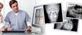
Dental X-ray is a method of examination that helps the dentist make a correct diagnosis and build a treatment plan. The photographs show the dentist what is hidden from view; with their help, he can easily see whether there is caries, gum disease, or the general condition of the teeth. This diagnosis is very valuable for both the patient and the doctor. It allows you to quickly determine the scope of work and carry out high-quality therapy, relieve pain and unpleasant symptoms. You can familiarize yourself with the features of this survey in a few minutes.
Why do you need a dental x-ray?
Radiation diagnostics is needed in most cases during dental treatment. Without accurate photographs of the oral cavity, the doctor cannot determine the entire affected area and the spread of the disease. Many problems cannot be assessed visually alone, such as nerve damage. X-rays reflect bone tissue well, so this is a very effective technique for examining teeth. Soft tissues also cast a shadow on the photo; using them, the dentist can detect an abscess, cyst or tumor.
Content:
- Why do you need a dental x-ray?
- How it's made
- In what cases is an x-ray needed?
- Where can I do it?
- Examination of children
- X-ray for pregnant and lactating women
- Frequently asked questions from patients
During a visual examination, the dentist can only guess the cause of pain, bleeding or other problems. For an accurate diagnosis, he needs an image of the internal structure of the oral cavity: gums, teeth, roots, canals. The procedure is always carried out before a diagnosis and treatment regimen are made. X-ray examination is necessary not only at the first stage; it is used to determine the effectiveness of therapy.
Be sure to take a series of photographs during cleaning and filling of the canals. They are used to determine the depth of each canal and the tactics for filling it. If the dental treatment is carried out incorrectly, for example, the canal is not completely sealed, this will lead to re-inflammation and various complications are possible, in the form of new dental problems. You cannot do without such technology after prosthetics, complex removal, or treatment of periodontal disease. That is, radiography of the jaw is needed to make a correct diagnosis and to monitor the progress of therapy.
Modern dental x-ray - what is it like?
Radiography does not stand still and today teeth are taken using a digital x-ray. For example, a radiovisiograph is used for targeted examination of individual teeth. This is exactly the device used in the Avicenna dental clinic.
The visiograph has a number of advantages:
rapidity;- safety;
- tracking treatment results and saving images for transfer;
- the image is taken using a computer sensor, and there is no need to bite a special card, as with an analog X-ray machine.
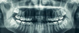
Some clinics use an orthopantomograph to take panoramic photographs of teeth. OTPG reflects the condition of the entire jaw, the placement of the dentition and even pathologies in the maxillary sinuses and soft tissues.
To hold an x-ray in a clinic, you must be licensed, follow clear instructions, and have the necessary knowledge and experience.
Our clinic has a license for an X-ray room. This means that X-rays at Avicenna Dentistry are performed in accordance with all safety rules.
How it's made
The process takes place in a separate room; most dental clinics have such X-ray areas. The walls and floor in this room are covered with lead material, which protects other rooms from the rays. The office is equipped with equipment, sometimes several types of equipment, as there are different types of dental x-rays.
No preparation is required before the procedure. The patient removes jewelry so that it does not distort the image. To protect other parts of the body, the patient is asked to put on a special cover from the neck to the waist; in medicine this is called a lead apron. Special fabric does not transmit radiation rays, so the radiation dose is significantly reduced. Then a series of pictures are taken or just one, depending on the doctor’s goals and the patient’s complaints. The procedure for obtaining images varies, depending on the type of equipment used in the X-ray room, as well as on the scope of tasks.
Bitewing radiography
This is the easiest and fastest way to obtain images of the internal structure of the jaw. This requires the simplest technique, which is why this technique is very popular in many dental centers. The patient sits on a chair, applies a small film to the gum in the desired area of the mouth, on which the image is then displayed. The film is placed on the side of the tongue and held there with the help of a bite plate. The laboratory technician presses a remote button and an image appears on the film. One such picture shows several teeth, the bone between the teeth, a canal, and periodontium. For complete information about the problem area, 4 photographs are taken. The results usually need to wait no more than 5-10 minutes; immediately after the procedure, the patient receives a series of images and gives them to his dentist.
A variation of this technique is a procedure called the “full set”. The picture is taken in exactly the same way, but in a larger quantity. To examine the entire jaw, the doctor needs 14 to 20 separate image films. From this “mosaic” a clear picture is formed. “The Complete Set” is recommended during your first visits to the dentist to examine all possible problems and prevent them.
Periapical radiography
This type of diagnosis is carried out to examine a separate part of the jaw; the image shows the entire tooth with the root and the gums near it, and the canals are clearly visible. The process is no different from the previous type of examination: a photo is taken on film, which is applied to the diseased tooth. In fact, this is another type of bitewing x-ray. This method is also used to control the treatment of a specific tooth, determine the cause of pain and bleeding. The advantage of the method is that the patient receives a lower dose of radiation.
Occlusal X-ray
This picture is taken when an image of an entire row of teeth is needed. In the office, the patient does everything the same, but the film is clamped with his teeth from a different angle. The plate is placed between the top and bottom rows and bitten. The photo shows all the teeth of the upper jaw or lower jaw, or both at once. This technique is indispensable for the treatment and diagnosis of malocclusion.
Orthopantomogram (OPTG)
A more informative method is a panoramic photo. For this we need a new generation device. The patient is still required to remove jewelry and wear a protective lead membrane.
Stages of the procedure:
- The patient is taken to an orthopantomograph and told how to position himself correctly.
- You need to hold on to special handrails with your hands; some orthopantomograph models also have a head holder. It is very important that the subject does not move during the examination, as the results will be distorted.
- There is a plastic tube in the chamber of the device; the laboratory assistant puts a disposable cover on it. The patient clamps the tube with his teeth and assumes a fixed position.
- The camera will begin to rotate around the patient's head and take pictures.
- Sometimes the radiologist may ask you to reposition your head or body for a different projection.
As a result, the dentist receives a panoramic image of the dentition, roots, and jaw bone. One such session replaces 14-20 bitewing radiography images.
Cone beam computed tomography
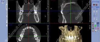
New technologies are increasingly replacing film X-ray machines. In dentistry, the most advanced diagnostic method is CBCT or 3D dental x-ray. The procedure is exactly the same as a panoramic shot. The difference is only in the results. CBCT provides comprehensive information about the patient's jaw. While the camera of the device rotates around the patient’s head, data from it is transmitted to a computer monitor. In this case, the image is obtained not in the form of photographs, but in the form of a three-dimensional model. The dentist can examine in detail the soft and bone tissue in any part of the oral cavity. Moreover, the result is transferred to a disk or flash card; when viewing, each individual section is enlarged many times over. Therefore, this method allows you to find even the smallest pathologies and prevent most problems. This method is not used often, since it is more expensive than all other types of examination.
What is a visiograph and how does it differ from an x-ray?
This one of the frequently asked questions is akin to the difference between a car and a traffic light... It seems that both concepts have some kind of connection, but it is somehow difficult to compare them. It's the same here. A radiovisiograph is a system that receives x-ray radiation, transforms it into digital form and displays the image on a computer screen. Roentgen (who is Wilhelm Conrad) is a long-dead German physicist who gained worldwide fame for his discovery of short-wavelength rays with enormous penetrating power. The physicist himself called these rays X-rays (in English today they are called exactly that - X-ray), but now we often call them X-rays, and in everyday life simply “X-rays”. The unit of radiation power was also called the x-ray. Now it is clear that a visiograph and an x-ray are completely different things. If we compare the visiograph with anything, it is with x-ray film, which it is universally replacing from all areas of medicine.
In what cases is an x-ray needed?
During a visual examination, the dentist can see approximately 40% of the oral cavity. However, most problems lie inside: in the canals, roots, interdental space. Therefore, most patient complaints can be resolved only after X-ray diagnosis.
Indications for examination:
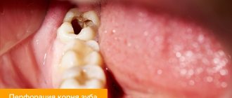
- caries in any part of the jaw, including under the crown;
- root destruction: fractures, cracks, bone depletion;
- periodontitis - destruction of bone, while the gums become inflamed and bleed, teeth begin to loosen and hurt;
- broken bite;
- pathology of the dental joint;
- abscesses and tumors, cysts;
- periodontitis is an inflammation of the root, accompanied by the appearance of a cyst that enlarges.
Painful sensations do not always appear, so patients often delay treatment. Advanced periodontitis leads to gumboil and tooth loss.
In addition, canal cleaning, prosthetics and implantation, bone grafting, and orthodontic treatment are not performed without a detailed examination.
Types of X-ray
After the examination, the doctor can prescribe the patient one of four possible types of x-rays
.
Prikusnoy
This method allows you to reflect the crown part of the tooth in the image. It is used to detect periodontitis and interdental caries. Bitewing can be used to obtain images of the upper and lower teeth.
Sometimes such a picture can be taken after prosthetics and crown installation to see how correctly the procedure was performed.
Sighting
With the help of a targeted image, it is possible to discern a specific affected tooth or several. Moreover, such a picture cannot include more than 4 teeth.
Panoramic
Using panoramic images, you can monitor the quality and effectiveness of the treatment already performed. They allow you to see a complete picture of the state of the entire dental system, and this is not only teeth with obvious problems (for example, caries, chips, etc.), but also roots, periodontal tissue, paranasal sinuses and the lower jaw joint.
On a panoramic image, the doctor will be able to see the presence/absence of filling material, hidden carious cavities, inflammation of the peri-root tissues, cysts, tumors, as well as teeth that have not yet erupted.
Digital or 3D X-ray
This type of x-ray is considered the most modern and safe. Using 3D X-rays, you can get a clear image of the entire row of teeth and a specific tooth. The result is a three-dimensional image that is displayed on the monitor.
Where can I do it?
Most dental clinics have an X-ray room where the patient can undergo this procedure without leaving the building and return to his dentist with the images. State clinics also have such installations, but they are exclusively of the old type. This means that you will have to go through the procedure several times and pay less. Old equipment also has high radiation doses. Unfortunately, many private dentistry are also equipped with such equipment. Sometimes a private doctor may refer you to another diagnostic center to obtain data. Cone beam computed tomography is only available in progressive private clinics, but the accuracy of the results fully pays for itself.
The price of the service will depend on the type of examination. Thus, bitewing radiography costs from 3 to 10 dollars, depending on the number of images. At the same time, prices are almost the same in both public and private institutions. A panoramic image will cost approximately $20-25. It can only be done in private institutions, but some clinics can provide this service for free if the patient is being treated by them. The most expensive diagnostic is CBCT, which is done in single diagnostic and dental centers. Its cost will be 50-60 dollars.
Are dental x-rays safe for human health?
In accordance with SanPiN 2.6.1.1192-03, the maximum permissible annual radiation dose during diagnostic x-ray procedures is 1000 μSv (microsievert) for an adult and 300-400 μSv for a child.
To receive it, within a year you must complete:
- 500 targeted dental examinations using a radiovisiograph;
- 100 procedures of conventional x-rays with image output on film;
- 80 digital or 40 film orthopantomographies (obtaining a panoramic image).
Thus, dental X-rays for almost any purpose do not exceed the permissible doses. Therefore, it does not have a negative effect on health. Even when producing film OPTG, the radiation dose is a maximum of 30 μSv. If it is necessary to take a targeted X-ray of the teeth and use radiovisiography, you can even get several images during one appointment. Without any risk to health, since a single dose in this case is only 2-3 μSv.
Examination of children
Dental x-rays are often prescribed for young children. You should not be afraid of this, since children sometimes need such examination more than adults. Baby teeth are often subject to carious problems, and caries appears in inaccessible places. As with an adult, the dentist cannot fully assess the condition and depth of the problem in a child. To accurately assess the situation, treat and prevent diseases, X-rays are needed. Also, a photo of baby teeth helps determine the process of eruption of molars, the quality of bone tissue, and the formation of a bite. In childhood, it is easiest to prevent improper formation of the dentition. If such problems are not resolved in time, in later life this will happen longer and more severely.
For small patients, only minimal doses of radiation are used. The doctor builds treatment and diagnostic tactics so as not to expose the child to radiation again. The procedure is the same as for adults: the rest of the body is covered with a protective apron, the session lasts a couple of minutes, no pain or discomfort occurs. In addition, some clinics use a collimator - a tube that is attached to the device. This device shrinks the X-ray beam and changes its contour, so children receive even less radiation.
X-ray for pregnant and lactating women
Women during pregnancy should see a dentist as part of a routine checkup. The fact is that destructive and inflammatory processes in the gums and roots of teeth can lead to negative effects on the entire body. This situation is extremely undesirable when carrying a child, so a pregnant woman is sometimes prescribed x-rays. To examine the expectant mother, only minimal doses of radiation are used; they try not to take panoramic photographs, replacing them with targeted ones. However, radiation is still present, which raises the question among patients: is it possible to do an X-ray on a pregnant woman?
During pregnancy, of course, it is better to avoid such diagnostics. Manufacturers of new technology claim that it is absolutely safe for women even during this period. But still, the opinions of doctors on this matter remain skeptical. Such an examination can be prescribed only when the disease threatens life more than the possible negative impact of radiography on the fetus. In the third trimester, such diagnostics can be carried out without harm to the fetus. In the process of obtaining images, the patient’s chest, neck, abdomen, and pelvic area are tightly covered.
After childbirth, women often experience dental problems, mainly due to the loss of beneficial microelements. Nursing mothers should not be afraid of x-rays. This does not affect the structure of milk at all (this does not apply to studies of the abdominal and thoracic cavity). During lactation, women undergo X-rays under a protective apron. You can feed your baby after the x-ray on the same day.
Dental clinic in Vladimir The whole range of dental services!
Many patients are faced with the need to receive some X-rays. This, of course, is necessary for proper treatment and prosthetics, but citizens are wondering how safe it is? X-ray radiation is a type of ionizing radiation. By the way, among other types of such radiation, x-rays are considered the safest and have the least negative impact on the human body.
The most important property of X-ray radiation, which is used in medicine, is its excellent penetrating ability. Subject to certain rules for the use of X-ray machines and the necessary protective measures, this is a fairly safe and convenient type of research. In any case, today there is no alternative to this instrument when treating teeth.
The principle of image formation during x-ray examination, including in dentistry, is based on the properties of different organic structures of the body to absorb this type of radiation differently. The resulting image is recorded on a special photographic film or on a sensor matrix using a computer. The second technology is more modern and has a number of advantages.
Why do we actually need x-rays in dentistry? More than half of the tooth surface is simply not visible to the dentist’s eye, not to mention the internal structure. Under these conditions, the only way for the dentist to look into the thickness of the gum is an x-ray. X-rays are used both for the initial diagnosis of diseases and for monitoring the results of treatment. Sometimes the use of X-rays for preventive purposes is allowed.
X-rays, in particular, are used in the diagnosis of caries in interdental and subgingival surfaces, and possible secondary caries under fillings and crowns. X-rays are indispensable for determining the depth of a carious defect, its relationship with the tooth cavity, for monitoring the quality of treatment of tooth canals, for assessing the condition of the bone tissue surrounding the tooth root (this is necessary for periodontitis). And of course, during tooth extraction, implantation and prosthetics. Today it is difficult to name areas of dentistry where x-rays are not used.
As already mentioned, image recording during an X-ray examination can occur in two ways: either on film or using computer sensors (radiovisiographs). But in both cases the radiation sources are no different. What is better than using “computerized x-ray”? First of all, reducing the radiation exposure to the patient by reducing the irradiation time. If for visiographs (computer x-rays) durations of 0.125 seconds or less are sufficient, then classical methods require a time of the order of 0.5 seconds or more. This, by the way, results in a reduction in radiation exposure to the patient by up to 90%.
Unlike other areas of medicine, X-ray machines used in dentistry have a very narrow beam of radiation, and the duration of the radiation is short, so the doses received by patients are very small. For comparison: the average dose received by a patient during a routine examination using a visiograph (computer X-ray) is approximately 0.001 MSV (Sievert is a unit of measurement for exposure to ionizing radiation). The radiation dose during a panoramic examination is -0.02 MSV. Over the course of a year, from the external environment a person receives radiation (natural background) of approximately 3.0 MSV. For preventive (not therapeutic) purposes, the annual radiation dose should not exceed 1.0 MSV. These numbers and their relationships are easy to analyze on your own.
In conclusion, it can be noted that the use of x-ray examination is absolutely necessary in dentistry today. In this case, the degree of possible harm to health can be neglected due to its insignificance. And for doubtful patients, we can recommend contacting reputable clinics that have the latest computer radiovisiographs.
Frequently asked questions from patients
Are x-rays harmful?
Radiography is based on radiation exposure. This sounds quite threatening to the average person. Meanwhile, every person receives a dose of radiation every day, even our bodies are radioactive. The background radiation level per day is 10 μSv (microsieverts). To obtain 4 photos with a bitewing X-ray, the patient receives 20-51 μSv. A panoramic image gives 5-25 μSv. CBCT is accompanied by a higher radiation dose; in one session a person receives from 20 μSv to 700 μSv. The level of radiation will depend on the settings and type of device, and the width of the area being studied.
Thus, there is no direct threat in the procedure. However, radionuclides can accumulate, so diagnostics are prescribed if necessary. After the session, the radiologist must write down how many sieverts the patient received, this will make it possible to calculate the next dosage with minimal harm to the person. After the examination, it is advisable to eat more carrots, apples, radishes, beans and citrus fruits. These products will help remove radionuclides from the body.
How often can it be done
Best materials of the month
- Coronaviruses: SARS-CoV-2 (COVID-19)
- Antibiotics for the prevention and treatment of COVID-19: how effective are they?
- The most common "office" diseases
- Does vodka kill coronavirus?
- How to stay alive on our roads?
The type of x-ray and its frequency depends on the condition of the oral cavity and the complexity of the treatment. It is better to do CBCT no more than 3 times a year. Bitewing film photographs are prescribed no more than 7 times a year; the new technology uses lower radiation doses, so there may be more digital diagnostics. The acceptable norm is 7 diagnostic studies per year. When changing dentists, it is not necessary to take new photographs; it is enough to take the ones you already have with you to the appointment. If digital data has been lost, it can be requested from the clinic that conducted the examination. CBCT results are stored for up to a year in the archives of medical centers.
Is it true that a visiograph is safer than a regular film photograph?
When asked about such a comparison, they mean the radiation exposure that the patient receives when using different techniques. In this sense, indeed, a visiograph is preferable, since its sensor is much more sensitive than the best film. Therefore, to obtain a high-quality image using a visiograph, much shorter shutter speeds are needed. To take a picture on film, the shutter speed is 0.5-1.2 seconds. To obtain the same image using a visiograph sensor – 0.05-0.3 sec. Those. 10 times shorter. As a result, the radiation exposure received by the patient when using a visiograph is reduced to an insignificant minimum.