How dangerous is x-ray?
In many cases, dental treatment is simply unthinkable without X-ray examination. When you can't do without it:
- when filling thin and curved tooth canals;
- to detect hidden carious cavities, caries under fillings;
- when the question arises whether wisdom teeth need to be removed (for example, if they are incorrectly positioned);
- to diagnose a tooth root fracture, cyst, determine the degree of inflammation of the tissues around the diseased tooth;
- when the issue of removing or restoring a tooth is being decided.
In the first three months, it is not advisable to take dental x-rays for pregnant women, since the fetus has not yet formed. Radiation can have an adverse effect on dividing cells, cause chromosome abnormalities and provoke intrauterine developmental defects. By the second trimester, at 16–20 weeks, the fetus is already fully formed - a tiny dose of radiation will not harm the baby in any way, so pictures can be taken without fear.
Dental X-rays during pregnancy are much safer than examinations of the pelvis or chest, because the mother’s belly is reliably protected by a lead apron, and dental examinations are always carried out using special devices with reduced radiation exposure. This applies to both old models and modern visiographs.
Is X-ray examination dangerous during pregnancy - MEDSI
Is X-ray examination dangerous during pregnancy?
X-ray examinations are carried out to determine the presence of any pathologies, tumors or diseases.
For this purpose, a directed beam of electromagnetic waves of a given length and frequency is used. It passes through human organs, the tissues of which reflect and absorb it in different ways. Thanks to this difference, various anomalies can be seen. The result is sent to the screen or recorded on a special film.
During a preventive examination, this examination is carried out in a special room. If the patient is injured, it can be used immediately in the emergency department, operating room or intensive care unit.
Effect of radiation on the fetus
X-ray radiation in general is not always beneficial for the body, since it destroys those cells that are in a state of constant division. This leads to the destruction or mutation of DNA chains.
There are not many such newly formed cells in the adult human body. But in the fetus in the early stages of development they are the basis. Therefore, such an examination is quite dangerous for him. Complications are most likely to occur when using x-rays during pregnancy in the first trimester (initial 12 weeks).
What problems can x-rays cause in early pregnancy?
Due to the gradual development of the fetus, the formation of future systems of the child’s body occurs in each week of its existence. Therefore, the exposure of the mother's body to large amounts of X-ray radiation at these stages can have various serious consequences:
- The first two weeks. Possible death of the embryo, miscarriage, ectopic pregnancy.
- Weeks three and four. Pathologies at an early stage of fetal development, miscarriage.
- Fifth-sixth week. Developmental disorders of a number of organs and systems: thyroid, thymus and gonads; immune, nervous, circulatory, endocrine systems.
- Seventh week. Damage to the liver, intestines. Metabolic disease.
- Eighth. Pathologies of the development of joints and limbs, the oral cavity.
- Ninth week. Damage to the respiratory and reproductive systems.
- Weeks ten and eleven. Problems associated with dental development. Heart disease.
- Twelfth week. Pathologies of the thyroid gland, immune disorders.
After this period, the effect of radiation on the fetus decreases, but it is still not recommended to do such a study until the end of pregnancy, unless absolutely necessary.
X-ray in early pregnancy
Doctors try not to prescribe x-rays to pregnant women, because even a minimal risk of harm from radiation always remains. It is especially great in the first twelve weeks.
The most dangerous types of tests for the fetus are:
- Abdominal x-ray
- X-ray of the pelvis and spine
- Mammography
- Fluorography
- CT scan
- Isotope scanning
The following types of x-ray examination are less dangerous:
- Chest (lungs, heart)
- Brain
- Limbs
Are there any non-hazardous types of examination?
The safest types of x-ray examination are:
- X-ray of teeth
- X-ray of the nose
In these cases, the impact occurs locally, and therefore the radiation dose is minimal.
The amount of radiation that a fetus can receive in two months is regulated by Sanitary Rules and Standards and should be no more than 1 millisievert (mSv).
There are other types of examinations that can be used instead of x-rays during pregnancy:
- MRI
- Visiography
- Ultrasound
Still, doctors try not to prescribe MRI in the first trimester of pregnancy, since statistical studies are insufficient to clarify its safety during this period.
What to do if you can’t do without an x-ray?
X-rays for pregnant women may be necessary in a situation where a disease or injury threatens the life and health of the mother and child, and it is impossible to use other diagnostic methods. And the harm from not using x-rays exceeds the potential harm from its use.
- If it is necessary to conduct an examination of an area that does not touch the pelvis, abdomen or spine, then they must be shielded with lead aprons and overlays.
- If in the early stages of pregnancy it is necessary to take an x-ray directly through the fetus, then the doctor may suggest terminating it to avoid mutations and miscarriage.
- A woman can refuse an abortion, but in this case she must understand the risk she is taking and the pathologies that may appear in the fetus.
In all these cases, after undergoing the study, it is recommended to go for an ultrasound to monitor the condition of the fetus and the appearance of certain pathologies.
If it is possible to postpone the use of x-rays until the last trimester or postpartum period, then it is necessary to do so.
At a very early stage, a woman may not be aware of pregnancy. Therefore, it is recommended to undergo additional examination before the x-ray.
Advantages of performing X-ray analyzes at MEDSI:
- 30 types of X-ray examinations
- The latest equipment with the ability to regulate radiation intensity
- Carrying out urgent examination in case of injury or other medical indications
- Technologies that are suitable for both adults and children
- Radiologists of high qualification categories, candidates of medical sciences
- Sign up for research and consultation by phone 8 (495) 7-800-500
- More than 20 diagnostic centers
Visiograph - an alternative to x-ray
A visiograph is a modern analogue of an x-ray machine. Its advantages:
- Allows you to take an image instantly, high-quality data is immediately displayed on your computer monitor.
- A narrow beam of rays is directed exclusively at the diseased tooth, does not affect adjacent tissues, and certainly cannot “touch” the abdomen.
- The duration of exposure (irradiation) is 10 times lower than when using old-style X-ray machines and is only 0.05–0.3 seconds. Radiation exposure is only 2 microsieverts (when using old devices - from 7 to 80 microsieverts).
For comparison, just sitting in front of the TV for three hours and enjoying your favorite series, a mother receives a radiation dose of 5 microsieverts. The safe radiation dose per year is 1,000 microsieverts (that’s 500 shots). Of course, you shouldn’t take the procedure lightly, but you shouldn’t panic and overestimate the danger to the baby either.
Is an x-ray always necessary before treatment? What are the indications for it?
The doctor will see little with the naked eye. Dental diseases are insidious and make themselves felt when they go deep enough, but before that they “sit quietly.” Therefore, a doctor can completely cure inflammation and remove infection only with a complete picture in hand. Of course, if there is no urgent need for an X-ray, the doctor will do without diagnostics - that is, the final decision, of course, rests with a specialist.
The best way to avoid serious treatment during pregnancy is to visit the dentist at the planning stage of the baby and eliminate dental problems in advance
How to minimize risks
What should an expectant mother do so as not to worry about the baby’s health?
- Not all clinics, especially municipal ones, have visiographs. It is better to inquire about this in advance.
- Be sure to use a protective apron and collar.
- You need to inform your doctor about the exact timing of your pregnancy. In case of a delay, even if the test shows a negative result, you must inform the specialist. In unclear situations, diagnosis (and treatment) are carried out as if pregnancy was definitely established.
- Severe pain, swelling, inflammation are good reasons not to delay a visit to the dentist, even in the first trimester. Unless absolutely necessary, the doctor will never prescribe dental x-rays during pregnancy and will try to do everything to postpone treatment to the 2nd, safe trimester or to the postpartum period. However, some acute conditions may require emergency intervention, such as purulent pulpitis or acute periodontitis. In this case, the mother needs to remember that a rapidly developing infection in the oral cavity will cause much more serious damage to the baby than 1-2 pictures on a visiograph.
- If there are concerns that the X-ray has somehow damaged the baby, you should consult with a geneticist and do an ultrasound. This will reassure those mothers who may have taken the photo without knowing that they were expecting a baby.
Since dental treatment during pregnancy is still associated with certain difficulties, the best thing an expectant mother can do is to go to the dentist and have all her teeth treated at least a month before the date of planned conception. And already in pregnancy, mommy should brush her teeth thoroughly and eat foods high in calcium.
Diagnostic results: interpretation of images
The contrast agent does not transmit x-rays, and the cavities filled with it are visible on the image as bright white spots. An indicator of good tube patency is the spreading of the contrast agent along the peritoneum to areas remote from the insertion site. If the contrast stops at any section of the fallopian tube, it means that there is an adhesion or other pathology in this place. Depending on the shape of the uterus and tubes, the doctor can determine which pathological process is preventing conception. Interpreting images of the uterus and fallopian tubes requires extensive knowledge and considerable practical experience. Self-diagnosis is a completely useless exercise.
If the results of the examination give reason to suspect the presence of uterine cancer, it is necessary to order additional examinations, including taking tissue for a biopsy. The diagnostic doctor's conclusion, along with the photographs, is transmitted to the attending physician who referred the woman for examination.
Interpreting images of the uterus and fallopian tubes requires extensive knowledge and considerable practical experience. Self-diagnosis is a completely useless exercise.
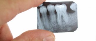
Analysis of targeted dental images –
Analyzing X-rays is not difficult if they are of good quality.
Almost any patient will be able to see signs of periodontitis or cysts in the image, and will also be able to determine how well his root canals are filled. All you need here is skill. But it is worth remembering that absolutely everything cannot be diagnosed using X-rays, for example, inflammation of the nerve of a tooth. You can diagnose from the image -
- caries (but not in full),
- periodontitis (granulomas, cysts),
- quality of root canal filling after treatment of pulpitis and periodontitis,
- perforations and root cracks,
- fragments of instruments in root canals,
- presence of periodontal pockets...
1) A group of images indicating the development of inflammation (periodontitis) at the apexes of the roots of previously untreated teeth. In this case, you will always see a clear or vague darkening at the apex of the tooth root, which can be of different sizes and shapes.
2) A group of photographs taken after root canal filling. The first 2 pictures (Fig. 14-15) show what well-filled root canals look like. The following images show poor quality treatment and complications that arose (read the descriptions in each image).
X-ray of fallopian tubes before conception
If you have not been able to get pregnant up to this point, your doctor may suggest obstruction of the fallopian tubes. And in this case, an x-ray examination must be carried out. After all, it is this factor that determines whether the long-awaited pregnancy will occur or not. If there is an adhesive process in the tubes, then there may be no talk of fertilization for now.
The doctor can find out what condition the fallopian tubes are in only after an x-ray. This procedure is carried out using a contrast agent, which allows you to display the condition of the pelvic organs on the pictures. In addition to the patency of the tubes, an X-ray examination provides information about the presence or absence of fibroids, polyps and other formations, which is of great importance not only for the health of the unborn child, but also for the woman herself.
Quite often it happens that after conducting such a study, a woman becomes pregnant.
There is an explanation for this. The contrast liquid used for the procedure, injected under pressure, promotes the divergence of adhesions. As a result, the patency of the fallopian tubes can be restored.
This point must be taken into account when planning pregnancy and going through a similar process. Conception should not be allowed to occur during the same menstrual cycle, since the egg has received a large amount of radiation exposure. What do we have to do? If an x-ray is scheduled for the middle of the cycle or the last third (it happens that you can get pregnant during this period), then you should take care of pregnancy protection at this time, despite the great desire to have a child.
Can you trust the equipment?
Today, modern digital equipment is used to conduct radiographic examinations. This, of course, does not eliminate X-rays, but many doctors believe that minimal radiation received after the procedure will not cause any harm when planning a pregnancy. In addition, X-ray itself is a fairly informative diagnostic method that allows you to identify many pathologies, so it should not be ignored.
However, many, especially people of the older generation, are sure that after an x-ray, the unborn child may develop various pathologies and developmental anomalies. However, if there is no child yet, where will they come from? If you are very worried about the upcoming fluorography or dental x-ray examination, then you can ask the doctor for a protective apron. By the way, they give it anyway, especially when conducting examinations using old-style equipment.
Individual X-ray protection equipment is used during the study.
Does penis size affect fertility?
If we are talking about normal, pathologically acceptable sizes of the penis, then no. If the penis is very short (the so-called micropenis), then during ejaculation most of the sperm do not reach the cervix, which reduces their chances of meeting the egg. However, this problem can be solved simply: immediately after sex, a woman needs to lie on her back and place something (for example, a pillow) under her tailbone. This position helps sperm flow into the cervix.
Speaking about whether the size of the penis affects pregnancy, we should also mention the insufficient size of the testicles. This may indicate congenital pathologies that are accompanied by hormonal imbalance.
Dental X-ray – price for 2022
The cost of one digital X-ray in Moscow will average from 250 to 350 rubles in different clinics. In addition, this price may only apply to a diagnostic initial image, while all other images taken during the treatment phase may cost less (about 150 rubles per image). Therefore, you should carefully read the clinic’s price list.
It should also be noted that there are a large number of clinics in which the cost of dental treatment is indicated on an all-inclusive basis. Accordingly, the cost of treating your tooth will already include the required number of x-rays (usually 2-4 pictures), for which you will no longer pay anything extra.
What else you need to pay attention to is that the price list of some clinics may state that the cost of a targeted x-ray indicated on the clinic’s website applies only if you are a patient of this clinic. Therefore, if an image is taken to be submitted to another clinic, its cost may be 100 rubles higher).
In addition, if you need a printout of a digital image, some clinics may also charge you about 50 rubles for this. The same applies to the description of the x-ray image: if you want to receive a written description of the image taken by a radiologist, then in some clinics they may additionally ask you for about 100-150 rubles.
Summary: important points
As a practicing dentist who knows the system from the inside, I want to draw your attention to the following points that are important for your safety. If the clinic has an X-ray machine, then a license must be obtained for it, the issuance of which presupposes the mandatory presence of a certified radiologist on the staff of the dental clinic. However, in reality, even in large clinics and public clinics, it is not always the case that x-rays will be taken by a trained specialist.
Even if he is, he may go on vacation or get sick, and a regular nurse (dental assistant) will take the pictures instead. This is a gross violation that leads to both the production of low-quality images and an increase in the radiation dose. In small clinics, the risks of receiving a poor-quality x-ray examination are much higher, and the first thing that makes you suspect a forgery is if the picture is taken not by a special employee, but by a nurse from the dentist you came to see.
The second very important point: if you see that the x-ray is not done in a special room, but the x-ray machine is located right next to the dentist’s chair, then you should change the clinic and the doctor. The fact is that all objects in this office (including the dental chair and doctor’s instruments) will have an increased background radiation, and this is no longer safe for health. We hope that our article on the topic: X-ray of the jaw and teeth was useful to you!
Sources:
1. Dental education of the author of the article, 2. Based on personal experience in therapeutic and surgical dentistry, 3. National Library of Medicine (USA), 4. “Digital and film radiography in outpatient dentistry” (Chibisova), 5. “X-ray diagnostics in dentistry" (Lutskaya I.K.).
FAQ about Fluorography
According to epidemiological indications (regardless of the presence or absence of signs of tuberculosis), preventive medical examinations are carried out 2 times a year:
– military personnel undergoing military service upon conscription; – persons in contact with sources of tuberculosis infection, including persons accompanying foreign citizens with tuberculosis; – persons removed from dispensary registration in medical anti-tuberculosis organizations due to recovery, during the first 3 years after deregistration; – persons who have had tuberculosis and have residual changes in the lungs, during the first 3 years from the moment the disease was diagnosed; – HIV-infected; – patients registered with drug treatment and psychiatric institutions; – persons who are members of the preventive drug treatment group in connection with the use of psychoactive substances and drugs; – persons under investigation held in pre-trial detention centers and convicts held in correctional institutions; – persons released from pre-trial detention centers and correctional institutions during the first 2 years after release; – persons who, by the nature of their professional activities, have contact with the contingent of persons under investigation and convicts; – persons without a fixed place of residence.
According to epidemic indications (regardless of the presence or absence of signs of tuberculosis), preventive medical examinations are carried out once a year:
– patients with chronic nonspecific diseases of the respiratory system, gastrointestinal tract, and genitourinary system;
– patients with diabetes mellitus;
– patients with oncohematological diseases;
– persons receiving corticosteroid, radiation and cytostatic therapy, TNF-a blockers, genetically engineered biological drugs;
– foreign citizens and stateless persons, including those carrying out labor activities on the territory of the Russian Federation, refugees, internally displaced persons;
– persons living in stationary social service institutions and social assistance institutions for persons without a fixed place of residence and occupation;
– employees of social service institutions for children and adolescents;
– employees of sanatorium-resort, educational, health and sports institutions for children and adolescents;
– employees of medical organizations;
– employees of social service organizations for the elderly and disabled;
– employees of organizations processing and selling food products, including milk and dairy products, organizations of consumer services, workers of water supply facilities;
non-transportable patients (examination is carried out using sputum microscopy).
The following undergo an extraordinary preventive medical examination for tuberculosis:
– persons who applied to medical organizations for medical assistance with suspected tuberculosis;
- persons who sought medical care in outpatient clinics, admitted for inpatient treatment, and persons admitted to children's medical organizations for the purpose of caring for children undergoing inpatient treatment, if more than a year has passed since the date of the last preventive examination for tuberculosis ( in case of emergency admission of patients for inpatient treatment, preventive examination for tuberculosis, if possible, is carried out in a hospital setting);
– persons from the environment of children with changes in sensitivity to tuberculin (“virgin” children), if more than 6 months have passed since the last fluorographic examination;
– persons coming from other territories of the Russian Federation for employment, permanent or temporary residence, if more than a year has passed since the last fluorographic examination;
– persons living together with pregnant women and newborns, if 1 year or more has passed since the previous fluorographic examination at the time of birth;
– citizens conscripted for military service or entering military service under a contract, if more than 6 months have passed since the last examination;
– persons who have been diagnosed with HIV infection for the first time, if more than 6 months have passed since the last examination, as well as those infected with HIV in the stage of secondary manifestations (4A-4B) or infected with HIV with a low level of CD4 lymphocytes (less than 350 cells/ µl);
– applicants upon admission to study, if 1 year or more has passed since the date of the last preventive examination for the purpose of early detection of tuberculosis;
– persons without a fixed place of residence – for any application to social protection or health care institutions, if there is no information about undergoing a preventive examination for tuberculosis or more than 6 months have passed since the last examination;
– persons who use psychoactive substances and drugs that are not included in the group of preventive drug treatment registration – when identified by employees of internal affairs bodies, in the absence of information about preventive examinations for tuberculosis over the past year;
– foreign citizens and stateless persons when applying for a temporary residence permit on the territory of the Russian Federation, residence permit, citizenship or work permit in the Russian Federation.
– The annual coverage of the population aged 15 years and older with preventive X-ray fluorographic studies should be at least 65% of the population,