Home / Articles / Impacted fang
It has long been known that teeth can fall out too early and humanity is trying to combat this problem in every possible way. Recently, another problem of a slightly different nature has appeared - teeth began to erupt ahead of time, and this leads to another problem - an impacted tooth. Most often, this happens in childhood, while adults are faced with impacted teeth at the time of the eruption of the so-called “wisdom teeth” or eights. What is an impacted molar or canine tooth, why does it need close monitoring, and should it be removed?
Causes of impacted teeth
- Due to injury or damage to the jaw;
- If the child’s primary fangs were pulled out prematurely;
- Incorrect location of the tooth germ;
- Dental crowding, which means teeth are too close together;
- Inflammatory diseases of baby teeth and gums;
- The presence of “extra” teeth (supercomplete);
- Poor nutrition;
- Rickets;
- Exhaustion or weakness of the body.
The main prerequisite for the appearance of retention is the peculiarities of the child’s development in the embryonic period (lack of minerals in the body, thickening of the mucous membranes, low fetal growth rate).
Possible complications
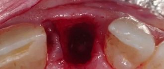
After removing the fang from the socket, various complications may develop, which in the vast majority develop due to infection of the wound.
Dry socket syndrome
The problem arises due to the patient’s failure to comply with the doctor’s recommendations or too active treatment of the oral cavity with antiseptic solutions. The actions lead to the fact that a blood clot does not have time to form in the wound, protecting the soft tissues from the penetration of pathogenic microorganisms.
The condition is not life-threatening, but can cause a number of consequences, for example, inflammation of a postoperative wound.
Alveolitis
The disease is an inflammatory process that spreads to the mucous membranes of the socket. Alveolitis causes infection to spread to bone structures. Pathology rarely develops after the removal of fangs on the upper jaw; more often, this condition appears after the extraction of molars located in the lower row.
Nerve injury
If the canine is positioned incorrectly or has a curved root, then during surgery damage to its nerve endings may occur. Characteristic symptoms of this condition are loss of sensitivity in the operated area, numbness of the cheek, and the appearance of “running goosebumps.”
Alveolar exposure
Even with a normal course of the postoperative period, problems with wound regeneration may occur. If pain occurs at the site of the hole when consuming hot or cold food, this indicates that the area of the bone is not covered with soft tissue.
Cyst on a tooth
The development of a pathological formation after canine removal is a rare consequence. A cyst is a growth filled with fluid. In this way, the human body independently separates infected soft tissue from healthy areas.
The tumor, increasing in size, can spread to neighboring areas and therefore requires immediate surgical intervention.
The occurrence of odontogenic phlegmon
The pathology develops against the background of osteomyelitis of the jaw structures. The problem manifests itself as pain and swelling of the cheek in the upper jaw area. It becomes difficult for a person to open his mouth and consume food. The pathology is accompanied by a deterioration in health and a rise in temperature.
Odontogenic periostitis
It develops against the background of alveolitis or osteomyelitis and is an inflammation of the periosteum. The condition is manifested by a number of the following symptoms:
- rise in temperature;
- toothache ;
- swelling on one side of the cheek.
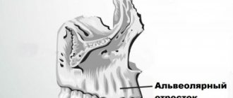
Removal of the maxillary canine is carried out taking into account the structure of this element of bone tissue. The tooth has only one root, which in 30% is located in the wrong position. The stages of surgery for this type of intervention are no different from removing the remaining elements located in the top row. Depending on the location of the canine and the condition of its crown, the doctor performs simple, complex or atypical tooth extraction.
Types of location of an impacted tooth
| Fine | The tooth has a vertical position and grows upward very slowly. Baby teeth and canines can grow properly. “Wisdom teeth” are rarely positioned normally. |
| Horizontally | The impacted tooth forms a right angle to the healthy one, located horizontally. This situation is detrimental to a healthy “neighbor”. In such a situation, it is necessary to remove the impacted tooth, which is performed only surgically. |
| Obliquely | An impacted tooth grows at an angle toward the cheek or tongue. If such an anomaly does not cause inconvenience to a person, then removal is not necessary. |
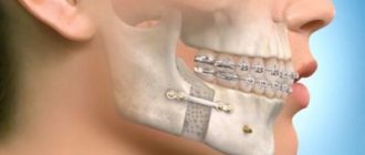
Why is the “vampire smile” formed?
In dentistry, the incorrect position of the eye teeth is called dystopia. It can manifest itself in different ways:
- fangs can protrude forward in relation to the dentition;
- hide behind adjacent teeth;
- be shortened or too long;
- unfold in the jawbone;
- be impacted, that is, not fully erupted.
Such anomalies arise because they erupt at the age of 9–12 years, later than other teeth, and in the jawbone, for various reasons, they may simply not have enough space:
- if the place is occupied by another tooth, the fang begins to grow “in the second row”;
- if the baby inherits large teeth from one of the parents, and a small jaw from the other, the fangs will become curved due to lack of space;
- If a change in bite occurs with a shift in timing, the eye tooth may protrude forward.
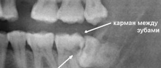
Symptoms of tooth impaction
In most cases, an impacted tooth is detected on an x-ray at the dentist or at an appointment with an ENT doctor. Retention in a child can be determined if one of the elements of the jaw row is absent for a long time or if a baby tooth is not replaced by a permanent “brother” for too long. The eruption of an impacted tooth is usually accompanied by inflammatory processes. The child may be bothered by swelling of the gum tissue and jaw, pain and aches, numbness and tingling in the retention zone.
On palpation of the gums, thickening and hardening are noted, reminiscent in shape and outline of an unerupted tooth.
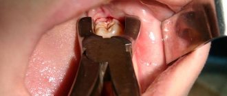
Removal of lower incisors
Removing the lower incisors is the easiest procedure. Due to the fact that the incisors have only one flat root. The presence of additional roots in these teeth is extremely rare. Removal occurs under local anesthesia. During this procedure, the patient takes a sitting position. The doctor's hands securely fix the patient's jaw. In such cases, the surgeon uses beak-shaped forceps. First of all, the tooth begins to dislocate in the labial side, and then in the lingual side, a slight rocking is allowed.
Removal of impacted teeth
If there are no complaints at all about the impacted tooth, and it does not cause any inconvenience (cysts do not form near it, the mucous membranes of the mouth are not injured), then removal may not be necessary. But if the impacted tooth is not removed, then it is necessary to undergo an X-ray examination of the mouth 2 times a year.
However, there are a number of cases when it is necessary to remove an impacted tooth:
- Retention causes constant inflammatory processes in the oral cavity;
- The occurrence of various paradental or follicular carpal formations, abscess, osteomyelitis in the gum tissue;
- Injuries and scratches of the oral mucosa that are caused by retention;
- An impacted molar or canine is also dystopic (improperly positioned in the dental arch) towards the cheeks or tongue.
The main factors on which the decision is made to remove impacted teeth are:
- The likelihood of injury during surgery,
- The location of the canine or molar and ease of access to them,
- Possibility of complications.
In most cases, the operation is painless and eliminates the possibility of pain even after the anesthesia wears off.
The removal of an impacted tooth itself takes place in several stages:
- pain relief by local anesthesia,
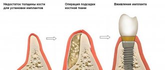
— an incision in the gum above the impacted canine and removal of a flap of tissue of the mucous membrane and periosteum,
- sawing out the bone to the dental crown using a drill,
- removal of a fang or molar from the bone using forceps (special biomaterials are left in its place).
Removal of premolars located on the upper jaw, canines and molars of the lower jaw has its own characteristics:
- Due to the fact that the roots of the teeth of the upper jaw are sometimes located close to the sinus, their removal must be carried out with the utmost care.
- In the case where the maxillary sinus has nevertheless been opened, medical treatment of the hole is not carried out, and the place where the bone has been broken is immediately sutured.
- Then a biomaterial is placed inside, which will promote rapid and painless overgrowth of bone tissue.
- The previously removed flap of mucous tissue and periosteum is returned to its place and sutures are applied.
- When removing impacted teeth in the lower jaw, the dentist must determine their location as accurately as possible, calculating their proximity to the mandibular canal. The safest way to access such teeth is through the vestibule of the mouth.
Types of tooth extraction
In medical practice, all removal procedures are divided into:
Easy removal
The doctor manages to extract the entire tooth using special forceps; its duration is up to 5 minutes. This is how single-rooted teeth or teeth affected by periodontitis are removed.
Difficult removal
To extract a tooth, the doctor uses auxiliary tools (most often an elevator), and the entire extraction process takes up to 15 minutes. It is most often performed when a part of the root or crown is broken during the removal of multi-rooted teeth.
Atypical removal
It has no clear boundaries in terms of time and extraction method and implies a complex and multi-stage procedure. For this, the doctor can use a bur, an elevator, forceps, a chisel, surgical sutures and needles to close the wound. Often such removal requires from 30 minutes to 2 hours.
This division is reflected both in the cost of the removal itself (due to the various labor costs of the doctor) and in the rehabilitation period:
Simple removal results in minimal damage to surrounding tissue. At the same time, squeezing them with an instrument (and stopping full blood circulation in this area) lasts no more than 5 minutes, and does not in any way affect the subsequent healing of the wound. If you follow the recommendations, the risk of complications is minimal.
Complex removal involves the use of more aggressive instruments and prolonged compression of the walls of the bone alveolus. After it, there may be difficulties with the formation of a blood clot and the development of alveolitis of the “dry socket” type, which is supervised by taking anti-inflammatory drugs and applying turunda with iodoform.
Atypical removal is a surgical intervention in which the doctor violates the integrity of the mucosa, periosteum, or a certain part of the bone. Pressure on tissue can last up to 2 hours, which greatly affects tissue microcirculation. Sometimes a hemostatic sponge is used to stop socket bleeding during such removal. The wound is sutured, which ensures the tightness of the hole and protects it from the development of alveolitis, but it is important to maintain a hygienic regime to avoid infection on the soft tissue.
The doctor’s further tactics, his prescriptions and recommendations for the recovery period depend on how the tooth was removed.
Removal of lower canines
A significant difference between the fangs is the presence of a particularly massive long root. In many cases, such a root has a slight curvature in its upper part. There are often cases when the root of the fangs has additional branches. After administering anesthesia to the patient, the doctor begins the tooth extraction procedure. Here, preference is given to tongs with wide cheeks. This tool provides a high-quality, reliable grip. The tooth is dislocated by swinging outward toward the cheek, followed by swinging inward toward the tongue.
How is upper jaw teeth removed?
Before the operation, an examination is carried out, if necessary, radiography, consultations with other specialists. Typically, tooth extraction surgery is performed on an outpatient basis. Local anesthesia is most often used. Before the operation, the mucous membrane and teeth are cleaned to prevent infection of the socket. For tooth extraction, forceps and elevators are used, and in some cases, the tooth root is cut out with a bur. Special forceps are used to remove the wisdom tooth of the upper jaw.
Removing the upper front teeth is usually simple. Certain difficulties arise when removing the upper canine, since it has a long, massive root, the apex of which is very often curved, as well as when removing molars, since they have three roots.
When removing the upper teeth, the patient sits in a chair with a slightly reclined back and headrest. The dentist is positioned to the right and in front of the patient.
Stages of tooth extraction:
- Separation of the gums from the edge of the alveolus and the circular ligament from the neck of the tooth (with a raspatory or a trowel);
- Applying and advancing forceps under the gums, fixing them;
- Rocking or rotation of the tooth, during which the periodontal fibers that connect the tooth root to the walls of the socket are torn;
- Extracting a tooth from the socket;
- Inspection of the surgical site, extraction of small pieces of bone and tooth roots.
The success of the operation does not depend on the physical strength of the dentist, but on the correct execution of all stages of the procedure. Minor bleeding from the hole stops after 2-5 minutes, the hole is filled with a blood clot, which protects the wound from infection. To avoid damaging the clot and causing bleeding, the patient is advised not to rinse the mouth or eat food for several hours. On the day of surgery, you should not consume hot food or drinks, or take any thermal procedures; it is recommended to refrain from heavy physical activity. If all recommendations are followed, the tooth healing process will proceed quickly.
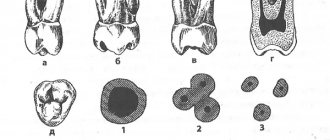
Molar removal
The lower molars have branched roots. One of the roots has a long, wide shape with a curved line. The second one is thin and has a deviation towards the back. There is a possibility that the roots will move closer to each other or move in different directions. Removing such teeth can be a difficult procedure. The surgeon uses forceps equipped with wide cheeks. To ensure a secure grip, the tongs have protrusions or spikes. Cases of tooth crushing and parts being extracted separately cannot be ruled out. It is not advisable to perform rotational movements during this procedure.
From medical practice, it is worth noting that wisdom teeth can sometimes have more than three roots. The roots may diverge to the sides and be twisted. In most cases, wisdom teeth are abnormal, which makes them difficult to remove.