Publication date: March 13, 2022.
Information on this page was updated on December 31, 2021.
TMJ dysfunction is a fairly common pathology these days, as it is largely caused by stress factors. Here it can be difficult to understand what is primary and what is secondary, because people with joint dysfunction usually come with bite pathology, pathology of the musculoskeletal system (curvature of the spine, neck). Therefore, joint treatment is a complex story. It happens that the primary pathology is a pathology of the joint, and sometimes it is the musculoskeletal system.
Comprehensive treatment of TMJ
When the doctor has determined the cause of the joint pathology, or causes, he determines the patient's readiness for a comprehensive treatment plan. In addition to the orthodontist, an osteopath or chiropractor may be involved, or even an orthopedist if more complex correction of the musculoskeletal system is required.
The patient should be aware that it is possible to straighten the jaw using a splint or splint, but this will not solve the problem of malocclusion. Orthodontic treatment will be required to correct the bite. If you have already had orthodontic treatment before, then it is more difficult to decide on repeated treatment.
Therefore, first the problem with the joint is solved using a splint or joint splint, then bite correction is carried out, and, if necessary, prosthetics. In parallel, work is underway with an osteopath to restore the muscular corset of the back and neck.
It happens that a patient refuses treatment with braces after the problem with the joint has been resolved. In this case, we warn him about the need to wear a joint splint constantly in order to avoid the recurrence of old problems with the TMJ. After all, a relapse can happen quite quickly due to stress.
Dentist-implantologist
Such a specialist is involved in treatment planning when there is a need for prosthetics.
If at the start of treatment some teeth are missing, then two tactics are considered.
- Tighten the spaces between teeth so that adjacent teeth replace the missing one. This is possible if, for example, the second molar (seven) was removed, but the third (eight, wisdom tooth) erupted. Functionally, these teeth differ little, so it is justified to move the figure eight to the place of the lost 7th tooth. But if we are talking about other teeth, then this treatment method can lead to asymmetry and an aesthetically unattractive result.
- Prepare the site for subsequent prosthetics. To do this, the teeth are not tightened, but rather, the space between them is expanded. During treatment with an orthodontist, you will have to spend some time with a noticeable gap in the row of teeth, and then contact an implantologist. This is a longer method of treatment, but the result is worth it: all the roots of the teeth will be in place, and an implant will be installed to replace the lost tooth.
This is what preparation for implantation looks like: with the help of braces, a place for the implant is prepared.
Sometimes the implantologist himself refers patients to the orthodontist. This happens if a patient applies for implantation, not knowing that malocclusion is an obstacle to prosthetics. In this case, the correct order of treatment is as follows: first correct the bite with orthodontic treatment, and only then install implants.
What may be the symptoms of TMJ dysfunction?
- Tenderness or pain in the area of one or both TMJs at rest or when opening the mouth.
- Crunching, clicking, crepitation and other noises in the area of one or both TMJs when opening the mouth.
- History of TMJ injuries (previous), incl. dislocation, subluxation, chronic subluxation.
- Restrictions in the mobility of the TMJ, restrictions in opening the mouth.
- Excessive tone of the masticatory muscles, bruxism (“grinding” of teeth in sleep, at rest).
- Asymmetry of the chin, lips, lip frenulum, asymmetry of mouth opening, S-shaped opening.
- Suspicion of a forced position of the lower jaw.
Structure of the TMJ
The presence of one or more of the above symptoms may indicate TMJ dysfunction.
Traditional orthodontic treatment does not address TMJ dysfunction. During orthodontic treatment, the severity of dysfunction may not change, decrease or increase. At the moment, in the world scientific orthodontic literature there is no convincing data on the connection between orthodontic treatment and the condition of TMJ. Deterioration of the joint after treatment may have nothing to do with this treatment.
Note! Even in the absence of visible clinical manifestations of joint dysfunction, hidden disorders may occur that require special diagnostics to identify them.
If there is a forced incorrect position of the lower jaw, its position may change during the treatment process with changes and complication of the treatment plan (the need to remove individual teeth, increasing the duration of treatment). A reliably forced position cannot be diagnosed by traditional orthodontic methods; to verify its presence, as a rule, a special analysis is required (manual functional analysis, determination of the central relationship of the jaws), the use of a special articular splint for a period of several months, which, however, does not give 100 % guarantees.
To conduct a detailed articular diagnosis, explain the specifics of your case, and further manufacture an articular splint, you can make an appointment with an orthodontist who deals with the issue of TMJ dysfunction.
TMJ dysfunction is a chronic condition that can be compensated, but not cured (i.e., it is possible to eliminate symptoms, however, pathological changes in the joints, if they have already occurred, will most likely persist).
How does TMJ arthrosis manifest?
Osteoarthritis of the maxillofacial joint can be caused by trauma, inflammation, the absence of a large number of teeth, or the use of dentures with poorly formed dentition. The causes of the development of pathology can also be metabolic disorders, connective tissue diseases or nerve conduction disorders.
Symptoms of arthrosis appear gradually as dystrophic changes develop. Most often patients complain of:
- aching pain that can radiate to the upper part of the skull - the area of the eyes or ears;
- characteristic crunching or clicking sounds that occur when the joint moves;
- decreased mobility of the bone joint, especially noticeable in the morning;
- displacement of the lower jaw to the right or left side from the center.
What happens if TMJ dysfunction is not treated?
If the dysfunction is not treated, the compensatory capabilities of the body may sooner or later be exhausted, the symptoms will worsen, the pathology will begin to progress, causing greater discomfort (sometimes for several years), thereby affecting the deterioration of the function of the dental system.
In order to try to prevent this and carry out treatment taking into account the individual characteristics of the structure and functioning of the temporomandibular joints, patients are usually offered the following approach.
How is an appointment with a gnathologist?
An appointment with a gnathologist begins with a conversation with the patient and collection of anamnesis. Then the specialist conducts a visual examination, checks the closure of the teeth, evaluates the functioning of the muscular system, the position of the head and the severity of facial asymmetry. During the examination, the doctor places the main emphasis on the temporomandibular joint - assesses the TMJ in a stationary position and during functioning.
The gnathologist also studies other elements of the muscular-ligamentous apparatus - checks the degree of tension in the muscles of the neck, back, and posture of a person. These measures help the gnathologist make a presumptive diagnosis and identify therapeutic tactics.
Mandatory additional diagnostic methods are computer and hardware studies. The doctor determines the list of relevant ones in a particular clinical case. Possible:
- orthopantomogram, survey x-ray of the temporomandibular joint - allow us to identify pathologies in the development of bone tissue of the ligamentous apparatus;
- CT scan – helps the gnathologist examine the joint layer by layer and detect degenerative changes in the tissues;
- magnetic resonance imaging – necessary to assess the condition of the periarticular soft tissues and articular disc;
- axiography, hardware recording of the movement of the lower jaw and graphic diagnostics;
- analysis of occlusal contacts;
- diagnostic jaw models made of plaster.
In the topic of functional diagnostics, myography occupies a special place - a method for diagnosing the masticatory muscles and the quality of teeth closure.
Computer programs help speed up diagnosis and increase the effectiveness of treatment for TMJ dysfunction. They allow us to offer the most optimal ways to correct the system.
Treatment method for TMJ dysfunction
1. Diagnosis of TMJ dysfunction.
- When diagnosing a joint in the clinic, a series of measurements and tests are carried out, all sensations in the joint area are recorded (discomfort, clicks, pain, deviation of the jaw when opening and closing), the difference in sensations in the right and left joint.
- The orthodontist also takes impressions of the jaws and takes photographs of the face and intraoral photographs, and also performs three-dimensional computed tomography of the face (3D CT); if necessary, the doctor can give a referral for an additional study - magnetic resonance imaging of the TMJ (MRI).
- Often, the orthodontist, in addition to manual functional analysis, conducts a visual assessment of: posture, symmetry of the shoulder girdle, shoulder blades, hip bone structures, etc., performs the necessary tests and photographs. Based on the results, it is possible to schedule a consultation with an osteopath or chiropractor to jointly manage the patient. Related specialists (orthopedist, surgeon, periodontist) can also be involved in drawing up a treatment plan.
What exercises are prescribed to patients to normalize the work and relax the masticatory muscles?
Exercise No. 1
Draw a vertical line on the mirror with a marker, stand opposite so that the line divides your face into the right and left halves, place your fingers on the area of the articular heads, lift your tongue up and back, open and close your mouth along the line (it may not work right away), 2-3 times /day 30 repetitions. There is no need to open your mouth wide (a comfortable width), the main thing is symmetrically (so that the jaw does not “move” in any direction). If there is a click, open until it clicks.
Exercise No. 2 (cycle)
Do it whenever possible, for example, in front of the TV, at the computer, or in a traffic jam while driving. Open and close your mouth without closing your teeth for 30 seconds, then alternately reach your right and left cheeks with your tongue for 30 seconds. Open - close your mouth again, then for 30 seconds move your tongue in a circle inside the vestibule (behind the lips), first in one direction, then in the other direction (clockwise - counterclockwise), open again - close your mouth, etc.. For this a half-hour cycle, the teeth should not touch, the lips should be closed. If you want to close your mouth or swallow, place your tongue between your teeth. Repeat the cycle for 20-30 minutes 2-3 times/day
What diseases are most often treated by an orthodontist?
Most often, patients come to this doctor to correct their malocclusion. Moreover, patients notice such pathologies
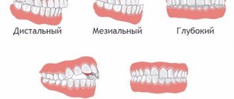
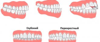
Among the pathological bite are:
- Mesial (progenic): in the anterior region, the lower incisors overlap the upper ones or form a direct closure. With this pathology, the patient experiences protrusion of the lower jaw.
- Distal: The upper front teeth protrude above the lower teeth. The front teeth do not close together; upon examination, a gap is visible between them. The lower jaw is behind the upper jaw.
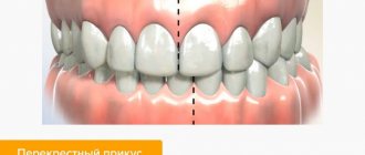
- Crossbite: there is a violation of the facial configuration, a displacement of the chin towards the lips.
Treatment of malocclusion must be started in a timely manner to avoid complications. We recommend that you consult with an orthodontist to determine if your situation has an overbite and how to treat it.
Occlusive therapy for TMJ dysfunction
After diagnosis, the patient is scheduled for an appointment with the orthodontist to determine the central relationship of the jaws (“true” position of the lower jaw, the position in which your joint and chewing muscles will be most comfortable).
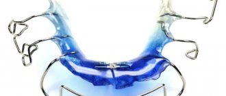
In order to more accurately establish and fix this position, an occlusal splint (splint) will be individually made for the patient from a special plastic, which is erased as it is worn. The splint must be worn constantly (sleeping, talking, eating in it if possible) - this is the meaning of occlusion therapy, which will help the joint and masticatory muscles rebuild into the most comfortable functional state.
Cleaning and caring for the splint is very simple - after eating (as well as while brushing your teeth), brush with a soft brush with toothpaste or soap.
Otolaryngologist
Cases where there is a direct connection between problems with the respiratory system and bite are not frequent, but sometimes a consultation with an ENT doctor is necessary. This is especially true for those who, before contacting an orthodontist, suffered from chronic ENT diseases with frequent exacerbations and impaired nasal breathing.
At the first appointment, the orthodontist collects information about the patient and asks about other diseases. A complete picture of your health condition will help you prescribe comprehensive treatment to achieve the best result and minimize the risk of regression.
Installation of a brace system for a patient with TMJ dysfunction
Installation of a brace system on the upper jaw is carried out on average after 3 months of occlusion therapy. The splint is adjusted once every 1-2 weeks, or at the discretion of the doctor, until the main complaints from the TMJ are eliminated (in parallel with the alignment of the teeth in the upper jaw), then a brace system is installed on the lower jaw with partial reduction (grinding) of the interfering parts of the occlusal tires, or complete removal. Here the patient needs to be patient - the process may take several months.
At the same time, the new position of the lower jaw is monitored: repeated manual functional analysis, photometry, bite registration is possible, computed tomography of the face during treatment, continuation of orthodontic treatment with a brace system.
Upon completion of orthodontic treatment, final monitoring of the position of the lower jaw follows (manual functional analysis, photometry, bite registration, 3D CT scan of the face upon completion (after) treatment).
Joint splint
Joint splint with braces
How does an orthodontist perform diagnostics?
An examination and diagnosis is required before starting treatment. During the consultation, the orthodontist conducts:
- collection of patient complaints and anamnesis data;
- examination of the patient’s face: symmetry, proportionality, and facial profile are assessed;
- when examining the oral cavity, the doctor fills out the dental formula, determines the bite, examines the frenulum of the lips, tongue, etc.;
If parents and a child come to the clinic for orthodontic treatment, the doctor conducts a survey of the parents about the child’s condition, its characteristics, etc.
Additional research methods in orthodontics include:
- Measuring plaster models of jaws: the doctor takes impressions of the jaws and casts plaster models. They are used to measure the dentition, diagnose their symmetry, and more.
- Orthopantomography (OPTG): the resulting image shows all the teeth, their roots, rudiments, and the structure of the bone tissue of the jaws.
- Teleradiography (TRG) in the direct and lateral surfaces: allows you to study the structure of the facial skeleton, establish an accurate diagnosis, and also predict the success of treatment.
During the consultation, the orthodontist will tell you what studies will be required in this case and what treatment methods are possible.
D
