until January 31
Free* consultation with dentists (generalist, surgeon, orthodontist) More details All promotions
Orthopantomogram
– a high-precision panoramic image of the teeth, allowing you to get a complete picture of the condition of the oral cavity. An orthopantomogram is done using a special device - an orthopantomograph, a type of X-ray equipment that has a circular rotating element. Compared to a traditional X-ray machine, the orthopantomograph provides a reduced radiation dose, so it can be safely used when the need for a panoramic image arises.
An orthopantomogram shows teeth, maxillofacial joints, facial bones of the skull, and nasal sinuses. The main advantage of an orthopantomogram is the ability to simultaneously obtain a panoramic image of the upper and lower jaws. It is the need to see the whole picture that immediately determines the need to use a panoramic photograph.
Orthopantomogram
Orthopantomogram - when you need a panoramic image of the jaws and what it gives
A panoramic photograph of teeth or orthopantomogram (OPTG) is a radiographic examination technique that makes it possible to study in detail the anatomical features of both jaws, the condition of soft and bone tissue, dental roots, maxillary sinuses and joints. The study is carried out using equipment with reduced radiation exposure and does not pose any threat to the health of the body. Today we’ll talk about what this is, why a panoramic shot is needed and how often it can be taken.
When is an orthopantomogram needed?
Orthopantomogram is used:
- in therapeutic dentistry. A panoramic photograph of the teeth allows you to identify periodontal problems, as well as carious cavities, and helps to create a comprehensive treatment plan;
- in surgical dentistry - before tooth extraction operations;
- in orthodontics. An orthopantomogram allows you to get a general idea of the state of your bite and select the right technique and hardware to correct it;
- in implantology – for planning implantation decisions.
What is OPTG and why is it performed?
Unlike a standard targeted photograph, an orthopantomogram allows you to obtain a fairly clear and detailed image of both jaws at once with the ability to study not only the condition of the teeth and their root system, but also the jaw bones, paranasal sinuses and other important anatomical structures. To understand what this diagnostic technique is, you first need to answer the questions: what does it do and why is it carried out. So, here's what the x-ray shows:
- deviations in the formation of occlusion - defects in bite, position, shape and size of teeth,
- early stages of caries and hidden foci of infection,
- periodontal pockets,
- neoplasms: cysts, granulomas, fibromas,
- hidden inflammatory processes and the extent of their damage,
- condition of the temporomandibular joint,
- impacted teeth that have not yet erupted - including wisdom teeth,
- condition of the root canals after treatment,
- diseases of the paranasal sinuses, for example, sinusitis.
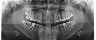
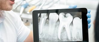
The procedure is mandatory before orthodontic treatment, prosthetics and, of course, implantation. In the latter case, the image gives the doctor the opportunity to carefully examine the condition of the jawbone, select suitable locations for implants, or prescribe a bone tissue augmentation procedure. To understand what the result of OPTG looks like, take a look at the example image below.
The photo shows a panoramic shot
Important! It should be noted that today, within the framework of one-stage implantation techniques, the preparatory stage involves undergoing a computed tomography scan. This is a more modern X-ray method that allows you to obtain a three-dimensional image of the patient’s jaws with the possibility of layer-by-layer study of tissues, without distorting the actual sizes and shapes of the teeth.
OPTG is also performed before treatment or removal of wisdom teeth or “eights”. The image will allow you to identify the exact location and position of the elements, detect hidden carious lesions and make a decision on the need for removal. In general, an orthopantomogram helps to carefully examine the clinical picture, make an accurate diagnosis and prescribe adequate treatment.
What equipment is used as part of the study?
This is not a new method - by now, OPTG has been actively used in dentistry for decades. And if earlier film cameras were used to obtain a panoramic image, today they have been replaced by more modern digital devices - orthopantomographs. The image is generated in a special computer program and, if necessary, can be printed on film.
Images taken using digital equipment are more accurate and detailed, which virtually eliminates the risk of making an incorrect diagnosis. Answering the question about how long a panoramic photograph of teeth is valid, it must be said that the information received will remain relevant for several months - a maximum of six months. If more time has passed since the examination, the procedure must be repeated.
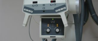
This is what an orthopantomograph looks like
Equipment for orthopantomography
A panoramic photograph of the jaw is performed using a special X-ray machine - an orthopantomograph -
which can be either film or digital. After dental x-rays, panoramic images can be printed using a digital orthopantomograph. At Dial-Dent, a panoramic image of the teeth is taken digitally, the image is entered into the patient’s computer record, and any clinic specialist can quickly view it to clarify the diagnosis and plan treatment. If it is necessary to consult with doctors in other clinics, a panoramic photograph of the teeth is printed on paper or the results are recorded on electronic media. The presence of a computed tomograph at the Dial-Dent clinic makes it possible to obtain a panoramic image of higher accuracy.
Indications and restrictions
The procedure is completely painless, carried out as quickly as possible and without any discomfort for the patient. OPTG is prescribed in the following cases:
- as part of implantation – carried out to study bone tissue, select implant models and places for their implantation. Based on the data obtained, the doctor will be able to draw conclusions about the advisability of building up the jawbone,
- to assess the quality of dental canal treatment, the condition of the roots - before prosthetics,
- before starting orthodontic treatment - before installing braces and other corrective structures,
- before an upcoming operation, complex removal, including wisdom teeth,
- to examine the dental system in children, assess the correctness of its formation and identify possible deviations,
- in order to determine the extent of damage to periodontitis and periodontal disease,
- to identify neoplasms in hard and soft tissues.
This procedure may be required in various clinical cases.
X-ray diagnosis is undesirable in the first and last trimester of pregnancy - it is carried out only in the presence of emergency indications and with the permission of a gynecologist.
Orthopantomogram: how is the procedure performed?
Painlessness, speed and minimal radiation exposure are what make this study so popular and in demand in dental practice. A panoramic dental photograph is taken for all clients of the Mira clinic who have appropriate indications.
An orthopantomogram may be ordered before and after the following procedures or problems:
- implantology and prosthetics (with the aim of studying the characteristics of bone tissue, the location and size of the maxillary sinuses, etc.);
- trauma (for a comprehensive assessment of the volume of the injured area);
- tooth extraction (for the purpose of complete visualization of the teeth configuration);
- some inflammatory diseases of the teeth and oral cavity (the appropriateness of the examination is determined by the clinic and the course of the disease);
- pathological formations in the oral cavity (granulomas, cysts, etc.);
- clinical situations requiring assessment of the degree of formation of tooth roots, visualization of joints and sinuses, assessment of the treatment performed.
Of course, dentistry also uses other instrumental methods for diagnosing diseases of the teeth and oral cavity. However, if we talk about the ratio of price and quality, not forgetting to take into account the information content, conditions and time required for the study, as well as the availability of such diagnostic systems, then the priority of performing these studies for patients becomes clear.
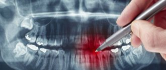
Usually, OPG of 2 jaws is taken at once, but images of each jaw can be taken separately. This image perfectly reflects the structure of the teeth and allows you to identify violations in the area of the roots and structure of the jaw. In addition, the study provides comprehensive information about the condition of soft tissues.
A panoramic photograph of the jaw is effective for preventive purposes, and is also indispensable in case of severe tissue damage. Common inflammatory diseases such as periodontal disease and caries, including under crowns and in the area of wisdom teeth, will be visible in the image. The condition of wisdom teeth can also be easily determined using a circular image. This way the doctor will be able to determine the boundaries of inflammation and prescribe effective treatment.
A detailed reflection of the situation in the oral cavity on a panoramic photograph of the jaw will allow you to make an accurate diagnosis. This procedure uses a small amount of X-ray radiation, so the patient will first be wearing a lead apron and collar.
Features of conducting OPTG for children
We figured out what is visible in panoramic photographs and why they are taken, and found out that this is a completely safe diagnostic method. Now it should be noted that an orthopantomogram for children is carried out taking into account some nuances. A radiation exposure of 55 μSv (1 shot) is completely safe for a child, so such a procedure is allowed from 5-6 years of age.
The examination requires the patient to stand still for some time, but for young children this task can be quite difficult. When it comes to small patients, the procedure is carried out in exposure mode - the area of exposure is reduced, and the sensor is adjusted to a trajectory of movement corresponding to the small size of the jaws.
“My son also recently had a panoramic photo taken. He's 10, everything went great. Now there are new technologies everywhere, everything is safe. The procedure took less than a minute, and the doctors assured me that there was nothing wrong with it. How else can you start correcting your bite? The orthodontist prescribed OPTG for us. So far I’ve made the plate, but in a couple of years we’ll get braces. There’s no place here without pictures!”
Lyudmila V., Chelyabinsk, from correspondence on the woman.ru forum
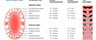
In pediatric dentistry, OPTG is used to detect the rudiments of permanent teeth, assess the correctness of their formation, to diagnose adentia and identify malocclusions. Most often, this procedure is prescribed by an orthodontist before starting to correct defects in bite and teeth position.
The procedure is completely safe for the child
What does an orthopantomogram show?
The overview photograph is a mirror image of the dental system: its right side is shown on the left, and the left side on the right. To facilitate orientation, the image is often marked: L and R (left, right).
To number the teeth, the image is divided into 4 segments. The 2 top ones - left and right, are “tens” and “twenties”, and the 2 bottom ones, the same left and right, are “thirties” and “forties”. The teeth are numbered starting from the center of the dentition, so in the center at the top there are incisors 11 and 21, and at the bottom - 41 and 31, and the row ends, as a rule, with wisdom teeth with numbers 18 and 28 at the top and 48 and 38 at the bottom.
In addition to the dentition, the following images are clearly visible in the panoramic image:
- mandibular canal with neurovascular bundle;
- maxillary sinuses and nasal passages;
- TMJ - temporomandibular joints;
- fillings and sealed canals (for this purpose, radiopaque components are introduced into the filling materials);
- wisdom teeth, supernumerary teeth, and in a child’s photograph - the rudiments of permanent teeth;
- hard palate with zygomatic bones, hyoid bone, nasal septum, conchae and other structures.
Harm and degree of radiation exposure
Let's start with the fact that radiation exposure is measured in micro- or millisieverts - μSv or mSv, respectively. According to the resolution of the Ministry of Health of the Russian Federation, the permissible radiation dose within the framework of OPTG for an adult patient should not exceed 55 μSv, and for children under 15 years of age - 24 μSv1. It should be noted right away that only digital X-ray equipment meets these requirements, while film devices provide a slightly higher load. To determine how many times you can take a panoramic photo, you need to take into account that the maximum permissible radiation dose for an adult patient per year corresponds to 1000 μSv.
However, it is still not worthwhile to use such procedures frequently, even despite their obvious safety and harmlessness. X-ray irradiation has a negative effect on a weakened body, especially with repeated examinations. The children's body is more susceptible to this kind of influence, therefore it is strictly forbidden to exceed the permissible dosages when examining small patients.
When contacting the clinic for OPTG, be sure to clarify what type of equipment is used and what the degree of radiation exposure from one image will be. Some private dentistry purchases cheap, outdated equipment, which can produce 2-3 times the recommended dose. The best option where you can take a panoramic photograph of your teeth is a specialized diagnostic center equipped with modern high-precision equipment.
Where to take a panoramic photo of your teeth
Of course, not every clinic can take a panoramic photo of your teeth in Moscow. If the dental clinic does not have its own orthopantomograph, then the question will arise of where to take a panoramic photograph of the teeth. You can find a clinic where they take panoramic images (orthomantomogram), but traveling around Moscow is a significant investment of time. When choosing a dental clinic, it is advisable to immediately pay attention to whether the clinic has its own orthomantomograph. Just as mentioned above, when being observed in one clinic, you know exactly the total radiation dose received during the research. Dial-Dent uses the most modern equipment to carry out panoramic photographs of teeth: an Orthophos orthopantomograph (Germany) and a Samsung Rayscan Symphony Alpha Pano 3D tomograph.
| orthopantomograph Orthophos | computed tomograph Samsung Rayscan Symphony Alpha Pano 3D |
Digital OPTG - description of the process
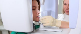
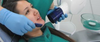
Now let's move on to consider the question of how a panoramic photograph of teeth is taken. To begin with, the patient is asked to remove all metal products from himself so that they do not affect the operation of the equipment and the final result. Next, the patient stands in the place indicated by the specialist, bites down on a special plate that ensures the immobility of the jaws, and after turning on the orthopantomograph, its moving part begins to rotate around the head.
The photo shows the process of performing an orthopantomogram
The procedure takes literally half a minute - the exact duration will depend on the type and newness of the equipment. Next, the resulting image is formed in digital form, that is, in a special computer program, or on film media (outdated version). A digital photograph can also be printed onto film if necessary.
What does an orthopantomogram show?
A panoramic photograph of the teeth helps to detect:
- dental anomalies;
- caries, including at the initial stage;
- periodontal pockets;
- tooth cyst;
- foci of inflammation.
Using an orthopantomogram it is possible to establish:
- the need to begin correcting malocclusion;
- condition of periodontal tissues;
- degree of formation of tooth roots;
- when installing implants (for example, the condition of the bone at the site of intended implantation);
- and much more.
Information content of the image and reading (deciphering) technique
If you undergo an examination at a specialized diagnostic institution, the finished image is usually accompanied by an explanation from a specialist with a list of detected problems. Many people are interested in how to decipher an image on their own, how to read it correctly, and whether it can be understood at all. It should be noted here that in order to correctly explain the results of OPTG, you need to have a good understanding of the anatomy of the maxillofacial apparatus, and without the appropriate education, little can be understood. It is better to entrust this task to an experienced, qualified doctor.
Orthopantomogram during implantation
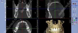
As part of implant treatment, a panoramic image is taken more than once. First, an X-ray examination is carried out at the preparatory stage - the doctor carefully examines the resulting image, assesses the condition of the bone tissue, selects places for implants, and makes a verdict on the need for bone grafting. If we are talking about one-stage immediate loading protocols, then patients are usually sent for a computed tomography scan of the jaw - a 3D image provides more detailed information about the clinical picture.
3D diagnostics makes it possible to study a more detailed three-dimensional image
You will have to undergo a repeat examination after implantation. In this case, OPTG will give the doctor the opportunity to assess the quality of the operation performed, monitor the process of engraftment of artificial roots, and recognize possible complications in the later stages of their development. Several photographs may be required during the osseointegration period of the implants.
What are the advantages and disadvantages of OPTG
An orthopantogram can significantly improve the quality of diagnosis, and today dental treatment is practically not carried out without radiography, except for minor problems. Among the undeniable advantages of the technique, experts highlight the following points:
- speed of the procedure - literally in a couple of minutes the image is ready for study,
- modern equipment makes it possible to adjust the height and trajectory of the emitter, which made it possible to undergo diagnostics for children and wheelchair users,
- minimal radiation exposure and absolute safety for the body,
- high quality and accuracy of the obtained images,
- the ability to zoom in on the area under study and study it in detail on a computer monitor.
An orthopantomogram uses minimal radiation exposure.
If we talk about the disadvantages, the main disadvantage of OPTG is obtaining a flat two-dimensional image. As a result, the image may slightly distort the dimensions of teeth, root canals and bone tissue. Such deviations can vary from 15 to 30%. To obtain a more accurate image, it is better to resort to computed tomography.
What is the advantage of digital panoramic dental photography?
- Reduced exposure time, since the digital camera takes less time to create an image. This results in a reduction in radiation exposure to the patient by up to 90%.
- There is no need to develop films, which ensures fast image acquisition, eliminates the need to work with chemicals, and also eliminates the need for repeated x-rays in case of poor-quality development.
- It becomes possible to further examine the resulting images by enlarging individual areas using software.
- Possibility of convenient archiving and quick access to the database with images.
- Immediate transfer to the attending physician located outside the clinic.
- Convenient storage in the form of files.
Approximate prices
How much a panoramic dental photograph will cost directly depends on whether the study will be carried out using film or digital equipment - in the latter case, the cost will be an order of magnitude higher. Approximate prices are given below:
- a photograph on a film camera – about 700 rubles,
- digital photo - from 1000 rubles and 150-200 rubles for printing on film.
We have already mentioned above that today, for a more detailed layer-by-layer study of the tissues of the jaw apparatus, computed tomography (CT) is performed. Essentially, this is the same panoramic photo, but much more detailed and in 3D format. An important advantage of this technique is the accurate transmission of the sizes and shapes of all elements of the dental system.
- Vorobyov Yu.I. New possibilities of X-ray diagnostics in dentistry, 2002.