Teeth of ancient people
The dentofacial apparatus of prehistoric and modern humans differs significantly. Ancient people had more than 36 teeth, protruding fangs and a massive jaw. This was explained by the need to chew rough food and raw meat. With the addition of thermally processed foods to the diet, the dentition began to change. The canines were the first to transform, becoming aligned with the bite line. Then the jaw arch narrowed, the interdental spaces disappeared, and the teeth themselves decreased in size. Currently, 32 teeth in humans are the norm, but third molars are considered to be an atavism.
Interesting fact!
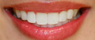
The teeth of ancient man cannot be called aesthetic, but they were healthy. According to scientists, cavemen never suffered from caries and other oral diseases.
Which teeth bite is not considered correct?
The presence of deviations from the norm is at least the basis for a dental diagnosis, based on the results of which a decision is made on the need for treatment. The best option is orthodontic correction in childhood, since the dentofacial apparatus, which is in the stage of formation, is more easily amenable to the targeted influence of a special machine and takes on the desired structure without surgical intervention. The most commonly diagnosed abnormal forms have certain signs that allow us to understand whether a person’s bite is correct or not, what is going wrong, and how this deviation can affect the further development of the jaw:
- Distal - diagnosed with excessive development of the upper jaw, noticeably protruding forward and distorting facial features.
- Mesial is a reverse pathological condition in which the mandibular row protrudes, almost completely covering the incisors and canines.
- Cross - characterized by the presence of free areas between the teeth, as well as a “scissor” intersection of elements in a random order.
- Deep - significant, more than 30%, overlap of the lower section.
- Open - contact during closure is observed only between individual chewing units, while in the incisal region there are pronounced gaps, creating the impression of a constantly slightly open mouth.
Any pathological manifestation requires timely medical intervention. Not all patients know what a correct bite in a person is and what it is needed for - but this indicator affects the quality of respiratory and speech function, ensures the conditions for normal food processing and the functioning of the gastrointestinal tract. By contacting Dentika dentistry, you can undergo a comprehensive diagnosis, receive recommendations from qualified specialists and, if necessary, resort to orthodontic correction services.
Name of human teeth
Depending on the location and structure, dental units have their own functional characteristics and are called differently.
- Incisors.
On both jaws there are four front teeth in humans - medial and lateral incisors, which are used for biting food. - Fangs.
Sharp teeth designed for chewing hard foods. - Premolars.
"Fours" and "fives" on the left and right sides of each jaw arch grind soft or small pieces of food. - Molars.
Three large outer teeth in each row are aimed at grinding coarse substances. - The canines
and incisors are part of the anterior group, or the “smile zone,” and the human molars are part of the chewing segment.
In addition, teeth are divided into temporary and permanent. In the first case, we are talking about dairy products that appear in children from the fifth month of life to three years. The second refers to the final bite, which is formed between six and thirteen years of age. Milk teeth differ from permanent teeth only in size, but in structure they are identical.
Prosthetics of frontal units
Prosthetics of anterior teeth is a complex process from a technical point of view. It requires the doctor to take special care in carrying out each manipulation, since as a result the patient hopes to receive an exact copy of a healthy tooth with the appropriate shape, shade and strength. Progressive prosthetic methods allow you to achieve ideal results
in recreating the integrity and beauty of the frontal zone.
For high-quality restoration of segments in the smile area, three crown options are used, which are the most acceptable, both in terms of strength and aesthetics, for replacing natural segments:
- Zirconium.
- Ceramic.
- Metal-ceramic.
If it is necessary to install dentures on the front teeth, crowns made with ceramic or zirconium are first considered. The fastest and most promising option is an orthopedic crown on a tooth using CEREC technology. You can get an attractive smile in just one visit. However, it is also the most expensive. A compromise in this case could be a metal-ceramic prosthesis.
A turnkey metal-ceramic tooth crown is an excellent combination of price, quality and aesthetics. The only drawback is the metal base; it can be visible through the ceramics. But the defect is noticeable if the crown is on only one segment. If you plan to install a crown on four or six front teeth
, it will be difficult to distinguish natural teeth from dentures.
Today, very often, patients no longer choose metal-ceramics, but e-max or cerec ceramics, which not only do not have the disadvantages of metal-ceramic crowns, but also look aesthetically more beautiful.
Classic dental prosthetics, regardless of the location of the crown, is carried out in stages.
How many teeth does a person have?
The number of teeth a person has depends on age and anatomical features. The child has a set of 20 primary teeth, which are replaced by a permanent bite of 28 teeth. Third molars erupt, as a rule, after twenty years or do not grow at all, which is not a pathology.
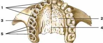
In dentistry, a single numbering of human teeth is adopted. Doctors classify teeth as lower and upper and distinguish the right and left segments of the jaws. Each of them includes two incisors, a canine, two premolars and three molars. The countdown starts from the first front tooth and ends, accordingly, with a figure eight. Sometimes a number is added to the serial number indicating the location zone. For example, the right canine of the top row is numbered 13. This order in the schematic representation is called the formula of human teeth.
Polyodontia
In rare cases, an anomaly such as polyodontia is observed - supernumerary, or extra teeth in a person. Dental units can appear in the primary and permanent dentition anywhere in the jaw, separate from or fused with the main teeth. The defect affects not only the aesthetics of the smile, but also leads to the formation of incorrect occlusion, impairs the quality of chewing food and diction. Most often, supernumerary teeth are removed in childhood or built into the dentition.
Edentia
There is also a deviation of the opposite meaning called edentia - congenital or acquired absence of dental units. The causes of the phenomenon include heredity or improper development of the embryo in the womb. People without teeth cannot fully eat and speak, have a deformed facial contour and weakened immunity.
Aesthetic rehabilitation of anterior teeth using direct composite restorations
D. Volkov
specialist in the field of endodontics, member of the RSO, participant in international congresses VDW Endodontic Synergy
K. N. Khabiev
Ph.D., specialist in aesthetic rehabilitation, group president
Harmony and naturalness of a smile are important factors for the successful social integration of a modern person, the key to his inner confidence when communicating with others. Increasingly, the dentist has to solve not only medical problems related to the prevention and treatment of various specialized diseases, but also eliminate the aesthetic problems of the patient’s smile.
The most popular method of restoring teeth is direct composite restoration. With its help, you can not only restore the volume of tissue lost due to a pathological process, but also correct the anatomical shape of the teeth to harmonize the patient’s smile ensemble. To successfully implement a rehabilitation plan using direct composite restorations, a material is required that meets many requirements, such as high mechanical strength and resistance to abrasive wear, optical characteristics characteristic of natural tissues, good polishability and durability of the “dry shine” of the surface over time. In this article, we will consider the nanofilled restoration composite CAPONatural (SchützDentalGmbH) as the material of choice.
The reasons for the choice are the flagship strength properties (Young's modulus - 11173 MPa; Vickers hardness 784 MPa; compression load 502 MPa), refractive index -1.62, corresponding to the natural refraction of enamel, a wide palette of colors adapted to the stratified tooth restoration technique. The low volumetric shrinkage rate (1.2%) due to the high filling of the polymer matrix makes it possible to significantly simplify handling techniques for introducing and distributing portions of the material.
Restoration requires a material that meets many requirements, such as strength and wear resistance, and optical characteristics characteristic of natural tissues. Using only three layers of material, you can achieve a full match of the color of the restoration to the color of the tooth being restored. The main task of a restorative dentist is to achieve equal transparency of the tooth being restored and the restoration. The first layer of dentin should be two shades darker than the base color. If the main color is A3, the first shade should be A4. We use it to restore 70-80 percent of the tooth. Then the main dentin shade is applied (up to 90% of the restoration). The remaining 0.3-0.5 mm are restored using an enamel shade according to Table No. 1.
Table No. 1
Tooth color according to Vita Dentin 1 Dentin 2 Enamel A1 A3 A1 Incisal white A2 A3.5 A2 Incisal medium A3 A4 A3 Incisal medium A3.5 A4 A3.5 Incisal clear A4 A4 A4 Incisal clear B1 A2 A1 Incisal white B2 A3.5 B2 Incisal medium B3 A4 B3 Incisal clear C2 A3.5 C2 Incisal clear C3 A4 C3 Incisal clear
Shades for creating a snow-white smile light Primary tooth color Bleach 1 Incisal white extralight Primary tooth color Bleach 2 Incisal white
Clinical case
Initial situation: the existing composite restorations, made about 5 years ago using the direct method, are aesthetically untenable: teeth 11 and 12 have a pronounced discoloration, teeth 11 and 23 show marginal depressurization and staining of the fillings. The surfaces of all restorations do not have a “dry shine” and are visually defined as matte.
On the palatal surface of teeth 12, 11, 21, 22, 23, marginal depressurization and staining of fillings and secondary caries are observed. There is deep caries on the proximal surfaces of teeth 13 and 12 (Fig. 1-3).
Rice. 1. Initial situation. Rice. 2. Initial situation from the palatal surface. Rice. 3. The patient’s smile before treatment.
After making the silicone key, the old restorations were removed; after removing the material, strong gray-brown pigmentation of the dentin of tooth 12 was observed. Additional translucent optical diagnostics were carried out to determine the contours of the dentinal body (Fig. 4-5).
Rice. 4. Making a silicone key. Rice. 5. View after removal of old restorations.
A layer of transparent enamel (incisal medium) is applied to the silicone key (Fig. 6). The palatal surfaces of the teeth were restored using a silicone key with an incisal medium enamel shade (Fig. 7-8).
Rice. 6. Applying a layer of transparent enamel (incisal medium) to the silicone key. Rice. 7. Restoration of the palatal surface using a silicone key. Rice. 8. The palatal surface was restored with incisal medium enamel shade.
Restoration of contact points using a transparent matrix and wedge (Fig. 9). The cervical area of the teeth was restored with Capo Slow flow composite material, color A4 (Fig. 10).
Rice. 9. Restoration of contact points using a transparent matrix and wedge. Rice. 10. Restoration of the cervical area with flowing composite material Capo Slow flow (A4).
Restoration of 70-80% of the tooth with a dark dentin shade (A4) (Fig. 11). Applying a shade of the main color (A3) and forming mamellons (A1) (Fig. 12) and the enamel layer (incisal medium) (Fig. 13).
Rice. 11. Restoration of 70-80% of the tooth with a dark dentin shade (A4). Rice. 12. Applying a shade of the main color (A3) and forming mamellons (A1). Rice. 13. Applying an enamel layer (incisal medium).
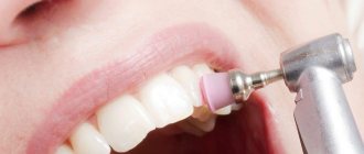
The final appearance of the restoration after polishing with PoGo heads (Dentsply) and DialogGlass micron paste (SchützDentalGmbH) based on aluminum oxide. Enhance polish cup (Dentsply) polishing sponges were used as a carrier (Fig. 14).
Rice. 14. Final view of the restoration after polishing with PoGo heads.
In Fig. 15-16 shows the view of the restoration the next day, in Fig. 17-18 - 5 years after the start of treatment.
Rice. 15. View of the restorations the next day. Rice. 16. View of restorations from the palatal side. Rice. 17. View of restorations after 5 years. Rice. 18. The patient’s smile 5 years after the start of treatment.
Using only 3 layers of material, you can achieve a full match of the color of the restoration to the color of the tooth. The main task is to achieve equal transparency.
Analyzing this clinical case, we can confidently state that a nanofilled restoration composite material, which has high physical and mechanical characteristics and optical parameters similar to natural tissues, can be successfully used not only for local aesthetic restoration of a specific tooth, but also for complex artistic rehabilitation of the patient’s smile as a whole.
Dimensions of human teeth
The upper central incisors are twice as wide as their antagonists. The remaining dental units of the same name have approximately equal parameters. The size is determined using special tables with the optimal size and permissible deviations. Experienced doctors calculate proportions by dividing the length of a person’s teeth by the width. A result of about 0.75 millimeters is considered close to ideal. For more detailed diagnostics, other professional formulas and techniques are used.
Size deviations from the norm occur due to improper formation of the jaw, fusion of tooth buds, or genetic predisposition. Teeth that are too large are called macrodentia, and abnormally small teeth are called microdentia. Pathologies are accompanied by problems with bite and chewing functions, but can be successfully corrected by a dentist.
Interesting fact!
The longest tooth in the world belongs to an Indian teenager. The size of its crown is almost four centimeters. About a year ago, the tooth was removed, and the young man was included in the Guinness Book of Records.
When do baby teeth appear?
The formation of a child’s teeth begins in the womb: this is partly why doctors recommend that pregnant women eat foods rich in calcium and other beneficial minerals. After birth, the first baby tooth (in the vast majority of cases, this is the lower incisor) appears when the baby is six months old. There are cases when the first teeth erupt already in the third month, and sometimes a child is already born with one or more teeth (among many peoples this is considered a lucky sign). Conversely, some children acquire their first teeth after reaching one year of age. Below you can see a table that shows the current timing of the appearance of baby teeth.
Milk teeth on the lower jaw
| Name of teeth | Age of appearance |
| Central incisors | 6 – 9 months |
| Lateral incisors | 10 – 14 months |
| Fangs | 17 – 21 months |
| Premolars | 13 – 16 months |
| Molars | 23 – 30 months |
Milk teeth on the upper jaw
| Name of teeth | Age of appearance |
| Central incisors | 8 – 11 months |
| Lateral incisors | 9 – 12 months |
| Fangs | 16 - 19 months |
| Premolars | 13 – 16 months |
| Molars | 25 – 32 months |
The structure of the human tooth
Anatomy
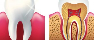
From an anatomical point of view, a human tooth consists of three parts.
- Crown.
The visible part protruding above the gum. It has four sides: the occlusal, or cutting edge, in contact with the antagonist teeth; contact wall adjacent to adjacent dental units; vestibular and lingual surfaces facing the lips and tongue, respectively. - Root.
Fixed in the socket by connective tissue, located in the recess of the jaw. As a rule, premolars have two roots, and molars have three, four or even five. The remaining dental units have one root canal. - Neck.
It is located between the coronal part and the root of a human tooth, surrounded by periodontium.
Histology
What are human teeth made of? Let's look at the cross-section of the structure of a human tooth.
- Enamel.
A transparent protective coating of the crown, almost entirely consisting of inorganic microelements. - Dentine.
The hard base of the tooth, containing 80% mineral components and 20% organic substances. The shade of dentin is responsible for the color of dental units, as it shines through the enamel. - Cement.
The bone tissue covering the tooth root. Plays the role of a fastening element connecting the tooth to the alveolus. - Pulp.
Soft tissue filled with bundles of nerves and capillaries. Painful sensations during caries are explained precisely by the presence of nerve endings.
Two methods of prosthetics for front teeth for caries:
1. The crown on the front teeth is attached to a pin
– a miniature screw rod is screwed into the sealed root canal. Filling material is applied to its upper part, ground to the required size, and a crown is installed. But again, pins are gradually being replaced by the more patient-friendly CEREC technology. And patients come to me with problems with previously installed pins with the goal of replacing them with a single dental module, as in the following video:
2. Crown on the front tooth using a stump inlay
– this element is made in the laboratory. Outwardly, it resembles a tooth stump. It is securely attached to the root canal and the prosthesis is securely fixed on its upper part (stump). But, despite its popularity, the stump inlay has one significant drawback - it forms an additional adhesive connection both with the tooth tissue and with the crown being installed. This can be avoided by restoring the front teeth using CEREC technology. When using Cerec technology and using “root + crown” type modules, we reduce the gluing points to just one:
Installation of dentures using “root + crown” modules on the front teeth increases the service life of the crowns. In addition, they allow you to create the ideal tooth shape, which is important when replacing teeth in the smile area.
Human wisdom teeth
A “wisdom tooth” is the third outer molar with three to five roots. In structure it is no different from its “neighbors”. To the question “How many wisdom teeth does a person have?” cannot be answered unambiguously. They erupt around the age of twenty, one on each side of both jaws. However, there are people without wisdom teeth. This is a variant of the norm, since in the process of human evolution the need for the “eight” disappeared, and the structure of the jaws underwent corresponding changes. Today, third molars are considered a vestigial organ.
Tooth development.
The formation of the rudiments of baby teeth in the fetus begins at 4–5 months of intrauterine development. That is why the mother’s illnesses during this period lead to disruption of various stages of dental development, for example, a violation of the mineralization of the tooth occurs. As a result, the enamel can become weak and brittle. Taking certain medications during this period can also affect the development of teeth. For example, if the mother was treated with tetracycline antibiotics, the child’s teeth will be dark yellow or even brown (so-called tetracycline teeth).
Enamel mineralization begins in the prenatal period and continues for 6 months after birth. Mineralization of the crown part of the teeth has time to take place in utero, while mineralization of the cervical region of the incisors, canines and molars continues after birth, in completely new and not always favorable conditions. The mineralization process can be negatively affected by the nature and diet, social living conditions and various diseases of the child (acute respiratory infections, intestinal infections and functional diseases). That is why the cervical area is the most vulnerable and typical place for the occurrence of “bottle” caries of primary teeth.