Anatomical structure of the pharynx
The pharynx, another name is the pharynx.
It starts at the back of the mouth and continues down the neck. The wider part is located at the base of the skull for strength. The narrow lower part connects to the larynx. The outer part of the pharynx continues with the outer part of the mouth - it has quite a lot of glands that produce mucus and help moisten the throat during speech or eating. When studying the anatomy of the pharynx, it is important to determine its type, structure, functions and risks of disease. As mentioned earlier, the pharynx is shaped like a cone. The narrowed part merges with the laryngopharynx, and the wide side continues the oral cavity. There are glands that produce mucus and help moisten the throat during communication and eating. From the front side it connects to the larynx, from above it adjoins the nasal cavity, on the sides it adjoins the cavities of the middle ear through the Eustachian canal, and from below it connects with the esophagus.
The larynx is located as follows:
- opposite 4 - 6 cervical vertebrae;
- behind - the laryngeal part of the pharynx;
- in front - formed due to the group of hyoid muscles;
- above - hyoid bone;
- lateral - adjacent to the thyroid gland with its lateral parts.
The structure of a child's pharynx has its own differences. Tonsils in newborns are underdeveloped and do not function at all. Their full development is achieved by two years.
The larynx includes in its structure a skeleton, which contains paired and unpaired cartilages connected by joints, ligaments and muscles:
- unpaired consist of: cricoid, epiglottis, thyroid.
- paired ones consist of: corniculate, arytenoid, wedge-shaped.
The muscles of the larynx are divided into three groups and consist of:
- thyroarytenoid, cricoarytenoid, oblique arytenoid and transverse muscles - those that narrow the glottis;
- posterior cricoarytenoid muscle - is paired and expands the glottis;
- vocal and cricothyroid - strain the vocal cords.
Entrance to the larynx:
- behind the entrance there are arytenoid cartilages, which consist of cornuform tubercles, and are located on the side of the mucous membrane;
- in front - epiglottis;
- on the sides there are aryepiglottic folds, which consist of wedge-shaped tubercles.
The laryngeal cavity is also divided into 3 parts:
- The vestibule tends to stretch from the vestibular folds to the epiglottis.
- Interventricular section - stretches from the inferior ligaments to the superior ligaments of the vestibule.
- Subglottic region - located at the bottom of the glottis, when it expands, the trachea begins.
The larynx has 3 membranes:
- mucous membrane - consists of multinucleated prismatic epithelium;
- fibrocartilaginous membrane - consists of elastic and hyaline cartilages;
- connective tissue - connects part of the larynx and other formations of the neck.
Pharynx: nasopharynx, oropharynx, swallowing department
The anatomy of the pharynx is divided into several sections.
- How is swallowing done?
Each of them has its own specific purpose:
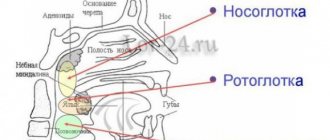
- The nasopharynx is the most important section, which covers and merges with special openings into the back of the nasal cavity. The function of the nasopharynx is to moisturize, warm, clean the inhaled air from pathogenic microflora and recognize odor. The nasopharynx is an integral part of the respiratory tract.
- The oropharynx includes the tonsils and uvula. They border the palate and the hyoid bone and are connected by the tongue. The main function of the oropharynx is to protect the body from infections. It is the tonsils that prevent the penetration of germs and viruses inside. The oropharynx performs a combined action. Without its participation, the functioning of the respiratory and digestive systems is not possible.
- Swallowing department (hyopharynx). The function of the swallowing department is to carry out swallowing movements. The laryngopharynx is related to the digestive system.
There are two types of muscles surrounding the pharynx:
- stylopharyngeal;
- muscles are compressors.
Their functional action is based on pushing food towards the esophagus. The swallowing reflex occurs automatically when muscles tense and relax.
The process looks like this:
- In the oral cavity, food is moistened with saliva and crushed. The resulting lump moves towards the root of the tongue.
- Further, the receptors, irritating them, cause muscle contraction. As a result, the sky rises. At this second, a curtain closes between the pharynx and nasopharynx, which prevents food from entering the nasal passages. The lump of food moves deep into the throat without any problems.
- Chewed food is pushed down the throat.
- Food passes to the esophagus.
Since the pharynx is an integral part of the respiratory and digestive system, it is able to regulate the functions assigned to it. It prevents food from entering the respiratory tract during swallowing.
What functions does the pharynx perform?
The structure of the pharynx makes it possible to carry out serious processes necessary for human existence.
Functions of the pharynx:
- Voice-forming. Cartilage in the pharynx takes control of the movement of the vocal cords. The space between the ligaments is constantly subject to change. This process regulates the volume of the voice. The shorter the vocal cords, the higher the pitch of the sound produced.
- Protective. The tonsils produce immunoglobulin, which prevents a person from becoming infected with viral and antibacterial diseases. At the moment of inhalation, the air entering through the nasopharynx is warmed and cleared of pathogens.
- Respiratory. The air inhaled by a person penetrates the nasopharynx, then the larynx, pharynx, and trachea. The villi located on the surface of the epithelium prevent foreign bodies from entering the respiratory tract.
- Esophageal. The function ensures the functioning of swallowing and sucking reflexes.
The diagram of the pharynx can be seen in the next photo.
- The structure of the human throat and larynx: photo
Diseases affecting the throat and pharynx
Diseases of the ENT organs can be triggered by an attack of a viral or bacterial infection. But pathology is also caused by fungal infections, the development of various tumors, and allergies.
Pharynx diseases manifest themselves:
- ARVI;
- sore throat;
- tonsillitis;
- pharyngitis;
- laryngitis;
- paratonsillitis.
Only a doctor can determine an accurate diagnosis after a thorough examination and based on the results of laboratory tests.
Possible injuries
The pharynx can be injured as a result of internal, external, closed, open, penetrating, blind and through injuries. Possible complication - blood loss, suffocation, development of a retropharyngeal abscess, etc.
First aid:
- in case of injury to the mucous membrane in the oropharynx area, the damaged area is treated with silver nitrate;
- deep injury requires the administration of tetanus toxoid, analgesic, antibiotic;
- severe arterial bleeding is stopped by finger pressure.
Specialized medical care includes tracheostomy and pharyngeal tamponade.
Human larynx: its structure and functions
The human larynx is a flexible, finely structured organ of the respiratory system that connects the pharynx to the trachea. It is extremely important for the breathing and digestion process, as it pushes out harmful elements trying to enter the respiratory tract. Sounds are also produced in the larynx; with the help of the vocal folds, the timbre, tone and volume of a person’s speech are regulated.
The device of the larynx
The larynx consists of dense tissue and is a short tube of nine cartilages, covered with epithelium characteristic only of the throat. The cartilages are connected to each other by special ligaments.
The human larynx is located in the area of the sixth and fourth vertebrae, behind the skin on the front side of the neck. The top of the organ approaches the nasal part of the pharynx, coming into contact with the bone located under the tongue.
The structural features of the larynx completely depend on the functions assigned to this organ. Externally, the tube of the laryngeal system schematically resembles two connected triangles touching at the vertices. The tube tapers towards the center but widens at both edges. The middle of the laryngeal system is the glottis - the uppermost fold of the vestibule of the vocal cords. The areas above and below the glottis are called supraglottic and subglottic, respectively.
On the sides of the organ between the vocal fold and the vestibule of the larynx there are deep pockets - the so-called Morganian ventricles of the larynx. These components of the larynx go up and forward to the arytenoid folds. When infected, they are the first to lose their original shape, which indicates the development of the disease. The vestibular parts of the larynx, which, if the functioning of the vocal cords is disrupted, can perform their function, sometimes become the center of inflammatory processes and swelling.
The pharynx is located at the back of the larynx; large blood vessels and nerve endings run along the sides. The pulsation of the carotid arteries can be easily felt on the neck on both sides of the throat.
The vocal cords are formed by a pair of yellowish-white parallel folds connected by muscles and stretched in the cavity of the larynx. One side of the vocal cords is attached to the angle of the thyroid cartilage, the other - to the arytenoid cartilage. Slightly above the sound gap is the vestibule of the larynx - the upper part of the cavity of this organ. It is surrounded by the edges of the plates of the thyroid cartilage, closed from below with folds, in front above the vestibule there is a corner of the thyroid cartilage (the commissure is the area of the vocal cords where the thyroid plates form an angle) and the epiglottis. Between the lateral sides of the vestibule of the larynx there are slit-like ventricles, stretching to the arytenopharyngeal folds.
The lower part of the larynx, located under the glottis and externally resembling a cone, is connected to the trachea. In a child at an early age, the elastic cone of the larynx consists of plastic connective tissue. This place is prone to increased swelling and the development of inflammatory processes.
Laryngeal cartilages
The anatomy of the larynx is quite complex. This organ is a framework of six forms of cartilage. Three paired and three unpaired cartilages support the overall structure. Let's look at each cartilage separately.
Paired cartilages:
- Horn-shaped - elastic formations shaped like a cone. This type of cartilage is found on the top of the two arytenoid elements.
- The arytenoids are areas of connective tissue that visually resemble triangles located on the plates of the cricoid cartilage. Consist of hyaline cartilage.
- Cuneiform - like horn-shaped, are elastic cartilages located near the top of the arytenoid plates.
Unpaired cartilages:
- Cricoid - consists of two parts of different shapes. The first part is a lamellar structure, the second part is formed from hyaline cartilage that forms the laryngeal border of the lower part, shaped like a thin arch.
- The epiglottis is an elastic tissue that creates groove-shaped cartilage. Its task is to raise the pharynx during food intake, or more precisely, directly at the moment of swallowing it. As it descends, the epiglottic cartilage completely covers the glottis.
- The thyroid is a cartilage formed by two plates located at an angle. This cartilage is called the Adam's apple. When the plates are connected at an angle of 90 degrees - typical for men - it protrudes noticeably on the surface of the neck. In women, the cartilages that make up the Adam's apple converge at an angle of more than 90 degrees, which makes it invisible under the skin. A special membrane connects this cartilage to the hyoid bone.
Muscles of the larynx
The structure of the human larynx involves the presence of various muscles. These muscles are divided into two types - external and internal muscles of the larynx. Internal muscles are responsible for changes in the length of the vocal cords, the degree of their tension and location in the throat. During their transformation, the sound produced is regulated. The extrinsic muscles act as a unit to perform the movements of the pharynx during eating, breathing, and voice production. The following types of muscles of the laryngeal cavity are distinguished:
- adductors (constrictors) - three types of muscles, two paired and one unpaired, compressing the glottis;
- Abductors (dilitors) are fragile muscle structures, problems with which can lead to paralysis of the laryngeal ligaments. The main task of this type of muscle is to expand and open the glottis - a function opposite to the purpose of the laryngeal adductors;
- cricothyroid muscle - when it contracts, the thyroid cartilage moves upward or forward, thereby regulating the tension of the vocal cords and maintaining their tone.
Functions
The anatomy and physiology of the larynx are entirely dependent on the functions of the larynx. Human life activity is directly related to its three main tasks - respiratory, protective and voice-forming. Let's look at each of them in more detail.
- Respiratory function: Without air, the human body cannot exist. The larynx, being part of the respiratory system, regulates the flow of oxygen into the throat. This activity is carried out due to the expansion and contraction of the glottis. Also, in the throat, too cold air warms up to pass into the lungs in this form.
- Protective function: carried out due to the work of many glands located on the epithelial layer. One of the ways of protection is the presence of so-called cilia - nerve endings. If pieces of food accidentally enter the respiratory system rather than the esophagus, the cilia immediately react and coughing attacks occur, allowing the foreign object to be pushed out. The epithelium directs any harmful element back into the external environment. When a foreign object hits the glottis, it completely closes access to the inside of the larynx and pushes it out using reflex actions (clearing the throat). The tonsils are located in the larynx - part of the immune system that fights elements of the pathogenic environment and does not allow them to enter the body. Porous tonsils trap germs and viruses with the help of special depressions - lacunae.
- Voice-forming function of the larynx (phonatory): the sound produced by a person is regulated here. The timbre of the voice depends on the structure of the human larynx and its individual characteristics. The length of the vocal cords determines the tone of the voice—the shorter the vocal cords, the higher the pitch. Therefore, high voices are typical for women and children with short vocal chords. In boys, by a certain age, a metamorphosis of the laryngeal structure occurs, and the voice begins to break. The phonatory function of the larynx is the most musical: the vocal cords allow us to sing and speak beautifully, subject to professional voice control. Interestingly, only a couple of octaves may be enough for singing, but up to seven octaves are usually involved in speech production.
The respiratory function is directly related to the protective function, since muscles and cartilage control the force and volume of inhalation and warm the air before it enters the lungs.
Let us focus on the voice-forming function of the larynx as the main one.
Voice-forming function
The structure of the throat and larynx may change depending on age. Babies have a short larynx, located three vertebrae higher than that of adults. The entrance to the larynx in children is much wider; they do not yet have corniculate cartilages and sublingual joints, which appear only at the age of seven.
In boys and girls under ten years of age, the structure of the larynx is practically the same. Next, age-related characteristics of the larynx are formed - in adolescence (after twelve years), the voice of boys begins to break. This occurs due to the increased production of male sex hormones and the development of the gonads, which leads to an increase in the length of the vocal cords. Transformation of the larynx is also typical for girls, but the change in the voice in women appears slowly and imperceptibly, and in men the voice can be significantly modified within one year.
The male larynx is about a third larger than the female, and the vocal cords are thicker and longer, so the stronger sex usually has a rougher and lower voice. The volume of speech depends on the width of the glottis, which is regulated by five muscles - the larger the gap, the louder the sound. When you exhale air, the vocal cords begin to move, this affects the change in the strength of the voice, its timbre, and pitch. In addition to the larynx, the lungs and chest muscles are involved in the process of speech formation - the sonority of the voice also depends on their strength.
The phonatory function of the larynx is a consequence of the coordinated work of the entire human body. The larynx is involved in the formation of sound; the oral cavity, lips and tongue transform it into speech. Many organs are connected to the larynx, and human health depends on their general condition.
This suggests that human speech - the timbre and tone of the voice - are a reflection not only of the structural features of the larynx, the mood of the individual, and an indicator of the activity of other body systems. A change in a person’s voice can indicate his physical condition or the presence of health problems. The timbre of the voice changes when a person has a cold, sore throat, or suffers from other throat diseases. Even taking hormones can lead to a temporary change in voice.
The vocal muscle provides the necessary tension on the vocal cords. It can be reduced either completely or partially. When the ligaments relax, speech becomes lower.
Due to the fact that the muscle creates local tension in the vocal cords, it becomes possible to reproduce additional sounds - overtones. It is their combination that determines the timbre of human speech.
Innervation and blood circulation
The blood supply to the larynx and thyroid glands is carried out using the carotid and subclavian arteries. The posterior laryngeal and thyroid arteries are also adjacent to the larynx.
Innervation of the larynx is the presence of nerve endings in the anatomy of the throat. Excitation and transmission of nerve impulses occurs thanks to the vagus nerve, consisting of parasympathetic, sensory motor fibers. The vagus nerve ensures the reflex function of the organ - the transfer of neurons to the cortical speech and sound centers. The nerve fibers form a pair of large nerve ganglia.
The first node consists of two types of fibers: external - innervates the lower muscle, responsible for contractions of the throat and cricothyroid cartilage, and internal - penetrates the mucous membrane of the larynx, located above the sound lumen, the mucous membrane of the epiglottis and the beginning of the tongue.
The recurrent nerve contains the same types of fibers; the right recurrent laryngeal nerve separates from the vagus nerve where it intersects the subclavian artery. On the left, the recurrent nerve splits off from the vagus at the height of the arched aorta. Two nerves surround the vessels and rise up on opposite sides of the larynx, cross under the thyroid gland and adjoin the subglottic cavity of the larynx.
The nervous system occupies an important place in the anatomy of the larynx; its damage can lead to serious consequences. When one side of the larynx is affected, only one side of the vocal cords and laryngeal cavity is affected. Damage to both sides leads to problems with the functioning of the respiratory system.
The structure of the larynx is extremely interesting and is determined by its functions. Studying the anatomy of this organ is necessary not only to broaden your horizons, but also to conduct self-examination in case of urgent need. With the appropriate knowledge, you will be able to take adequate measures for diseases of the larynx and do not miss the moment to consult a doctor to choose proven methods of prevention or begin effective treatment.
Anatomy of cartilage
When studying the structure of the larynx, special attention should be paid to the cartilage present.
They are presented as:
- Cricoid cartilage. This is a wide plate in the form of a ring, covering the back, front and sides. On the sides and edges, the cartilage has articular areas for connection with the thyroid and arytenoid cartilages.
- Thyroid cartilage, consisting of 2 plates that fuse in front at an angle. When studying the structure of a child’s larynx, these plates can be seen to converge in a rounded manner. This happens in women too, but in men it usually develops an angular protrusion.
- Arytenoid cartilages. They have the shape of pyramids, at the base of which there are 2 processes. The first, the anterior one, is the place for fastening the vocal cord, and the second, the lateral cartilage, is where the muscles are attached.
- Horn-shaped cartilages, which are located on the tops of the arytenoids.
- Epiglottic cartilage. It has a leaf-shaped form. The convex - concave surface is lined with mucous membrane, and it faces the larynx. The lower part of the cartilage extends into the laryngeal cavity. The front side faces the tongue.
Indications for the procedure
The most common indications are:
- aphonia (the voice loses its sonority due to organic and functional disorders - acute and chronic laryngitis, paresis and paralysis, tumors, etc.);
- noisy breathing;
- change in timbre, etc.
Other signs also indicate the development of the pathological process. For example, in the presence of large tumors in the larynx, the patient’s voice becomes muffled; in order to continue talking, the patient often needs to take deep, slow breaths, thus making pauses. Sentences can break up into separate words, leading to hyperventilation.
A rough and hoarse voice often occurs as a result of compensation of vocal function due to the work of the folds of the vestibule, which occurs in chronic disorders.
What is the throat, larynx and pharynx
A common misconception is to call the throat only the small area behind the tongue that turns red and hurts with sore throats. In this case, what is located below is often excluded. For example, there is a difference between the throat and larynx because it is part of this system below and is connected to the pharynx and trachea.
- Sore throat and fever, weakness in the body - what could it be? What to drink for these symptoms
The throat is not an anatomy term; it is the common name for the part of the upper respiratory tract from the hyoid bone to the level of the clavicle or manubrium of the sternum. The throat contains:
- pharynx or oropharynx - begins in the visible part of the mouth, the entrance gates are the tonsils, they are also tonsils that do not allow infections below;
- nasopharynx - cavities located above the palate;
- swallowing department - a small area behind the epiglottis that pushes food and liquids into the esophagus;
- The larynx is a cartilaginous tube for air, lined with mucous membrane and blood vessels.
Important to know: How to treat laryngeal paresis? We understand the aspects of the disease.
The epiglottis serves as a valve that prevents food and water from entering the larynx, at the top of which are the vocal folds (cords). Their closing and opening gives us the opportunity to make sounds. The larynx is protected in front by the thyroid cartilage, and behind it is the esophagus.
DISEASES AND THEIR SYMPTOMS.
LARYNGITIS
inflammation of the mucous membrane of the larynx, manifests itself in acute and chronic forms. Often laryngitis becomes a consequence of respiratory diseases. The development of the disease is promoted by:
- hypothermia;
- long stay in a dusty room;
- breathing through the mouth;
- overstrain of the larynx;
- smoking and drinking alcoholic beverages.
Symptoms of the disease in acute laryngitis:
- hoarseness of voice;
- sore throat;
- dry cough, especially at night;
- pain when swallowing;
- slight increase in body temperature;
- complicated wheezing, observed mainly in children, with severe swelling of the larynx.
For chronic laryngitis
patients complain of hoarseness, rapid voice fatigue, a feeling of constriction, a sore throat, which causes constant coughing. With an exacerbation of the inflammatory process, all these phenomena intensify.
TONSILLITIS (sore throat)
- an infectious disease
caused by streptococci or staphylococci,
manifests itself locally in the form of inflammation of the tonsils
. Acute tonsillitis and exacerbation of chronic tonsillitis are called tonsillitis.
The main cause of sore throat is infection.
Provoking factors are:
- general fatigue;
- decreased immunity;
- contaminated air;
- dampness;
- hypothermia of the body;
- sudden change in temperature.
Main symptoms of sore throat:
- high temperature 38-39 degrees;
- chills;
- headache and malaise;
- general fatigue;
- sore throat (most often occurs when swallowing);
- enlargement and redness of the tonsils;
- soreness and enlargement of the submandibular lymph nodes.
Laryngospasm
- sudden involuntary contraction of the muscles of the larynx. It is more common in early childhood, with rickets, spasmophilia, hydrocephalus, or due to artificial feeding. It is explained by an increase in reflex excitability of the neuromuscular apparatus of the larynx. In adults, it can be the result of reflex irritation of the larynx by a foreign body or inhalation of irritating gases. In children, it manifests itself as periodic attacks of convulsive closure of the glottis with prolonged noisy inhalation, twitching of the limbs, constriction of the pupils, sometimes with cessation of breathing, and rarely loss of consciousness. The attack usually lasts a few seconds and breathing is restored. In adults, an attack of laryngospasm is also short-lived and is accompanied by a severe cough, facial flushing, and then cyanosis (bluish discoloration of the skin).
TRACHEITIS
- inflammation of the trachea.
Occasionally, tracheitis occurs in isolation, most often it joins rhinitis, pharyngitis, laryngitis, bronchitis, forming rhinopharyngotracheitis, laryngotracheitis, tracheobronchitis. The main symptoms of tracheitis are:
- increase in body temperature (minor);
- concomitant symptoms of other respiratory tract diseases (symptoms of rhinitis, pharyngitis, laryngitis);
- dry cough (especially at night and in the morning, as well as with strong inspiration; with chronic tracheitis this is the main symptom);
- pain in the throat and behind the sternum.
PHARYNGOMYCOSIS
is a lesion of the pharyngeal mucosa by the Leptothrix fungus.
On the surface of the mucous membrane of the posterior wall of the pharynx, lateral ridges, and in the lacunae of the palatine tonsils, whitish dense formations appear in the form of spines, tightly seated at the base. These spines are clearly visible during pharyngoscopy. Pharyngomycosis is promoted by:
- long-term irrational use of antibiotics;
- chronic tonsillitis;
- hypovitaminosis.
The course is chronic, not bothering the patient; the disease is often discovered by chance during examination of the pharynx. Only sometimes does the patient indicate an unpleasant sensation of something foreign in the throat. During laboratory research, leptothrix mushrooms are found in dense thorns.
SCLEROMA
is a chronic infectious disease that affects the mucous membrane of the respiratory tract.
The causative agent is the Frisch-Volkovich bacillus.
Symptoms appear gradually and are associated with the stage of the pathology. At the initial stage, scleroma has common symptoms characteristic of many diseases, such as:
- weakness;
- lack of appetite;
- headache;
- fast fatiguability;
- muscle hypotension;
- increased feeling of thirst;
- decreased sensitivity of the nose and respiratory tract.
The disease is characterized by a slow course that progresses over many years.
In the initial stages, dense infiltrates are formed in the form of flat or tuberous elevations, which, as a rule, do not ulcerate, and are located mainly in places of physiological narrowing: in the vestibule of the nose, choanae, nasopharynx, subglottic space of the larynx, at the bifurcation of the trachea, at the branches of the bronchi. At a later stage, the infiltrates become scarred, thereby causing narrowing of the airway lumen and respiratory distress. Typically, scleroma simultaneously affects several sections of the respiratory tract. Less often, the process is localized in one area.
EPIGLOTTITIS -
is an inflammation of the epiglottis and surrounding tissues, which can lead to severe obstruction of the airway.
Most often occurs in children under 4 years of age. The most common cause of inflammation of the epiglottis is the bacterium Haemophilus influenzae, type b. This type of bacteria also causes pneumonia and meningitis. This microbe can enter the respiratory tract through airborne droplets.
Epiglottitis is sometimes preceded by an upper respiratory tract infection
.
The disease can progress rapidly and within 2-5 hours completely block the airways
as a result of inflammation and swelling of the epiglottis.
The main symptoms of epiglottitis:
high temperature, noisy whistling breathing, sore throat, irritability, anxiety, exhaustion; difficulty swallowing. To alleviate his condition, the child stretches his neck, sits down and leans forward with his mouth open and his tongue hanging out; the nostrils flare when trying to inhale.
In case of inflammation of the epiglottis caused by the bacterium Hemophilus influenza, fever and severe sore throat are noted.
Possible diseases
Diseases of the nasal cavity are divided into four categories.
Allergic. Symptoms of such diseases manifest themselves through redness and sore throat, lacrimation, itching, and nasal discharge.
Inflammatory. With such diseases of the nasopharynx, general intoxication of the body is most often observed:
- chills,
- apathy,
- febrility,
- appetite and sleep disturbances.
And with tonsillitis - an increase in the size of the nasopharyngeal tonsils.
Traumatic. This category includes diseases characterized by bleeding, bone crepitus, sharp pain, redness and swelling of the affected area.
Oncological. Symptoms characteristic of this group of diseases include the presence of a malignant neoplasm, difficulty swallowing or breathing, a decrease in body weight by 7–10 kg over a month, general weakness of the body, an increase in the size of lymphatic formations, persistent low-grade fever for more than half a month.
Most of the causes of nasopharyngeal diseases can be corrected with medication or by leading a healthy lifestyle. However, a predisposing factor in the occurrence of oncological and allergic pathologies of this organ is burdened heredity, which in no way can be neutralized.
More dangerous pathologies
Any diseases of the nasopharynx are under the supervision of an otolaryngologist. The most common and dangerous pathologies are:
- Sore throat and complications caused by it (inflammation of the tonsils).
- Abscess is a purulent inflammation of the tonsils (a complication of tonsillitis).
- Pharyngitis is an inflammation of the mucous membrane of the pharynx.
- Adenoid vegetation - an increase in the size of the nasopharyngeal tonsils. With this pathology, breathing through the nose is completely impaired.
- Laryngitis is an acute inflammation of the mucous membrane of the larynx.
You can protect yourself from diseases of this organ by taking the following preventive measures:
- Rational and proper nutrition.
- Consumption of mineral and vitamin complexes.
- A healthy lifestyle is partly sports and physical exercise.
- Daily ventilation of living spaces.
WHAT SYMPTOMS SHOULD YOU CONSULT AN OTOLARINGOLOGIST?
Symptoms of diseases of the ENT organs cannot be ignored. You should consult an otolaryngologist if you are concerned about the following symptoms:
- breathing complications;
- whistling when breathing;
- pain when swallowing;
- redness of the throat;
- enlarged lymph nodes under the jaw;
- hoarseness and sore throat;
- fatigue, dizziness;
- cough;
- pain in the ear when swallowing;
- sensation of a foreign body in the larynx;
- hemoptysis;
- pain in the throat area.
Anatomical structure of the larynx
The larynx (larynx) is lined with various tissue structures, blood and lymphatic vessels, and nerves. The mucous membrane, covered from the inside, consists of multilayered epithelium. And underneath there is connective tissue, which in case of illness manifests itself as swelling. When studying the structure of the throat and larynx, we observe a large number of glands. They are absent only in the region of the edges of the vocal folds.
See the photo below for the structure of the human throat with a description.
The larynx is located in the throat in the shape of an hourglass. The structure of the larynx in a child differs from that of an adult. In infancy, she is two vertebrae higher than normal. If in adults the plates of the thyroid cartilage are connected at an acute angle, then in children they are at a right angle. The structure of the larynx in a child is also distinguished by a long glottis. In them it is shorter, and the vocal folds are of unequal size. The diagram of a child’s larynx can be seen in the photo below.
What does the larynx consist of?
The structure of the larynx in relation to other organs:
- superiorly, the larynx is attached to the hyoid bone by thyroid ligaments. This provides support for the external muscles;
- below, the larynx is attached to the first ring of the trachea with the help of the cricoid cartilage;
- on the side it borders on the thyroid gland, and on the back on the esophagus.
The skeleton of the larynx includes five main cartilages that fit tightly together:
- cricoid;
- thyroid;
- epiglottis;
- arytenoid cartilages - 2 pieces.
From above the larynx passes into the laryngopharynx, from below into the trachea. All cartilages found in the larynx, except the epiglottis, are hyaline, and the muscles are striated. They have the property of reflex contraction.
What functions does the larynx perform?
The functions of the larynx are determined by three actions:
- Protective. It does not allow third-party objects into the lungs.
- Respiratory. The structure of the larynx helps regulate air flow.
- Voice. The vibrations caused by the air are created by the voice.
The larynx is one of the important organs. If its functional activity is disrupted, irreversible consequences may occur.
Diagnosis of sore throat
If the patient is bothered by a sore throat, then it is necessary to contact an ENT doctor as soon as possible. Untimely or incorrect treatment can lead to the development of a chronic course of the disease.
The doctor will conduct a detailed survey about how long your throat has been hurting, what other symptoms are bothering you, whether you have undergone any independent treatment, and so on. A detailed medical history must be collected in order to determine the cause of a sore throat, make a correct diagnosis and prescribe effective treatment.
The doctor conducts a thorough examination of the throat and may examine the larynx.
Diagnostic methods:
- pharyngoscopy is an examination of all parts of the pharynx visually and with the help of special instruments that allow you to carefully examine the pharynx and nasopharynx
- laryngoscopy is an examination of the larynx with a special mirror
- endoscopic examination - examination of the pharynx and larynx using special endoscopic equipment (fiberoscopy)
- computed tomography (CT), if necessary with contrast
- Magnetic resonance imaging (MRI) is performed for soft tissue pathologies
Depending on the cause of the disease, the patient may be sent for consultation to doctors of other specialties, such as a gastroenterologist, neurologist, oncologist, dentist, and others.
The doctor may prescribe various additional laboratory tests (complete blood count, ESR, C-reactive protein, flesh culture, etc.).
