Visiography of a tooth is
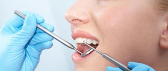
Dental visiography is the most informative method for diagnosing pathologies of the teeth and jaw, carried out using a special device that operates on the basis of radio frequency radiation.
The procedure is absolutely safe and not contraindicated even in childhood. The radiation exposure to the body is minimal (10 times lower than with classic x-rays). The procedure is also called “radiovisiography”. The main difference between the procedure and classical x-rays is the ability to instantly obtain an image of the area under study not on film, but on a monitor screen. In addition, experts confirm the high quality of the image and the possibility of obtaining a targeted image.
A dental radiovisiograph is used to identify any pathological processes in the teeth or jaw, including surrounding soft tissues and temporomandibular joints. As a result of diagnosis, a specialist can determine hidden processes (including hidden caries), inflammation, maxillofacial injuries, pathologies of the maxillary sinuses, and the condition of the jaw bones. Visiography is also used in dentistry to monitor the progress of treatment and its results.
Advantages and disadvantages
The main advantages of using the radiovisiography method include:
- The device is compact, easy to use;
- the pictures are informative;
- the image is immediately ready for examination, it can also be viewed on a PC screen using multiple magnification;
- the procedure is safe, radiation exposure is insignificant;
- manipulations take a minimum of time, without causing discomfort to the Patient;
- To improve the visiogram, you can use special editing tools.
The technique has almost no disadvantages, but the low spatial resolution should be noted. The sensor cannot provide fine detail to evaluate the difference between unequal densities and structural changes. To eliminate this disadvantage, tools to increase brightness and contrast are used.
What does the photo show?
Clinic Dr. Martin successfully uses digital radiovisiography for diagnostic purposes when indicated for therapeutic or surgical treatment.
Computer radiovisiography is increasingly used in endodontics, which deals with the treatment of dentin and pulp. The visiograph images clearly determine the shape and length of the root canals, which makes it possible to flawlessly perform mechanical treatment and filling.
Targeted images of a visiograph allow you to obtain accurate information about the condition of soft and bone tissues for high-quality implantation. Thanks to a detailed study using computer technology, the orthodontist can select the correct size and shape of the implant and monitor the result of its installation, as well as the healing process of the pin.
Dental radiography is performed in dentistry according to indications. The patient is referred by a specialist for diagnostics before planned implantation, installation of braces or any other design/products. The visiograph is able to identify any orthodontic products in the dental system. They are identified in the image in white. Foci of inflammation (granulomas, cysts, etc.) are displayed in dark color. The pathological zone of connective or bone tissue is highlighted in gray shades in the images.
What does a visiograph look like and when is it used?
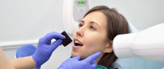
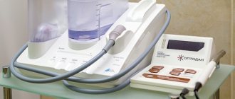
Dental radiovisiographs are small in size. To take an image, a small sensor of the device is placed in the patient’s oral cavity and transmits the result of the image directly to the computer.
The visiograph consists of several components:
- The basis is the high-frequency apparatus in the photo above, which sets the desired direction of particle movement;
- Small wired or wireless sensor;
- Analogue-digital system providing image digitization;
- A PC or laptop that allows you to view, store, and send images via email.
The wired visiograph is equipped with a special sensor that is applied to the tooth. Every radiologist should know how to take targeted photographs of teeth using such equipment. Using a wired connection, the image is transferred to the computer screen. A wireless radiovisiograph has a scanner capable of reading an image and displaying it on the screen.
The radiation dose during the examination is small and amounts to 2-3 μSv. Compared to this device, fluorography creates an exposure of 500-800 μSv.
The resulting image can be stored in an archive and on a hard drive. A visiograph belongs to the category of diagnostic and treatment dental equipment and allows you to obtain a clear image of a diseased tooth.
Advantages
Classic X-rays involve exposure to a significant dose of radiation to the body, so the procedure is allowed only a limited number of times during the year, observing time intervals. In addition, the image on such a device is printed on film, which does not exclude distortion of the image.
The operating principle of the radiovisiograph offers significant advantages:
- low level of radiation exposure;
- displaying the image on a computer monitor immediately after the start of the procedure;
- storing data in a computer database;
- transfer of data to a specialist or patient in electronic format, including sending an image using modern methods;
- high quality image with the ability to zoom in and view the desired area in detail;
- availability of using special filters without introducing a contrast agent into the body;
- the ability to take a targeted shot;
- Absolute safety for the patient’s body with no side effects.
Diagnostics using a visiograph is many times faster than when examined using standard X-ray equipment. The photo is taken in a few minutes. And immediately after the examination is completed, the images will be ready.
Difference from CT and radiography
Computed tomography is used to obtain three-dimensional images of tissues and jaw apparatus for analysis. The method requires the use of special equipment, the cost of the research is not the lowest. With the help of visualization, you can examine each unit in detail separately. This has several advantages if a tooth needs to be assessed before therapy. In addition, the technique is more convenient for monitoring treatment results.
X-ray allows you to obtain only flat images, which is not suitable for identifying a number of pathologies. For example, it is necessary to determine the presence of neoplasms at the initial stage. Radiovisiography can cope with this task, radiography cannot. The differences also include the greater sensitivity of the equipment’s CCD sensor than a standard X-ray film can provide.
How is the diagnosis carried out?
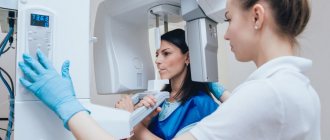
The visiograph is a modern diagnostic device consisting of three main parts: a high-precision sensor, an analog-to-digital converter and a cord connecting the parts of the device. The operation of the visiograph is ensured by a whole complex of parts, including x-ray equipment with a special guide (tube). The device itself has an external resemblance to a tomograph.
The sensor is a silicone chip (digital sensor) that records all the signals that come to it. It is the sensor that transmits the signal to the device, which is formatted into an image with 16-bit quality. The analog-to-digital converter is a monoblock equipped with a USB port for connecting to a computer and a special port for connecting to a sensor. The data received by the sensor passes through the connecting cable to the computer. All information is converted using a built-in program and displayed on a computer monitor.
The diagnostic procedure takes on average 3–5 minutes. The patient sits down on a chair near the visiograph, the specialist adjusts the direction of the X-ray tube in the desired direction and places the sensor against the area being examined. Then the device turns on and a photo is taken.
What is a radiovisiograph?
A visiograph is a compact sensor that records x-ray radiation. The signal is then converted, digitized and transmitted to a computer, where the user can observe the final image.
Reference. In modern medicine, visiographs are used to create control images after installing a filling or onlay, as well as during endodontic treatment.
It consists of three parts - a sensor that receives radiation, a connecting cord and software on a disk.
Note: Sometimes the kit includes an analog-to-digital converter (ADC), located between the sensor and the computer, connected by a cord.
Diagnostics at the clinic Dr. Martin
Visiography is recognized as the best method of modern diagnostics in dentistry when it is necessary to obtain a targeted image that allows one to examine in detail a separate area of the dentofacial structure and joints of the lower jaw.
Clinic Dr. Martin offers dental imaging at an affordable price. Diagnostics can be either a stage of treatment by our specialists or a separately provided service. The cost for the procedure and the image is indicated in the price list on the clinic’s website.
You can make an appointment with a specialist, a consultation or an examination by calling the contact number, or using the website service by ordering a call back from a consultant (“Make an Appointment”).
Advantages of using a visiograph
Let's start with the fact that the big advantage of this device is the ability to store all received digital images on any electronic media and transmit them electronically. Equally important is the ability to edit pictures and enlarge them, which allows you to view the image more clearly and in detail.
The speed of identifying a variety of foci of infection, inflammation, often not noticeable during a normal visual examination, detection of cysts, granulomas, foreign bodies in the canals of teeth and, of course, caries (even at the initial stage), neoplasms, pulpitis, periodontitis is impressive. Just a few seconds and the photo is ready.
An obvious advantage for both patients and doctors is the safety of dental examinations using a radiovisiograph. The radiation from this device does not exceed the dose we all receive from the daily use of household devices such as a scanner or digital camera. In this case, there is no need to talk about the advantages of a visiograph over an x-ray machine - the radiation dose of the former is several times less!
It has already been said that the resulting images are convenient to store and transmit electronically, but it would not be superfluous to mention this advantage again, because some types of dental treatment take place in several stages, which involves comparing the initial situation with the current one.
It turns out that examination using a radiovisiograph
- this is a real opportunity to quickly and accurately diagnose the problem and timely prescribe effective dental treatment.
Mechanical damage
In addition to the fading of the detection layers, a fairly common failure of sensors is mechanical damage to the connecting cable . Quite often, the cable bends, frays, or gets under the chassis of mobile devices: as a result, breaks and damage to the cores or insulation appear on the wire. Possible short circuits.
Mechanical damage to the manipulation arm of the radiovisiograph - the manipulation arm of the scanning head ceases to be fixed. This happens when mechanical components break down due to impacts or falls.
Advantages of a visiograph in dentistry
When considering the capabilities and characteristics, the following advantages stand out:
- radiation exposure is 90% lower when compared with x-rays due to the sensitivity of the sensor;
saving time - an informative and clear image is displayed to the doctor directly on the screen, no need to wait until printing is done;
- comfortable conditions for the patient, since there is no need to run from office to office, photographs are taken at the dentist’s chair;
assessment of the state of the oral cavity occurs in real time, which allows you to establish the most accurate diagnosis; digitized images are in the database and can be sent to colleagues at any time to agree on certain points.
With the advent of a visiograph in dentistry, the process of dental treatment has been significantly simplified, since only a few minutes pass from the moment the procedure is prescribed to the receipt of the image.
In the foreseeable future, the visiograph will undoubtedly displace traditional radiography from medicine. The picture is not only informative - it is easy to scale on a computer monitor.
Rules of service
To maintain hygiene conditions, the sensor must be placed in a special polyethylene case before being placed in the patient’s mouth.
For normal operation, the visiograph also requires cleaning using a soap solution or isopropyl alcohol. Before cleaning, you must turn off the power to the device. Since some disinfecting liquids can form explosive compounds when evaporated, in order to avoid ignition when turning on the visiograph after cleaning, some time is required to allow the vapors of these liquids to evaporate from the room.
IMPORTANT. It is extremely dangerous for the device if moisture gets inside the sensor or control unit. If liquid gets on the device board, it can cause a short circuit.
What does visiography tell you?
Visiography has become widespread in various areas of dentistry, in particular in therapeutic and implantology. But in endodontics this diagnostic method is especially highly valued.
Endodontics is a field of dentistry associated with the treatment of pathologies of the root canals and tissues that surround the teeth.
A visual image helps to visualize everything that is not visible to the naked eye and improve the quality of procedures: accurately calculate the length of the root canal, select the appropriate pin, ensure high-quality filling, notice a broken fragment of the endodontist’s instrument.
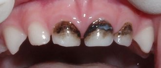
The image obtained by the visiography method clearly shows:
- dense structures painted white are fillings, pins and prostheses that block x-ray rays;
- loose structures painted gray - bone and connective tissue;
- inflammatory processes and hidden caries are painted in more intense, almost black shades.
Visiographs use ultra-sensitive sensors that transmit images in high quality even at low radiation levels. A 16-bit image makes it possible to make a correct diagnosis even with minor inflammations and carious lesions.