Publication date: 03/26/2020
The Axioma Dental clinic has its own visiograph and orthopantomograph - this is digital X-ray equipment that allows you to achieve high efficiency in diagnosis and treatment. A visiograph is an ultra-sensitive technique for creating clear, accurate images of teeth. You no longer have to wait for the X-ray film to be developed; the image is instantly visualized on the screen. This saves the patient’s time without harming health.
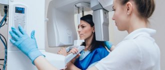
An orthopantomograph is a safe alternative to X-ray machines. The technique takes a panoramic photograph of the patient’s entire dental system. The equipment is indispensable in any dental field for developing a treatment plan. The dentist can quickly make a diagnosis and obtain detailed information about the condition of the dental system.
The principle of operation of the visiograph
A dental visiograph is a system of equipment that includes an ultra-sensitive touch sensor, an X-ray, and a computer for digitizing data. The principle of operation of the technique is similar to x-rays, but it is a safe technique. If the film unit directs rays onto the film, then the visiograph imaging device sends data to an ultra-sensitive matrix, a CCD sensor. The microcircuit chip consists of photodiodes that can capture and visualize the smallest details.
The main advantage is that to create an image you need an ultra-low dose of X-ray radiation, only 2-3 μSv. This is safe for humans, and the doctor has the opportunity to reduce the time for diagnosis.
How to take pictures using a visiograph:
- The lead apron is protection for the patient, who wears it before the procedure.
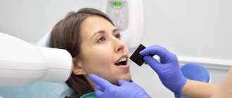
- Placement of the sensor in the oral cavity - it can be wireless or wired. An individual protective cover is put on the sensor and placed in the oral cavity. The sensor is applied to the area where pathology is detected, the condition of the bone tissue is clarified for successful implantation, treatment of caries, pulpitis.
- The sensor of a dental touch imaging device is connected to a digital device for data visualization, digitization and analysis of results.
- Snapshot.
Within a few seconds, the doctor sees the full clinical picture on the PC screen, makes a diagnosis and quickly prescribes treatment.
Working with a visiographer – choosing successful clinics
The domestic market offers a wide variety of visiographs from different manufacturers. Therefore, you can choose a device taking into account the structure of your clinic, its capabilities and preferences. Working with a visiograph requires special skills and specialization, and each individual device requires a detailed study of the individual instructions for the visiograph, because the devices differ in technical parameters, ease of use, operating and maintenance conditions. The accuracy of diagnosis and obtaining a high-quality image required to recognize a dental problem depends on correct work with a visiograph.
The difference between a visiograph and an x-ray
A visiograph is a device that does not independently emit rays, unlike an X-ray machine. Its task is to receive the rays onto an ultra-sensitive sensor and convert them into an accurate image for data analysis. At the same time, modern equipment does not need to produce a wide beam of radiation. A radiovisiograph is a very thin targeted beam of light that is captured by a sensor.
The system’s sensor is able to recognize and capture the smallest particles of radiation in just a fraction of seconds, which reduces the procedure and risks for humans. Modern digital equipment differs from x-rays in the speed of obtaining an image; shutter speed:
- visiograph - 0.06 seconds.
- film apparatus - 0.6.
Conclusion - innovative technology allows you to achieve maximum safety for the patient and reduce procedure time.
The Legend of the Visiograph - Prevention of Radioactive Misconceptions
Several years ago, Russian dentists received a visiograph for use. Everything that you definitely need to know about him was not written in Russian and, for the most part, remains a secret to this day. Literacy of the population is growing, visiographs are used everywhere in the economy, but some questions about their essence remain unanswered.
Is the
- No, a visiograph is not an X-ray, since Roentgen (Wilhelm-Conrad) died in 1923 and there is no way he can exist anymore.
An x-ray is also called a unit of radiation power, that is, this is the amount of radiation that, when absorbed in 1 cm3 of air, produces 2.08 × 10⁹ pairs of ions. This is not it either. Here, really, what is the question, is the answer.
Is X-ray radiation used in radiovisiography?
Yes, dental x-rays with digital image processing use x-rays with a wavelength of 0.17-0.19 angstroms.
How much safer is a visiograph than a regular x-ray?
By itself, a visiograph can do no more harm than a digital camera or an office scanner. The visiograph receives the radiation. It consists of three parts - a sensor, an analog-to-digital converter and a cord that connects them. A sensor or, as it is sometimes called, a transducer, is essentially a silicone chip, most often based on a CCD matrix (Charge Coupled Devise), which captures the incoming signal and transmits it to an analog-to-digital converter (ADC). The ADC consists of a board equipped with a specialized sensor port and a USB port. The ADC can be built into the computer system unit or used as an external device represented by a candy bar. The information passed through the ADC is an original digital image, which is processed using a special computer program, and as a result, an automatically (by default) converted image corresponding to the concept of “digital radiograph” appears on the monitor screen.
The visiograph can only function as part of a visiographic complex, which includes an x-ray machine with a full-fledged x-ray tube. For some reason, many people call him a visiograph and tell patients that “this is not an x-ray.” It’s not an X-ray - that’s true, of course, but it’s not good to deceive patients, and if it’s impossible to explain, but you have to deceive, then we can say that radiovisiography is based on the same principles as conventional radiography, but different equipment is used, therefore, the burden on the patient is reduced to a minimum.
What reduces the burden on the patient?
— The equipment has become more advanced. Firstly, modern X-ray dental devices (in the first half of the 20th century, they were called dentografs) imported generate a narrower, smoother and cleaner beam than Soviet, and now Russian-made devices. Secondly, the digital image receiver, which is the visiograph sensor, is more sensitive than any film. Accordingly, digital radiography requires less radiant energy to pass through the patient and form an image. For example, if we take a good high-quality film and a modern dentograph (for brevity we will use this convenient archaism), then to take a high-quality image of, for example, tooth 36 with a standard current and voltage, a shutter speed of 0.6 seconds is required. And for the same thing with digital radiography with a 5th generation sensor, 0.06 seconds is enough. The sensor adequately perceives the signal at exposure from 0.3 tA/sec to 1.8 mA/sec. On Soviet devices it is impossible to set such a shutter speed, and at large values the pictures turn out black, and the CCD matrix can deteriorate.
What dose will I receive?
- If the study was carried out using a visiograph, you can answer - 2 microsieverts for radiography of the teeth of the lower jaw (or 5 µSv for the upper jaw). This answer will satisfy a microscopic part of the population, since for the majority the word “sievert” is not associated with anything. If the question is formulated without specifying units, it can be assumed that the patient is not very knowledgeable about radiation exposure. To measure the amount of radiant energy applied to living tissue, various units are used - joule per kilogram, gray, rem, sievert, etc. The rem, the biological equivalent of an x-ray, is a non-systemic unit, equal to 0.01 sievert and is not currently used. In medicine, during intrascopic procedures, the dose received by the entire body during one procedure is usually assessed - the effective equivalent dose, measured in sieverts. When film radiography using low-dose devices and highly sensitive film, these values are 7 µSv and 14 µSv, respectively (Chibisova M.A. 2004), and when working with old domestic equipment and low-quality film they can reach 20 and 30 µSv, including using the 5D1 device can reach up to 80 μSv (Stavitsky R.V. 1991)…
How many pictures can you take?
— By law, every modern X-ray diagnostic device must be equipped with a dose counter and the dose received must be recorded in the medical history. This is ideal, of course. But if you try to find a dose counter for a dental X-ray unit, you will find that such a thing does not exist. Now in our country we use both old 5D1 type devices and ultra-modern low-dose units, therefore, it is extremely difficult to regulate the number of x-ray examinations in dentistry. Accordingly, it is necessary to make calculations for each type of device and for each tooth. In such a situation, it is logical to adhere to the maximum permissible effective equivalent dose for a person per year, excluding its excess. According to SanPIN, when carrying out preventive medical x-ray procedures and scientific research, this dose should not exceed 0.001 sievert (1 mSv (milisievert), 1000 µSv (microsievert)) per year. This is a load equal to approximately three chest X-rays. It is quite difficult to ensure that each application of X-ray radiation results in the appearance of a high-quality informative radiograph. This requires a good level of specialist training and certain experience. On the other hand, if the doctor clicks 20 exposures per pulpitis, this indicates a lack of understanding of what is happening.
Where can a visiograph be placed?
- Anywhere, if the SES allows placing a dental X-ray next to him. The fact is that almost anyone can now buy a visiograph and an x-ray tube, as well as a TV. Put it - at least in the kitchen. But, unlike a TV, which you can immediately turn on and watch, to use equipment that generates X-ray radiation, you must obtain a license to use it.
Should you leave the office while the image is being taken or can you stay with the patient?
— This question is very sensitive and touches on the ethical side of the problem. You can answer as in a joke: in principle - yes, but in general - no. If you have a state-issued protective screen and it is placed at the right distance and in the right place, there is no need to leave the office during dental x-rays with digital image processing. If you use double-sided medical protective aprons with a lead equivalent of 0.35 and can be at a distance of 2.5 m to the side of the emitter (not directly along, not behind, but to the side), you can also stay indoors. In this case, individual forced ventilation must be turned on to disperse ionized air, which is not at all useful (this is not a Chizhevsky chandelier). In all other cases, where to be, decide for yourself. Radiation is a thing that doesn’t smell, doesn’t squeak, and doesn’t sting the eye, so it feels like it’s not there. But it exists and acts, and selectively and cumulatively. Translated into Russian, this means that ionizing radiation affects different organs differently, and the severity of the damaging effect depends on the duration of exposure.
Is it possible to take dental photographs of pregnant women?
- It is forbidden. Or rather, in principle it is possible, but it’s better not to. Especially at a private reception. Even when working with a visiograph, because this issue is extremely delicate and controversial. The patient’s signature on the card in this case has no legal value, and the patient will probably have a husband who will declare that the patient, by definition, cannot be of sound mind, because she is in a different situation. The last word is his, and he “will arrange it for you all” if “God forbid”! “God forbid” something may happen through no fault of yours. The patient may, according to her personal understanding, engage in body fitness and eat anabolic steroids during pregnancy, live near a chemical plant, have a family history and not know about it, etc. In any case, if there is something seriously wrong with the newborn, you will most likely be remembered. The picture was taken, the fact of the use of X-ray radiation took place and was documented. Was? Was! There can be quite a bit of trouble.
Is it possible to take photographs of pregnant women if they wear two aprons?
— The number of aprons does not matter! See above. In contact radiography, the apron essentially protects not from direct radiation, but from secondary, that is, reflected. To X-rays, the human body is an optical medium, just as a glass cube is to a flashlight beam. Point the beam of a flashlight at one of the faces of a meter-long glass cube, and, regardless of the thickness and direction of the beam, the entire cube will be illuminated. It’s the same with a person - you can swaddle him completely in lead and shine only on his head - at least a little, but it will reach every heel. So, under two aprons with a good lead equivalent, it will simply be harder for a pregnant woman to breathe.
Is it possible to take photographs of nursing mothers?
- Can. X-rays are not the same as radioactive waste. By itself, it does not accumulate in the biological environment. If you give a loaf of bread a lethal dose, it will not mutate, get radiation sickness, or begin to “foul.” X-rays differ from light rays only in wavelength and, under certain conditions, have a direct damaging effect. If you shine a flashlight into a bucket of water and turn off the flashlight, the light is unlikely to remain in the bucket. The same is true in a protein-fat solution, which are many biological fluids; at a wavelength of 17-19 angstroms, the radiation undergoes true coherent scattering. That is, simply put, it flies through, weakening in denser tissues. So, with such a load, which is necessary to work with a visiograph, the milk itself is unlikely to do anything. As a last resort, to calm down, you can recommend skipping one regular feeding. Another thing is that the breast tissue itself during lactation is, of course, more susceptible to the harmful effects of radiation. But, again, at a dose more powerful than is necessary for digital radiography (of course, subject to all protective measures and without “shooting” 20 times anywhere).
Advantages and disadvantages of a visiograph
Pros of sensory equipment:
- Minimum radiation dose.
- High image accuracy.
- Maximum safety for the patient.
- The procedure can be performed for pregnant women and children - consultation with a doctor is required.
- Compact device, able to work in a small office.
- Instantly visualize your photo without the need to develop film.
- The doctor gets the opportunity to see each tooth - multiple magnification of the image, storage of data on a PC, transmission via e-mail, local network.
- The manipulation takes several minutes.
- Editing a picture - the dentist uses different filters to improve the visual perception of the picture.
- The procedure is painless and does not require special preparation.
The disadvantage is the high price of equipment and procedures. But in modern dentistry, the priority is accurate diagnosis, effective treatment, and patient safety. Therefore, reputable clinics invest money in purchasing equipment.
The essence of the X-ray machine
During treatment, prosthetics or implantation, it is necessary to take dental x-rays many times: before, during and after the intervention. The devices familiar to many caused irreparable harm to the patient’s body, because the radiation dose in them is very high. This was necessary so that the reflected X-rays could be imprinted on the film, thereby creating an image.
“Classic” X-ray also required a staff member who knew how to develop images, as well as an entire darkroom. And patients had to wait a long time for examination results.
Features of the orthopantomograph
Dental orthopantomograph is equipment for creating high-resolution panoramic images. Devices of various types have an overview imaging system, which makes it possible to visualize the entire jaw, the patient’s teeth, and jaw joints. The principle of operation is that the X-ray emitter and sensor in digital modifications rotate around the head, forming a clear three-dimensional image of the jaws.
Digital orthopantomographs make shooting as safe as possible for humans - the radiation level is 2-3 times less than with film units. Innovative technology does not require laboratories to develop images; the picture will be ready quickly and visualized on the computer screen without waiting for results.
If the orthopantomograph has a cephalostat, the doctor can take a frontal profile photograph of the skull, which is needed to build orthodontic structures.
Innovative technology works with different shooting programs:
- Standard overview.
- Separately upper and lower jaw.
- Photo of the maxillary sinuses.
- Children's photography.
- Image enlargement mode.
- Other programs for different clinical cases.
Safety, speed, high-resolution image quality, and information content are the characteristics of equipment used by clinics all over the world.
Disadvantages of radiovisiographs - is the radiation dose dangerous?
It must be said that radiovisiography has only one drawback
It consists in a lower (1.5-2 times) spatial resolution than that of film.
The sensor cannot as clearly convey the difference between structures that differ markedly in density.
True, this minus is compensated by another opportunity - to adjust the brightness and contrast of a digital image.
IMPORTANT: Are you an expectant mother? Do not forget to warn your doctor about this before treatment.
Is the radiation dose dangerous?
Here are the doses dental patients “receive” in studies:
- 1 image of a tooth: on the lower jaw - 2 μSv, and on the upper jaw - 3 μSv.
- 1 panoramic (digital) image - 15-20 µSv.
COMPARED WITH AN X-RAY : 1 fluorogram will “give out” 800 μSv.
Modern dentistry with visiograph and orthopantomograph
The technical equipment of the dental clinic is a guarantee of accurate diagnosis, effective treatment, and error-free diagnosis. Without the latest generation equipment, a medical institution cannot guarantee the patient a high level of service.
Axioma Dental Clinic is a center that invests money in purchasing innovative equipment. Doctors have a Sirona orthopantomograph and a visiograph at their disposal. This provides benefits to our patients:
- Examinations in one clinic - no need to waste time, sign up in advance for x-rays at clinics.
- Safety - there is no risk of receiving a high dose of X-rays.
- Treatment of children, adults, pregnant women - innovative equipment does not cause harm to health.
- Instant results - the photo will be ready in a few seconds.
The Axioma Dental clinic is the right approach to treating teeth, gums, correcting row defects, and implantation using safe equipment.
Sources:
- Yarnova E.A. Possibilities of radiation diagnostic methods in visualization of the temporomandibular joints in cases of dentofacial anomalies
- Goncharov I.Yu. Planning the surgical stage of dental implantation in the treatment of patients with various types of missing teeth, defects and deformations of the jaws, 2009
- Robustova Tatyana Grigorievna. Dental implantation: surgical aspects, 2003
Expert author:
Makanina Lina Viktorovna
Dentist-therapist
The information presented in this article is provided for reference purposes and does not replace the advice of a qualified specialist.
At the first signs of illness, you should consult a doctor.
Tell us about us:
How is a pregnant woman's dental x-ray performed?
- The patient is covered with a lead apron, she and the baby in the stomach are reliably protected.
- Each tooth has its own exposure, and the staff is careful not to exceed it.
- There is also a special super-sensitive class E film that allows you to reduce exposure.
- There are also visiographs that emit such microdoses that they are equal to the normal radiation background, and their beam is very precisely and narrowly directed only at a specific tooth. The sensitivity of their sensor is many times greater than that of a film camera, which allows you to take several pictures at a time without any harm to health. After all, sometimes you have to take up to 5 pictures to treat a tooth.
Photo of teeth: types
Dental X-ray is a study of the soft and bone tissues of the oral cavity using an X-ray image. The image informs the doctor about what he does not see during a visual examination. X-rays are prescribed to identify the degree and depth of inflammation, detect pathologies, defects and diseases.
There are many types of dental x-rays. Our clinic uses targeted radiography. A targeted photograph of teeth is a simple and accessible technique in dental research. This is a 2D image that shows the doctor 1-2 teeth located nearby. Dental images are taken using a digital radiovisiograph.
Sight radiography allows the doctor to:
- Identify inflammation in the early stages in the area of the apexes of the roots of the teeth.
- Check the quality of tooth root filling and bone tissue restoration before prosthetics.
- Identify carious lesions, pulpitis, periodontitis.
A targeted photograph of teeth, depending on the equipment used, can be of two types:
- X-ray on film.
- Electronic image (the image is transferred to a digital medium, which allows you to enlarge the required area for detailed diagnostics).
What does a radiovisiographic image reveal?
A digital radiovisiograph allows you to identify any foci of infection, the location of which is difficult to determine visually. A visiogram is done in case of possible occurrence of the following diseases:
- Caries;
- Pulpitis, which causes inflammation of the tissues surrounding the tooth;
- Small neoplasms in the oral cavity;
- Periodontitis.
The image allows you to detect hidden foreign bodies that have entered the canal. Any foci of inflammation, granulomas and cysts are painted black, which stands out sharply against the gray background of bone and connective tissue. With the help of detailed visual images, the dentist will be able to make an accurate diagnosis and prescribe appropriate treatment.
Radiovisiography allows you to determine the depth of tissue damage and control the quality of filling.
Difference from CT and radiography
Computed tomography is used to obtain three-dimensional images of tissues and jaw apparatus for analysis. The method requires the use of special equipment, the cost of the research is not the lowest. With the help of visualization, you can examine each unit in detail separately. This has several advantages if a tooth needs to be assessed before therapy. In addition, the technique is more convenient for monitoring treatment results.
X-ray allows you to obtain only flat images, which is not suitable for identifying a number of pathologies. For example, it is necessary to determine the presence of neoplasms at the initial stage. Radiovisiography can cope with this task, radiography cannot. The differences also include the greater sensitivity of the equipment’s CCD sensor than a standard X-ray film can provide.
What needs to be done before use?
Before you start working directly with a visiograph, it is important to prepare. What does it include?
Firstly, the initial installation and configuration of the device. Such work must be performed by a qualified specialist - an employee of a company for the repair and maintenance of medical equipment.
After checking the device for external damage, installation and configuration, you need to check all working documentation, including carefully studying the instructions for the specific model of the visiograph. If necessary, you can use the service of training employees to work with equipment, which is usually offered by companies for the repair and maintenance of medical equipment.
Indications for radiography using a visiograph
Dental X-rays are taken so that the dentist can analyze the condition of both individual teeth and the patient’s jaws. A visiograph takes control and intermediate images during therapy and filling of dental canals. Endodontic treatment is not carried out without radiographic examination.
An intermediate series of photographs is needed to measure the working length of the dental canal and determine its patency. A control x-ray is indicated after completion of therapy to monitor filled roots. Digital visiography will allow you to avoid serious complications, monitor the stages of dental canal filling and adjust the dentist’s work.
