Cyst and foreign body in the maxillary sinuses - this is a characteristic cause of pain and inflammation in the two paranasal sinuses. The sinuses are paired air planes in the bones of the skull, communicating with the nose and occupying almost the entire space of the upper jaw. These cavities perform many important functions, including supplying the nose with mucus, which is so important for maintaining immunity. Therefore, diseases of the maxillary sinuses can lead to very sad consequences.
What is a maxillary sinus cyst?
A maxillary cyst is a voluminous benign formation that originates from the mucous membrane of the maxillary sinus and has the appearance of a bubble containing fluid inside. At the same time, the contents of its cavity vary, depending on the mechanism of appearance and the nature of the ongoing pathological process.
Depending on this, there are several types of maxillary sinus cysts:
- mucous membranes (mucocele);
- serous (hydrocele);
- purulent (pyocele).
According to their structure, cysts can be true (with an internal epithelial lining in a thin wall) or false. A pseudocyst in the nose does not have clearly defined walls and looks like cavities in the tissue; they are usually formed in the thickness of the overgrown mucous membrane and can consist of several chambers of different sizes.
Cysts do not have a tendency to malignant degeneration, however, they cannot be classified as harmless formations. They can fester and burst, compress adjacent vessels and nerves, and gradually destroy the bone walls of the sinus.
Cystic formations negatively affect ventilation and drainage of the entire system of paranasal sinuses and impair nasal breathing. Therefore, surgery to remove a cyst often becomes a prevention of chronic recurrent inflammation of the ENT organs.
Complications
Rupture of the maxillary sinus is a serious consequence of dental procedures, the treatment of which most often occurs in an inpatient setting. What is the risk if the tooth root has grown into the maxillary sinus?
Self-treatment of the problem using traditional medicine methods can lead to even more serious complications, including:
- Severe inflammatory process in the sinus cavity with further infection of adjacent tissues. Then osteomyelitis develops in the upper jaw.
- Transition of the inflammatory process to other cranial sinuses, including the sphenoid, frontal and ethmoid.
- Loss of healthy teeth located in the perforation area.
- Formation of foci of pus, phlegmon and abscesses.
Due to the immediate proximity of the brain, and the fact that the tooth root has gone into the maxillary sinus and the latter has ruptured, the occurrence of an infectious lesion of the meninges cannot be ruled out. The next stage of serious complications may be meningitis or meningoencephalitis, which are life-threatening for the patient.
Why does a cyst appear in the nose?
The main reasons for the appearance of maxillary cysts include:
- Recurrent or chronic maxillary sinusitis (sinusitis). Inflammatory changes in the mucous membrane lead to blockage or scarring of the excretory glandular ducts. The produced secretion has no outflow, accumulates and stretches the gland. This is how true mucous and serous cysts are formed.
- Chronic non-infectious inflammation (usually of an allergic nature), accompanied by hyperplasia of the mucous membrane of the nose and paranasal sinuses.
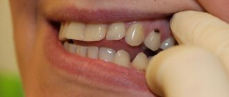
- Dental pathology, the resulting cysts are called odontogenic. The most common reason for their appearance is inflammation around the root of a carious tooth or near the tooth germ of the upper jaw. The purulent process leads to atrophy and destruction of bone tissue, spreading to the walls of the maxillary sinus. More rare causes include abnormally deep tooth roots and excessively traumatic tooth extraction.
- Predisposing factors are injuries to the facial part of the skull, congenital anomalies with asymmetry of the hard palate and nasal bones, and immunodeficiency states. Occasionally, a cyst in the sinus is formed against the background of a congenital defect in mucus production, when the secretion of the glands has an excessively viscous consistency.
Causes
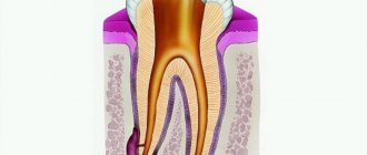
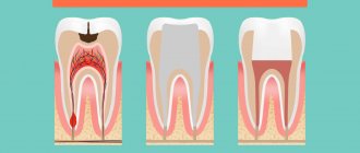
The process of cyst formation is not complicated. The mucous membrane of the sinuses consists of glands that produce mucous masses. Each gland has a duct through which mucus exits to the surface of the mucosa. If due to unfavorable factors such a duct is blocked, the mucus cannot come out. The same amount of mucus is formed as usual, but it does not find a way out. As a result, the walls of the gland stretch, fill with secretion, and a cyst forms. The following factors contribute to blockage of gland ducts and the formation of cysts:
- chronic diseases of the nasopharynx (chronic sinusitis, chronic rhinitis);
- allergy;
- acute infectious diseases;
- nasal polyps;
- carious teeth, disorders associated with teething;
- anatomical features of the structure of the nasal cavity.
What are the symptoms of a cyst in the nose?
A cyst of the upper jaw does not lead to strictly specific symptoms, and sometimes even occurs without obvious manifestations. Complaints from patients with this pathology include:
- Nasal congestion, clinical signs of chronic rhinitis.
- Unpleasant sensations, a feeling of fullness and pressure in the projection of the maxillary sinus (on the side of the cyst). Discomfort often increases when bending forward.
- A feeling of pressure from the inside on the eyeball, and with large neoplasms, transient double vision may occur.
- Recurrent headaches with a predominant localization in the forehead and bridge of the nose. Their appearance is mainly associated with impaired ventilation of the paranasal sinuses.
Reliable diagnosis is based on visualization of the maxillary sinus and its contents. Traditional examination methods include radiography of the facial part of the skull. Currently, CT scans of the paranasal sinuses are widely used.
Symptoms
When perforation and entry of the tooth root into the maxillary sinus occurs during dental resection procedures, characteristic signs of rupture appear, including:
- Bleeding from the tooth socket with small air bubbles, the number of which increases when you try to exhale sharply through your nose.
- Bloody nasal discharge from the side of the ruptured sinus.
- A sharp change in the timbre of the injured patient’s voice, characterized by nasal sound.
If there is a tooth root in the maxillary sinus, the symptoms cannot go unnoticed. Sometimes, after resection, the patient begins to complain about difficult passage of air through the resulting hole, as well as pressure and heaviness in the projection of the maxillary sinus.
When perforation occurs during implantation or endodontic therapy, signs of such a complication include:
- Specific failure of the instrument or material for implantation after some effort to move it into the jaw.
- Changing the position of the instrument used in the resulting wound.
- The appearance of small air bubbles and blood discharge from the hole.
Perforation may not be detected and repaired immediately after a sinus rupture, which leads to infection of the sinus cavity and is accompanied by signs of acute sinusitis or sinusitis.
The following symptoms are typical for such pathologies:
- Acute intense pain in the sinus area of the upper jaw.
- Swelling of the nasal mucosa on the side of the injury, accompanied by difficulty in nasal breathing.
- Discharge of purulent secretion from the nose.
Systemic complications and symptoms of intoxication appear, which are characterized by chills, headaches, weakness and increased body temperature. But how to detect the root of a tooth in the maxillary sinus?
When is surgery needed to remove a maxillary sinus cyst?
Formed cysts are often not prone to regression, and conservative therapy is also usually not able to lead to their disappearance even after the elimination of inflammation. Therefore, the only effective treatment method is surgical removal of the cyst in the sinus.
Indications for surgery include:
- Signs of suppuration of the contents of the cyst.
- Progressive growth of the tumor.
- The diameter of the cystic cavity is more than 6 cm.
- The appearance of double vision (diplopia) and blurred vision, which is a sign of excessive pressure of the formation on the bottom of the orbit.
- Often recurrent or continuous rhinosinusitis, chronic nasal congestion.
- Significant discomfort experienced by the patient even with a small tumor size.
Surgery to remove a cyst is usually performed as planned. The decision on surgical treatment is made by the ENT doctor after examining the patient and assessing the clinical situation.
Why is a dental cyst dangerous?
Despite the fact that being the result of a positive struggle of the body and a reaction to negative processes, reduced immunity, a dental cyst can lead to serious side effects and cause complications. The most dangerous and common of them:
- Flux is the concentration of a purulent process in the periosteal jaw area under the gum. A type of complex disease externally manifests itself in the form of a tumor, and a sharp throbbing pain is felt.
- Periodontitis is an advanced form of a purulent cyst that provokes the spread of infection to bone tissue.
- Tooth loss. A side effect that occurs extremely rarely, but occurs if the patient does not consult a dentist in a timely manner and does not receive drug treatment. The damaging effect extends to nearby hard areas of bone tissue, and there is a danger of losing several adjacent teeth.
Possible more complex exacerbations and consequences:
- inflammation of the lymph nodes;
- purulent abscess;
- fracture of the jaw due to decay of bone tissue and its complete destruction in a certain area;
- sepsis and osteomyelitis.
Even with minor signs of a dental cyst, treatment should be started. The sooner you see a dentist with a problem, the greater the chance of a complete recovery. While maintaining oral hygiene, make regular appointments with your dentist, and do not neglect the importance of medical examinations. You will not have to deal with the consequences of a dental cyst, since a qualified doctor will be able to recognize it at the initial stage. Take good care of your mouth at home and follow your dentist's advice.
Make an appointment with a therapist by phone+7(985)532-21-01
Treatment of caries between the front teeth
Hypoplasia of dental enamel, diagnosis and treatment
Caries in the white spot stage
Do fissure dental caries need to be treated?
Tooth root caries treated or removed
Tetracycline teeth treatment, whitening, veneers
Removal of a wisdom tooth cyst
How is surgery to remove a cyst in the nose performed?
For a long time, surgery to remove a sinus cyst was carried out only by opening the wall of the maxillary sinus through the oral cavity or through the cheek. This manipulation is called maxillary sinusotomy; it is quite traumatic and requires a long recovery period.
Currently, classical surgery is rarely performed, with preference given to minimally invasive interventions. But for extensive purulent cases, just such a radical intervention is used. The dangerous formation is completely removed, the sinus is washed with an antiseptic and drained.
In other cases, surgery on the maxillary cyst is performed endoscopically, with minimal disruption of the integrity of the sinus walls.
If necessary, additional plastic surgery of the anastomosis is performed and adjacent polypous growths are removed.
Endoscopic surgery to remove a maxillary sinus cyst is performed on an outpatient basis and usually lasts 20–40 minutes. After its completion, the patient remains in the clinic for 1–2 hours under the supervision of a doctor, then returns to his normal life.
Using an endoscope allows you to remove the maxillary cyst in a gentle way. This operation does not disrupt the drainage and ventilation of the sinuses, reduces the risk of chronic rhinosinusitis and does not require long-term recovery for the patient.
Diagnostics
For an experienced dentist, specific testing may not be necessary to confirm a maxillary sinus perforation.
If we are talking about tooth resection, then such a complication as soft tissue rupture is accompanied by a typical clinical picture. If the dentist is in doubt when perforation occurs during endodontic treatment, the following additional examinations may be required:
- Probing the hole formed after tooth extraction, as well as the root canal. For this purpose, a thin probe is used. In this way, it is possible to establish the absence of a bone bottom in the wound. During the study, the instrument passes through soft tissues without hindrance.
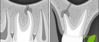
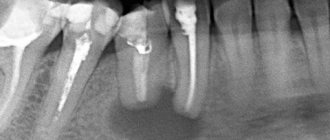
- Carrying out an X-ray examination of the sinuses. The resulting images will show darkening in the sinus cavity, indicating the accumulation of blood in this area, as well as filling material and fragments of teeth and implant. In some cases, contrast radiography may be required with the introduction of a special substance into the perforation cavity.
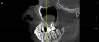
- CT scan. This diagnostic method makes it possible to detect ruptures and the presence of foreign bodies. In this case, the image is as accurate and informative as possible.
- If the perforation is old, it is recommended to donate blood for a general examination, based on the results of which it will be possible to draw a conclusion about the presence or absence of an infectious focus in the body.
After carrying out the necessary diagnostic measures and confirming the fact of a rupture of the maxillary sinus, the dentist will prescribe appropriate treatment. So, a person has a tooth root in the maxillary sinus - what to do?
Removal of a maxillary sinus cyst at the ENT clinic of Dr. Korenchenko
See also Treatment of ENT diseases Cyst in the maxillary sinus Treatment of a cyst in the maxillary sinus Surgery to remove a cyst in the maxillary sinus
Endoscopic ENT surgeries are not performed in all clinics. After all, they require modern equipment, the doctor having the appropriate skills and certificates. Dr. Korenchenko’s ENT clinic is a modern, specialized and well-equipped medical center. Our specialists are highly qualified and have rich clinical experience, all the necessary certificates and skills. When treating patients, we use only modern, clinically proven and highly effective techniques.
Endoscopy at Dr. Korenchenko’s Clinic is an important and widely used therapeutic and diagnostic procedure. It is included in the basic examination of all patients who apply and are observed, which allows doctors to receive reliable and accurate information about the current condition of the ENT organs. Our specialists also perform removal of maxillary cysts and most other operations endoscopically, with high results and without long-term rehabilitation of patients.
Treatment at Dr. Korenchenko’s ENT clinic is a modern and competent approach, using effective technologies and effective therapeutic regimens.