What kind of disease is this
In general, a cyst is a benign formation, similar to a cavity, with contents inside the walls.
In addition to the maxillary, it is often found in the sinuses near the nose, but under no circumstances does it extend beyond these zones. The cyst can be of any size - from small formations in the initial stages to large ones that expand if treatment is started.
There are two mechanisms for the development of education:
- Retention , which occurs and develops if the excretory duct of the glands in the mucous membrane is blocked.
- Odontogenic , progressing as a dental pathology, i.e. due to diseases of teeth and gums.
Both single and multiple cysts can also be detected. In some people this formation is acquired, while in others it is present from birth.
This is a very common disease: according to statistics, in every fifth person, a detailed examination of the nose can reveal a small maxillary sinus cyst (MSC). Such formation does not make itself felt for a long time, or manifests itself in the form of pain and unpleasant symptoms.
The patient who has been diagnosed with a cyst is being monitored dynamically. If his condition worsens sharply, surgical intervention is possible.
ENT about a cyst in the sinus:
ICD code
There are two disease codes according to the ICD:
- 8
- K09
Each of them is assigned depending on how the cyst was formed and what type it acquired.
What is a cyst in the maxillary sinus called?
A maxillary sinus cyst is a benign formation that has a hollow structure containing fluid inside. Cysts of the maxillary sinuses are quite common, but are almost always found at random. Their location and origin may differ, which will determine the characteristics of the clinic and even treatment.
Odontogenic cyst
Among the many benign tumors of the jaw tissue, the cyst is found most often and represents a pathology quite dangerous for the bone structure. Odontogenic formations in the vast majority of cases remain undiagnosed for a long time, and therefore reach enormous sizes. The growth of the cyst provokes atrophic processes in the bone tissue, which thins, becomes fragile, and then disappears completely.
Such formations are characterized by a long latent course until the cavity grows to gigantic proportions. Then a characteristic crunch begins to be noted in the jaw, especially during chewing loads. Ignoring the problem leads to spontaneous fractures of the jaw, compression of the maxillary sinus with disruption of its functions, exacerbation of the inflammatory process with damage to the structures of the maxillary sinus and the formation of sinusitis.
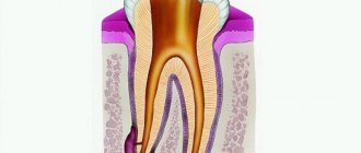
Tooth cyst growing into the maxillary sinus
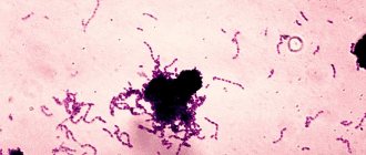
Damage to the dental canal involving the root and surrounding tissues is a consequence of an infectious process of bacterial origin. In the absence of adequate and timely treatment, a gradual delimitation of the inflammatory reaction from healthy tissues by a wave of cellular structures occurs.
This creates a cavity called a dental cyst. It gradually increases and destroys the bone tissue of the jaw. When localized in the anterior part of the upper jaw, gradual penetration into the maxillary sinus occurs.
Retention
The most common form of neoplasm in the maxillary sinus, which is also called a true cyst. It is located in the lower wall of the sinus and has an epithelial lining - a distinctive feature of true cystic cavities. The formation is clearly determined by X-ray examination randomly, as it does not manifest itself clinically.
When the cyst reaches a significant size, problems begin with the functioning of the maxillary sinus. The structure and physiology of the microvasculature is disrupted, resulting in the formation of edema. This symptom is manifested by nasal congestion, often without pronounced discharge from it. Due to the lack of specificity of the clinical picture, even the presence of subjective signs of the disease is not a reason to consult a doctor.
Main symptoms
This problem makes itself felt with a whole range of symptoms.
| Symptom | Description |
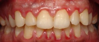
| Nasal congestion | The primary symptom that signals clogged ducts. A person experiences difficulty breathing through the nose and discomfort in the upper jaw. Rapid irritability and poor performance appear. There is a risk of exacerbation of chronic sinusitis or rhinitis. Manifests itself in everyday life or during physical activity. People, for example, who go diving, may experience problems. When diving under water, they will feel unpleasant pressure and pain in the area where the cyst has formed. If swelling appears or fluid begins to leak out, this leads to the appearance of ENT diseases. |
| Headaches, cheek and eye pain | Symptoms characteristic of both cysts in one and two sinuses. All this affects the overall performance of a person, appetite, drowsiness, as well as the respiratory process. In special cases, the patient suffers from vision problems (double vision). Sometimes a cyst located on one side can manifest itself with unilateral symptoms. For example, headaches and nasal congestion are felt mainly on the side where the tumor appeared. Pain and swelling in the cheeks can spread to healthy teeth. There is a risk of unpleasant sensations in the forehead area. Regular headaches and intoxication of the body occur. Clogged sinuses cause severe pain when tilting the head. |
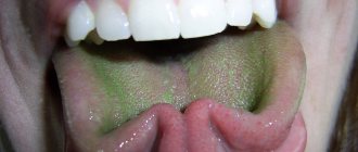
| Mucus and pus in the throat | The patient notices a strong discharge of clear yellow liquid from the nose. It can leak from one or two nostrils, depending on the location of the cyst. This phenomenon appears as a result of film rupture and leakage of a cyst-like formation. This will not last long, but the risk of contracting an infection during this period increases significantly. |
| Temperature | A natural reaction of the body, signaling the fight against an infection in the sinus. Cyst-shaped tumors take a long time to form, so elevated temperatures can also persist for a long time. |
Sinus lift techniques
Author: Daniel Spagnoli
Loss of teeth in the lateral part of the upper jaw and subsequent resorption of bone tissue and pneumatization of the sinus prevent the installation of implants (Fig. 1).
Figure 1. A computed tomogram shows a strong degree of pneumotization of the sinus and significant atrophy of bone tissue due to the absence of teeth.
Over time, certain techniques have been developed to increase bone volume for subsequent implant placement. Boyne and James were the first to describe a sinus lift technique using autogenous bone tissue.
Anatomy and physiology of the maxillary sinuses
The sinuses of the upper jaw are located lateral to the nasal cavity and have an asymmetrical shape. They are usually very small in size before the permanent teeth emerge. Until this time, the main volume of the jaw is occupied by the rudiments of permanent teeth. After the eruption of all permanent teeth, the sinus forms its final shape and size. These sinuses are the largest, having a volume of about 15 ml each. From an embryonic point of view, the paranasal sinuses are formed from the groove of the lateral wall of the nose at the 16th week of embryonic development.
Basically, the maxillary sinus consists of a single chamber, but sometimes it can be divided into several small bony septa that grow from the bottom or lateral wall of the sinus. The upper wall is the lower wall of the orbit, the lower is the hard palate and the alveolar part of the jaw, the lateral is the zygomatic process, the posterior border is the pterygopalatine fossa, and the medial border is the nasal cavity. The opening of the maxillary sinus passes between the inferior and middle turbinates (Fig. 2).
Figure 2. Anatomical relationship of the nasal cavity and adjacent structures.
The maxillary sinus is lined with pseudostratified ciliary semilunar epithelium with goblet cells. The mucosa of the respiratory tract is thin and the subepithelial layers are composed of loose connective tissue with a rich vascular system (see Fig. 12b). The complex of epithelium, connective tissue and periosteum together is called the sinus membrane - Schneiderian membrane.
The mucocellular apparatus is a major player in the protective function of the sinuses. The secretion produced by goblet cells is carried by the cilia of the epithelium into the choanae. Inhaled bacteria are evacuated from the sinuses at a rate of 3 to 25 mm/minute. Ciliary movement may be disrupted by inflammation and may be difficult to assess clinically. It is necessary to remember the functions of the sinus mucosa during the operation. Damage to the membrane can cause inflammation, which will impair drainage from the sinus. Patients with a history of maxillary sinusitis should consult an otolaryngologist if there are any changes after surgery.
The blood supply to the maxillary sinus is carried out by the branches of the external carotid artery. The main ones are the posterior superior alveolar artery and the infraorbital artery, which originates next to the maxillary artery. These two vessels create an anastomosis within the sinus, supplying the lateral wall of the sinus and part of the alveolar process. The posterior superior alveolar artery passes to the maxilla and its periosteum and divides into two branches: (1) the gingival branch, which supplies the mucosa in the area of premolars and molars; (2) dental branch. Both these
Causes
Maxillary sinus cyst on MRI
A cyst usually forms if the excretory ducts of the gland are blocked. If they are clogged, a special substance is released that stretches the walls of the duct, causing them to gradually fill with serous fluid. The substance inside the tumor continues to be produced. The longer they remain in this state, the larger the cyst becomes.
There are several reasons for the appearance of such formation:
- Injuries to this area, due to which the ducts are blocked (for example, due to blows to the face, falls, etc.).
- Due to hereditary predisposition, abnormal structure of ducts and bones in this area.
- The presence of sinusitis (chronic processes in this area).
- For problems with the upper teeth and gums (usually due to caries, periodontal disease, etc.).
- As a consequence of an allergic reaction.
- Due to the location of the roots of the teeth.
The last reason is related to the structure of the root system: the roots of the upper teeth (5 and 6 on one side or the other) can extend to the lower wall of the sinus, or be separated from it by a small partition.
If the pathology in this section has been neglected, ondogenic protrusions may form. There are two types of them: radicular (coming from the roots of the teeth) and follicular (formed when the tooth germ is displaced).
A neoplasm can also arise as a result of pathogenesis - when an infection enters this area and affects healthy tissues, as a result of which they are enclosed in a formation hidden by a membrane.
Such protrusions form in most of the population, but can safely resolve on their own. However, having reached a certain size, they begin to cause concern, and treatment is no longer possible.
Conservative treatment: when possible
Conservative treatment is only possible for small root cysts. At this time, the canals of the tooth are treated, on the roots of which a cyst has formed. The treatment consists of thorough cleaning and filling of the dental canals and is carried out under a microscope, which ensures the accuracy of all manipulations and allows you to find all the canals in the tooth and fill them. Most often, after endodontic treatment, the cyst disappears, which is recorded on an x-ray taken a few weeks after the operation.
Classification
Cysts are classified according to several criteria:
| Criterion | Varieties |
| Due to the occurrence | In medicine, there are three types:
|
| By content | Neoplasms may contain a number of substances inside:
|
| By location | It is formed in one or more places:
|
A small cyst can only be detected by x-ray
When the neoplasm is small, less than 10–15 mm, it is almost impossible to find out about its existence, since no alarming signs are observed. At this stage, the only option to identify pathology is to do a computed tomography scan. A dentist may also suspect its presence during a routine examination or other doctors if a person needs to visit them.
At the initial stage, the problem can only be detected on an x-ray
Alveolar cyst
We are talking about odontogenic neoplasms that are localized in areas near the alveolar bay. The negative impact of such a cyst, if left untreated, can damage the root system of the upper teeth. It can only be removed through surgery.
Maxillary sinus cyst
This variety practically does not manifest itself in any way. Only with a certain location and significant size can it lead to pain.
Most often, it is discovered by chance when the doctor suspects another disease. To do this, a CT, MRI, or x-ray is performed in the area of the maxillary sinus.
How does a sinus cyst appear?
The mechanism of development of paranasal sinus cysts is quite simple.
Let's start with the fact that in the mucous membrane of the paranasal sinuses there are glands that produce mucus. Each such gland has its own excretory duct for secreting secretions, which opens on the surface of the mucous membrane of the sinus.
Under the influence of various unfavorable factors, the mucous membrane thickens and the excretory ducts become clogged. At the same time, the gland continues its work with the same force, mucus is formed, but the exit for it is blocked. Due to wall pressure, the glands begin to fill with mucus, stretch and form a cystic neoplasm. Approximately the same effect is obtained if you put an inflatable balloon in a water tap and open the tap - water will flow and inflate the balloon.
There are several UNFAVORABLE FACTORS that will contribute to the development of this process, namely changes in the thickness of the mucosa and blockage of the ducts:
- exposure to an allergen and the occurrence of allergic reactions,
- polyps,
- infectious diseases,
- chronic diseases developing in the nasal cavity (rhinitis) or in its paranasal sinuses (sinusitis: sinusitis, sinusitis, etc.),
- diseases of the teeth of the upper jaw, anatomical features of the structure of the nose.
Dimensions
The size of the cyst does not affect the severity of symptoms. Large formations may not cause inconvenience. It is much more important where exactly the tumor is located :
- Those located in the lower section of the sinus cause less discomfort.
- But even a small formation located in the upper wall can lead to complications and headaches.
This is due to the close location of the cyst to the branches of the trigeminal nerve.
Recording of a webinar from a doctor of sciences on the topic:
Diagnosis of sinus cysts
In addition to the standard questioning and examination of the patient, all doctors at the Lor-Plus clinic are proficient in endoscopic examination techniques. Using an endoscope - a long thin tube with a miniature video camera, the doctor can conduct the most complete examination of the ENT organs, determine the presence of additional structural features of the nasal cavity, changes in the nasal mucosa in the deep sections that may interfere with normal breathing.
Currently, the gold standard for diagnosing sinus cysts is computed tomography . This method allows you to determine the size of the cyst and its location in the sinus with millimeter accuracy, which is very important for choosing a method for removing the cyst.
Diagnostics
Typically, such a pathology is discovered by chance when a person is tested for the presence of another disease.
To do this, they go to a dentist (if the disease concerns the jaws and teeth) or an otolaryngologist, who then issues a referral for an x-ray or another type of examination. ENT specialists can be involved in the diagnosis.
Laboratory tests will not accurately evaluate a cyst.
The doctor may do the following:
- Examine your nose.
- Refer him for endoscopic examination.
- Prescribe an x-ray or computed tomography scan of the patient’s sinuses and skull.
X-ray
X-rays reveal blockage of the sinuses.
Large tumors will appear in black on the image. An odontogenic cyst is identified by adjusting the x-ray to a specific projection. The x-ray will show the size of the cyst:
- Light areas indicate the sinus.
- Dark ones signal a cyst.
Tomography
Currently it is the most effective diagnostic method.
Thanks to it, it is possible to determine with high accuracy where the cyst is located and what the thickness of its walls is, what substance accumulates inside it. Based on the tomography results, the doctor obtains a layer-by-layer section of the patient’s skull. This helps to assess the structure of the formation, which is especially important when selecting a further treatment method.
Puncture
The method involves puncturing the cyst.
The color of the fluid that flows from the wound determines the appropriate diagnosis. This method is only suitable for identifying large tumors. For this, local anesthesia is used. The procedure is not only diagnostic, but also therapeutic. After pumping out the fluid, the person’s breathing returns to normal. The cyst shrinks and the patient's condition improves for a while.
Sinuscopy
An accurate way to study clogged areas and determine their location. The doctor inserts an endoscope into the excretory anastomosis.
With its help, it is possible to examine the tumor in detail and study it for other pathologies.
conclusions
The most objective way to diagnose dense space-occupying formations of the upper jaw is computed tomography (CBCT or MSCT).
If signs of osteoma are detected in the cavity of the upper jaw, the patient must be recommended to undergo surgical excision of the formation. Morphological verification of the removed material is mandatory.
The combination of an endoscopic transnasal approach and navigation equipment in the removal of osteomas of the upper jaw is the most modern and optimal option for treating the pathology.
MestaMidin-nos has proven itself in local therapy as a means to prevent possible postoperative infectious and inflammatory complications. The use of MestaMidin-nos allows you to avoid a course of systemic antibacterial therapy.
The publication was carried out with the support of Solopharm in accordance with internal policy and current legislation of the Russian Federation.
Treatment
Since such a tumor is not malignant, it is easily and without consequences surgically removed.
As long as the cyst has not developed to a large size, it can actually be cured without surgery. But the doctor must determine the symptoms, then order an examination, based on the results of which he will select an individual course of treatment.
Medication (without surgery)
A good method of treatment if the cyst has not reached a large size.
In this case, after some time you can reduce the size of the formation. The patient is given an appointment:
- Saline solutions used to rinse a clogged nasopharynx.
- Drugs that increase the outflow of mucus from the ducts.
- Nasal medications aimed at narrowing blood vessels (use drops and sprays).
- Antibiotics (local and general use).
Only taking special medications is suitable. Prescribing physiotherapeutic procedures and a course of warming up the sinuses is strictly prohibited - such actions accelerate the growth of the cyst and cause side effects.
If there is a risk of developing acute sinusitis, the doctor will prescribe a course of antibiotics. Plus, he recommends rinsing clogged sinuses and nasal cavity with antiseptic solutions.
How is the operation to remove a cyst performed?
Surgical intervention
An effective method, however, is resorted to only if it is impossible to overcome cyst formation with medications . An asymptomatic tumor does not need to be operated on - it is usually small and can resolve on its own with proper drug treatment and rinsing.
Reference! Only a doctor can give a referral to a surgeon. However, the procedure is prohibited if the disease has acquired a more acute form.
Operating methods
If the pathology can harm the patient’s health, the doctor decides to perform a surgical operation to remove it. The operating method is determined based on the size and location of the cyst:
| Method | Description |
Endoscopic removal | At the moment, endoscopy is considered one of the most modern and gentle methods. Surgery is performed under local anesthesia. No additional incisions are required and the risk of injury is minimal. Thanks to this, the patient undergoes a short recovery period. |
Puncturing | The area where the substance has accumulated breaks through. As a result, the accumulated liquid comes out. The procedure is risky and is used only for large formations that urgently need to be reduced. After the operation, you will need to comply with the conditions of the recovery period. In parallel, medications are used (mainly for general effects). |
Denker's maxillary sinusotomy | The most traumatic method of surgery, used when a tumor needs to be removed in a hard-to-reach place. The operation is performed only under general anesthesia: an incision is made under the upper lip, and then one of the walls of the cyst is opened. Then it is necessary to comply with the conditions of postoperative recovery - the patient must remain at rest for a week, after which the stitches can be removed. |
Caldwell-Luke operation | Gentle method of surgery: local anesthesia is used for removal. An incision is made under the gum to expose the walls of the cyst. The tumor is carefully removed so as not to harm healthy tissue. The infected mucous membrane along with damaged tissues is scraped out, after which the oral cavity is watered with a 3% solution of hydrogen peroxide. The incision is sutured and the sinus is temporarily packed. |
Treatment of maxillary cysts
There are no conservative methods for treating cystic formations in the maxillary sinus. The doctor chooses one of the options for surgical intervention, which depends on the type of cyst, its location, the professional qualities of the specialist and the individual characteristics of the patient.
In modern medicine, the following operations are widely used to treat cysts:
- Open surgery. A manipulation that involves general anesthesia and has many complications and risks. The anterior wall of the maxillary sinus is opened, and the cyst is removed through the resulting hole. The main problem is the overgrowing of the access with scar tissue. This completely contradicts the physiology of the body and does not allow sinus lifting and dental implantation in this area in the future.
- Endoscopic treatment. One of the most advanced and successful in modern medicine. Using a special instrument, the doctor enters the maxillary sinus through its natural opening without damaging the bone tissue. The cyst is then sucked out or desquamated.
- Classic removal. A typical open sinus lift approach is performed, through which the cyst is removed. The operation is performed in cases where the cause of the development of the formation is a diseased tooth.
Since cystic formations are most often discovered during the preliminary examination for sinus lift, the doctor must choose a treatment method that will not interfere with further bone grafting. The final decision always belongs to the patient, so he needs to be told the advantages and disadvantages of each therapeutic technique.
Consequences of deletion
For successful recovery after removal of the cyst, the patient is hospitalized. It lasts no more than 2 weeks:
- Every day the nose and sutures are treated and washed with an antiseptic.
- The patient takes antibiotics (broad-spectrum), antihistamines and, if necessary, painkillers.
- The patient will be offered to undergo a course of physical treatment aimed at resolving the swelling.
If the incision heals at a normal pace, the stitches will be removed after 7 days. But in general, rehabilitation will take up to 4 weeks.
During this period, the patient must follow a number of rules :
- Maintain oral and nasal hygiene.
- Rinse your mouth with plain water every time after eating.
- Do not pick your nose or put your fingers in your mouth.
- Reduce physical and thermal stress on the body. Therefore, stop playing sports for a while, do not bathe in hot water, and do not eat hot and spicy foods.
Important! At first, swelling of the lips and cheeks, numbness, and poor sensitivity in this area may persist. It happens that the sense of smell deteriorates for a while, breathing through the nose becomes difficult, and discharge appears. This is a temporary post-operative effect that will go away within a week.
Perforation discovered after the fact
If the patient suffered discomfort in the first 2-4 weeks and did not contact the dentist, who, in turn, did not identify the problem at the time of its occurrence, the perforated wound turns into a permanent fistula that is not prone to healing.
Typical signs of chronic sinusitis appear:
- the nose on the side of the fistula is constantly stuffy;
- the parasinus area gives off a dull pain, its waves can roll to the nearest eye and temple;
- pus is discharged from the nostrils and in the mouth (from the fistula);
- possible swelling of the cheek on the side of the fistula with visible deformation of the face.
In most cases, a fistula is felt as air passing between the nose and mouth while talking or sneezing. Pronunciation of a number of sounds becomes more difficult. Liquid food may enter the nose from the mouth. Therapy for an old fistula shows rather weak results, and relapse with such a problem is not uncommon. There is no alternative to surgical intervention - it is necessary to open the maxillary sinus, remove foreign objects and non-viable tissue. The fistula requires excision throughout its entire thickness, the defect is closed with healthy tissues of the patient. After the operation, an antibiotic course lasting one and a half to two weeks is prescribed; in parallel, antihistamines and anti-inflammatory drugs must be used.
Possible complications
Since this formation is not malignant and does not form metastases, it does not lead to disruption of the functions of other organs. Only a large tumor can lead to complications.
If the cyst has been damaged, then there is a chance of contracting ENT diseases, infections, purulent infection of oral tissues, bones, and also the loss of several teeth. Otherwise, with proper treatment and recovery process, the risk of relapse is minimal.
Could she burst?
If the size is significant and there is overload in this area, the tumor may burst. The liquid will begin to flow out and cause inconvenience.
Material and methods
We analyzed data from cone beam computed tomography (CBCT) of 100 patients who sought help in dental clinics in Minsk from 2012 to 2022. The average age of patients was 37.6±15.4 years. The inclusion criteria for the study were: 1) the presence of premolars and at least 2 molars in the upper jaw; 2) absence of surgical interventions in the area of the maxillary sinus and alveolar process of the upper jaw.
X-ray examination was performed using ProMax 3D Max devices (Planmeca Oy, Finland) in the Planmeca Romexis program and Galileos GAX5 (Sirona Dental Systems, Germany) in the GALILEOS Viewer program using standard programs for visualization of the upper jaw.
The traditional method of studying tomographic slices in the multiplanar reconstruction window was supplemented by image analysis in randomly constructed reformats (corrected slices), oriented according to the detected septum, as well as using the volumetric rendering option.
The most informative from the point of view of detecting vertical septa were axial and sagittal sections. Horizontal bone septa were reliably visualized in frontal and sagittal sections. The use of randomly constructed sections was due to the fact that the position of the septa, especially the upper parts of the sinus, rarely corresponded to standard planes.
Preventive measures
During the postoperative period, it is better to eat non-solid food at room temperature, so as not to irritate the nervous system and not fully restored tissues.
After and before the course of treatment, it is allowed to use traditional medicine.
Here are a few of them:
- The leaf of any plant must be washed and placed in the refrigerator, keeping it there for at least 3 days. Then the juice is squeezed out of it, mixing the resulting substance with water and instilling 3-5 drops of the resulting mixture into the nostrils several times a day.
- 2 gr. mumiyo and 5 ml. glycerol dissolves in warm water. Place this solution in the nostrils 3-5 times a day.
- Make an infusion of eucalyptus, tea and honey (in equal parts). The nasal passages are instilled in the morning, afternoon and evening.
Their individual use will not give great results, but together with other methods they provide a significant effect, reducing the size of the tumor and clearing the ducts. Plus, they must be agreed upon with the attending physician.
After removal of the cyst, you need to adhere to restrictions
Treatment of tumors in most cases involves their surgical removal. And naturally, after surgery the body needs time to recover. Usually 7–14 days. During this period, swelling, soreness, and difficulty breathing are normal. To make unpleasant symptoms go away faster, you must follow these tips:
- use antibiotics, antiseptics and vasoconstrictors prescribed by your doctor,
- avoid actions that provoke active use of facial muscles: long conversations, laughing, yawning, blowing your nose,
- do not visit saunas, steam rooms, do not take hot baths,
- follow a gentle diet with a predominance of soft and not too hot food,
- reduce the intensity of physical activity as much as possible; it is better to avoid visiting gyms altogether for a while,
- buy a high pillow: when sleeping, your head should be higher than your body level.
Sleeping on a high pillow promotes a speedy recovery after treatment.
Doctors consider the main measure to prevent maxillary sinus cysts to be timely treatment of dental problems and ENT diseases, sanitation of the oral and nasal cavity, as well as correction of jaw and nasal abnormalities (for example, the nasal septum), if they occur. be.
Notice
: Undefined variable: post_id in
/home/c/ch75405/public_html/wp-content/themes/UltraSmile/single-item.php
on line
45 Notice
: Undefined variable: full in
/home/c/ch75405/public_html/wp-content /themes/UltraSmile/single-item.php
on line
46
Rate this article:
( 5 ratings, average: 3.60 out of 5)
flux
- Sirak S.V. [and others] Surgical treatment of odontogenic cysts of the upper jaw penetrating into the maxillary sinus, based on clinical and morphological research data // Medical Bulletin of the North Caucasus. – 2014.
Expert “In advanced dental diseases, such as pulpitis and periodontitis, there may be several mechanisms for the development of a maxillary cyst. The first is that the formation appears at the root of the diseased tooth, and then grows into the sinuses. The second is that the infection spreads upward through the root canals, and a cyst begins to form in the sinus. In this case, it is possible to prevent the development of pathology if “dental problems” are solved in a timely manner. Dentist-therapist Elena Vladimirovna Orlova
Consulting specialist
Dzhutova Aida Vladimirovna
Doctor rating: 8.8 out of 10 (5) Specialization: Implant surgeon, periodontist Experience: 10 years
Comments
The other day I visited LORA, because... I'm worried about constant nasal congestion. HE examined and diagnosed “odontogenic sinusitis” and prescribed symptomatic treatment. He said that in order for everything to return to normal, you need to go to the dentist and look for the cause. No cyst was found. Explain what could be the reason and why this is dangerous?
Tanyusha (06/03/2020 at 17:53) Reply to comment
- Dear Tanyusha, the diagnosis of odontogenic sinusitis indicates that the problem should be looked for in the oral cavity. The cause of its development is often periodontitis, perforation of the sinuses during dental treatment, impacted and dystopic teeth. If the exact cause is not identified and eliminated, then the formation of a cyst will not take long to occur.
Editorial staff of the portal UltraSmile.ru (06/08/2020 at 09:16) Reply to comment
Hello. Today I had an MRI about pain on the right side of my face. A cyst was discovered in the maxillary sinus on the right. Tell me where to run and what to do? Thank you.
Victoria (08/17/2021 at 04:57 pm) Reply to comment