Lip cancer is a malignant neoplasm that consists of elements of the integumentary epithelium of the red border of the lips. A tumor on the upper lip appears rarely. Over time, lip cancer spreads to the bones of the lower jaw. Atypical cells are transported with lymph to the lymph nodes, resulting in the appearance of new malignant foci.
Doctors at the Yusupov Hospital carry out early diagnosis of lip cancer using modern research methods. Oncologists provide radiation therapy and perform gentle surgical interventions. In the later stages of lip cancer, complex therapy is used. Early diagnosis of a malignant lip tumor and adequate therapy can not only save the patient’s life, but also avoid cosmetic defects after surgery.
Lip cancer - what is it?
Depending on the type of tumor growth, the following forms of lip cancer are distinguished:
- Papillary;
- warty;
- Ulcerative;
- Ulcerative-infiltrative.
A malignant tumor of the lip in 95% of cases is represented by keratinizing squamous cell carcinoma. In 5% of cases, histologists have squamous cell non-keratinizing cancer of the lip mucosa. A tumor of the lip, which from the inside, on the mucous membrane, is characterized by a malignant course (infiltrative growth and early metastasis to regional lymph nodes).
A tumor of the lower lip most often affects the border of the lower lip. A crack, ulcer or swelling forms on it, which looks like a wart. The disease is predominantly diagnosed in people over 60 years of age.
Cancer of the upper lip occurs less frequently than the lower lip, but the tumor is more aggressive. Upper lip cancer has a high risk of metastasis and spreads quickly. This is explained by the fact that the malignant tumor is located close to the nasal cavity, where the blood supply system is developed. A tumor on the inside of the lip is dangerous because it quickly grows into the soft tissue.
Melanoma on the lip is 10 times less common than squamous cell carcinoma of the lip, but is characterized by a high degree of malignancy. Basically, the tumor is located on the red border of the lower lip. It penetrates deeply into tissues, quickly transfers to nearby tissues and gives metastases.
At the Yusupov Hospital, oncologists make a diagnosis of lip cancer using the following methods:
- Examination with the naked eye and using stomatoscopy;
- Cytological examination of impression smears from a tumor ulcer;
- Histological examination of lymph node punctate.
After establishing the primary diagnosis, a comprehensive examination of the patient is carried out.
Causes of lip cancer
Lip cancer develops for many reasons:
- From smoking;
- After a long stay in the sun;
- As a result of inflammatory processes of an infectious and non-infectious nature;
- Under the influence of high temperatures;
- In the presence of microtraumas;
- Due to prolonged exposure to chemicals.
Lip cancer often develops from smoking. Men get lip cancer much more often than women. Currently, the trend is changing, because many women suffer from tobacco addiction. Doctors believe that lip cancer develops due to smoking strong cigarettes for a long time.
The average smoker smokes at least ten cigarettes a day. In this case, the paper surface is constantly in contact with the lips. The skin here is especially delicate and sensitive. Microcracks appear on its surface. They are invisible to others and do not cause problems for the smoker. Damaged areas of the epithelium are affected by tobacco smoke. It contains a lot of harmful substances. Skin cells begin to degenerate.
The cause of mechanical trauma that causes lip cancer can be improperly made dentures, the habit of holding various objects with the lips (nails, the mouthpiece of a smoking pipe), or biting the lower lip. The mechanism of development of lip cancer is as follows: a long-term non-healing crack, wound, inflammation on the lip, papilloma develops into leukoplakia, Manganotti cheilitis, keratoacanthoma, warty form of dyskeratosis or other precancerous diseases. Against this background, lip cancer occurs.
Causes of warts
A question that worries even those who have not encountered this: why do warts appear? The main method of infection is contact. If you come into contact with a person who already has HPV in their body, there is a high probability that it will be transmitted to you, especially if there are associated factors. The main method of transmission is through handshakes. You can also become infected through common household items, such as bed linen, towels, slippers, etc. There is a high risk of catching the virus in public places - in swimming pools, baths, gyms, etc.
The risk of contracting warts increases under the influence of various unfavorable factors
:
- There are small injured areas on the arms and legs. People who do physical labor at work are at risk. It is extremely difficult for such employees to keep their hands from injury while performing work tasks. Cracks, scratches, and cuts appear on the hands, through which HPV can easily penetrate.
- Decreased protective immune functions of the body. You can determine the problem yourself. Just keep track of how often you get colds throughout the year. An indicator of 4 times or more should cause concern.
- Violent sweating in the hands and feet.
These factors make it easier for the papilloma virus to enter the patient’s body and cause the appearance of warts. The infection is characterized by a long incubation period - the first rash in some cases appears only 1.5 months after infection.
Precancerous diseases of the lips
Lip cancer does not occur on healthy mucous membranes. Malignant neoplasms develop against the background of obligate or facultative precancerous diseases. Obligate precancerous diseases include:
- Abrasive precancerous cheilitis Manganotti;
- Warty precancer of the red border;
- Limited precancerous hyperkeratosis of the red border.
Facultative precancerous diseases with greater potential for malignancy are:
- Erosive and verrucous leukoplakia of the lip;
- Papilloma;
- Keratoacanthoma;
- Cutaneous horn.
Malignant neoplasms of the lip can develop against the background of optional precancerous diseases with less potential malignancy:
- Flat leukoplakia of the lip;
- Chronic ulcers;
- Ulcerative and hyperkeratotic forms of lupus erythematosus and lichen planus;
- Chronic cracked lips;
- Post-X-ray cheilitis;
- Meteorological and actinic cheilitis.
Background conditions that are precursors to lip cancer include scars after burns, trauma, surgery, and benign neoplasms.
Heilith Manganotti
Abrasive precancerous Manganotti cheilitis occurs in older people. Small, round erosions appear on the lips, which do not heal for a long time. They have a smooth surface of yellow-red or bright red color. In some cases, a bloody or serous crust appears on the surfaces of erosions. If you remove it, the opened wound bleeds a little. Touching erosions does not cause pain. Once erosion appears, it does not heal for several weeks or months. After disappearing, soon enough new erosions appear in their place or nearby.
Manganotti cheilitis is diagnosed by external examination and questioning of the patient. This disease has symptoms similar to those of herpes, leukoplakia, lichen planus or lupus erythematosus. For differential diagnosis, oncologists scrape the affected area of the lip and send it for a thorough histological examination. This scraping makes it possible to detect emerging cancer cells in a timely manner and prevent the development of a malignant neoplasm of the lip.
Leukoplakia and lip hyperkeratosis
Leukoplakia of the lip is a lesion of the mucous membranes with keratinization of the epithelial tissues. Unfavorable factors that contribute to the development of leukoplakia may be alcohol abuse, smoking and eating very spicy foods. Leukoplakias of the lower lip most often develop in the mucous membranes at the corners of the mouth.
Hyperkeratosis of the lips appears as a limited area from 0.2 to 1 cm in diameter. Its surface is smooth, covered with thin, tightly packed grayish-white scales. Scraping cannot remove them.
Symptoms of lip cancer
There are local and general signs of lip cancer. Local symptoms of a malignant neoplasm can often be seen on the lower lip. When the pathological process is located on the mucous membrane of the lips, facing the vestibule of the mouth, the tumor has a pronounced malignancy. General signs of lip cancer can develop if the tumor is not detected in a timely manner and treated inadequately in the later stages of cancer.
First symptoms
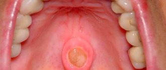
The first signs of lip cancer usually go unnoticed. First, you can determine the enlargement of the mental lymph nodes. You can notice this by feeling the lower jaw. The next early sign of lip cancer is a swelling of a dense consistency. Itching occurs in it. This neoplasm is usually mistaken for a herpetic rash.
A small ulcer with a crust forms in the center of the swelling, which does not cause pain. If it is removed, the patient feels quite severe pain, and upon closer inspection, he may find a bleeding base, which is formed by tubercles.
Local signs of tumor
Symptoms of lip cancer are:
- Dyskeratosis of the lips;
- Papilloma;
- Erosion;
- Cheilitis.
In most cases, dyskateriosis looks like cracks and ulcers. The erosions are covered with a crust and resemble herpes in appearance, but, unlike it, they do not heal after a certain period of time. Some patients have no ulcers or erosions. Instead, a small compaction appears, which over time grows and becomes covered with a crust.
On the red border of the lower lip, away from the midline, a patch or formation may appear that protrudes above the surface. An erosion or ulcer with a granular surface and a roll-like edge forms in the center of the tumor. The formation has a dense consistency and gradually increases in size, eventually acquiring an irregular shape. Its boundaries are unclear.
Exophytic lip cancer predominantly develops from a warty form of productive diffuse dyskeratosis of papilloma. With exophytic growth, the tumor has a dense consistency, often covered with flat scales. Endophytic growth of a cancerous tumor is characterized by the formation of an ulcer with uneven, dense edges. It often appears against the background of destructive dyskeratosis, quickly infiltrates the soft tissues of the lip and is prone to metastasis.
Symptoms of the disease should be a signal to immediately contact oncologists at the Yusupov Hospital. Lip cancer, treatment of which is started on time, is completely cured in 90% of cases.
Stages
Currently, the generally accepted classification of lip cancer is TNM (T-size of the tumor, N-damage to the lymph nodes, M-metastases). Based on the size of the tumor, there are 4 stages of lip cancer.
Table 1. Stages of lip cancer
| Stage | Tumor size |
| T1 | Less than or equal to 2cm |
| T2 | More than 2-4cm |
| T3 | More than 4cm |
| T4a | The tumor grows into the cortical layer of the bone, tongue muscles, maxillary sinus and skin |
| Т4в | The tumor grows in the bed of the masseter muscle, the pterygoid process, the internal carotid artery and the base of the skull |
If on the affected side there are single enlarged lymph nodes, the size of which is less than 3 cm, this is stage N1 lip cancer. At stage N2, enlarged lymph nodes are detected on the affected side, the diameter of which is more than 3 cm. If the patient has single enlarged lymph nodes on the affected side measuring 3-6 cm in size, this is stage N2a of lip cancer. At stage N2, oncologists determine multiple metastases to the lymph nodes. Their size is equal to or greater than 6cm. In the presence of bilateral metastases in the lymph nodes measuring 6 centimeters, they speak of the N2c stage of lip cancer. If the diameter of the lymph nodes exceeds 6 cm, this is stage N3 of the disease.
In the absence of distant metastases, oncologists determine stage M0 of lip cancer, if there are distant metastases - M1, in the case of distant metastases that cannot be assessed - MX. The diagnosis of “early stage lip cancer” is made in the presence of a tumor less than or equal to 2 cm, the presence of single enlarged lymph nodes less than 3 cm on the affected side and the absence of distant metastases. This is T1 N1 M0.
Classification
To classify the disease, the international TNM system is used, where:
- T (tumor) stands for tumor;
- N (nodulus) are lymph nodes that are affected by a tumor;
- M (metastasis) – metastases outside the lymph nodes.
T symbol gradation:
- Tx – there is no data to evaluate the primary neoplasm;
- Тis – preinvasive carcinoma (invasion of the lamina propria or intraepithelial invasion);
- T1 – tumor size up to 2 cm in greatest dimension;
- T2 – tumor size in greatest dimension is 2-4 cm;
- T3 – neoplasm in greatest dimension – more than 4 cm;
- T4a – the neoplasm spreads to neighboring tissues and organs (inferior alveolar nerve, skin of the nose or chin, floor of the mouth, etc.);
- T4b – the neoplasm affects the pterygoid processes, the masticatory zone, the base of the skull, and the carotid artery.
Gradation of the N symbol, which indicates metastases (or lack thereof) in regional lymph nodes (L/N):
- NХ – there is no data for assessing regional l / y;
- N0 – regional lymph nodes are not affected;
- N1 – in one lymph node on the affected side there are metastases up to 3 cm in size in the greatest dimension;
- N2 – in one lymph node on the affected side there are metastases 3-6 cm in greatest dimension; in several lymph nodes on the affected side in the greatest dimension there are metastases up to 6 cm; contralateral or bilateral involvement of lymph nodes with metastases up to 6 cm in greatest dimension;
- N2a – in one lymph node on the affected side there are metastases 3-6 cm in greatest dimension;
- N2b – on the affected side there are metastases in several lymph nodes up to 6 cm in the greatest dimension;
- N2c – contralateral or bilateral involvement of lymph node metastases up to 6 cm in greatest dimension;
- N3 – there are metastases in the lymph nodes more than 6 cm in greatest dimension.
M symbol gradation (distant metastases):
- M0 – no distant metastasis;
- M1 – distant metastases are present.
Stage according to TNM classification
| Stage | T | N | M |
| I | T1 | N0 | M0 |
| II | T2 | N0 | M0 |
| III | T3 | N0 | M0 |
| T1-T3 | N1 | M0 | |
| IV | T4 | N0 | M0 |
| Any T | N2,3 | M0 | |
| Any T | Any N | M1 |
Diagnosis of a tumor by a doctor in a hospital
What does lip cancer look like? It could be a small ulcer or a widespread tumor. Doctors at the Oncology Clinic of the Yusupov Hospital make a diagnosis of lip cancer based on the patient’s complaints, the results of an external examination and additional studies. The oncologist carefully examines and palpates the lips, cheeks, gums and regional lymph nodes. When examining the red border of the lips, skin and mucous membrane, use a magnifying glass.
How to identify lip cancer? Further examination is carried out using instrumental and laboratory diagnostic methods:
- Ultrasound examination;
- X-rays of the lower jaw;
- Panoramic tomography;
- Cytological examination of material obtained by taking fingerprint smears from the surface of the ulcer or histological examination of tissue obtained during a biopsy.
For lip cancer with lymphatic metastasis, a biopsy of the lymph nodes is performed. To exclude hematogenous metastases, chest X-ray and ultrasound examination of the abdominal organs are used. When the diagnosis of lip cancer is confirmed, an X-ray examination of the chest organs, general clinical and laboratory examination (electrocardiography, blood and urine tests) are performed.
A PET-CT study (positron emission computed tomography) is prescribed for the following purposes:
- Determining the stage of lip cancer;
- Assessment of response to treatment;
- Detection of disease relapse during the observation period.
PET-CT is an innovative method that combines the capabilities of computer technology and radiology. At the Yusupov Hospital, it is used not only to diagnose lip cancer, but also to monitor tumor development and evaluate the outcome of treatment. Thanks to PET-CT, radiologists have the opportunity to very accurately carry out radiation treatment, significantly reduce the irradiation area, and minimize the impact on healthy organs and tissues.
In many cases, the use of PET-CT excludes a number of additional studies in the future. This allows patients of the Yusupov Hospital to save time and money. The issue of the need to use this method is decided individually for a specific patient at a meeting of the expert council with the participation of professors and doctors of the highest category. A comprehensive examination of patients using the latest diagnostic equipment, the use of modern methods of performing laboratory tests using high-quality reagents allows oncologists at the Yusupov Hospital to obtain reliable results and provide adequate therapy for lip cancer at an early stage.
Why do they appear?
Like warts, papillomas are not dangerous
.
Their appearance is associated with the human papillomavirus (HPV),
which is transmitted through contact with a carrier. It is also possible to transmit HPV from an affected area of skin to a healthy one in one person. High humidity and microcracks in the skin increase susceptibility to viral particles. In this regard, you should always use personal hygiene items when visiting swimming pools, saunas and showers.
Important:
a large number of papillomas may indicate problems with the immune system.
For women in such cases, it is necessary to take a smear for HPV, since some of its types can cause cervical cancer
Treatment of lip cancer
How to treat lip cancer? When treating the disease, oncologists at the Yusupov Hospital take into account many different factors: the patient’s age, histological type of tumor, features of the spread of the tumor. Doctors at the Oncology Clinic provide multidisciplinary treatment for lip cancer:
- Surgical interventions;
- Radiotherapy;
- Chemotherapy.
Regardless of the chosen technique, they affect the lesion or tumor, areas of regional metastasis. For grades I and II of lip cancer, radiation treatment is performed, which includes external radiotherapy and surgery.
Treatment in the initial stages
Squamous cell carcinoma of the lower lip is a common complex tumor process. Its treatment consists of two stages: removal of the main focus and the fight against metastases that have spread to neighboring tissues and organs. Radiation treatment can suppress the development of a tumor focus. The radiologist selects the method of radiation therapy individually, depending on the size of the tumor, stage of the disease, and age of the patient.
Removal using the Krail method
In the initial stage of the tumor, surgeons perform a wedge-shaped excision of the entire thickness of the lip under local anesthesia. In order to remove a defect on the lip, plastic surgery is performed - skin flaps are transferred from the patient's cheek. Removal of the lesion is performed using the Krail technique. The operation is performed at the first and second stages of lip cancer, when the tumor has not spread to nearby tissues. Surgical excision of carcinoma makes sense in the absence of metastases.
The decision to remove a lymph node is made by an oncologist surgeon if the primary tumor process has already been cured or if the lymph node is removed along with the removal of the main lesion. In some cases, surgeons remove the primary cancerous tumor, the affected lymph nodes and the vessels that connect them. The scope of the operation is determined collectively by the clinic's oncologists.
Due to the fact that metastasis can occur in a cross way, the lymphatic apparatus of the suprahyoid region on the left and right is removed. If the oncologist sees that there are cancer metastases in certain lymph nodes, he decides to remove them, and those that are located along the lymph outflow.
Treatment tactics for lip cancer depend on the stage of the disease:
- Preventive surgery at stages I and II of lip cancer is carried out exclusively in cases where it is not possible to control the dynamics of the disease and there are unfavorable prognoses regarding the spread of cancer;
- At stage III, in the absence of metastases, treatment is carried out using a combined effect on the lesion and adjacent areas;
- In case of spread of cancer cells and the presence of single metastases in the lymph nodes, oncologists at the Yusupov Hospital perform combined treatment of lip cancer with subsequent surgery, plastic surgery and surgical correction of the lips;
- In stage IVC, palliative chemoradiotherapy is performed.
Candidates and doctors of medical sciences use innovative methods of treating lip cancer. The presence of modern equipment and competent medical personnel who are attentive to all the wishes of patients allows us to achieve good results in the treatment of lip cancer for many years. Lip cancer, treated in the early stages, is cured in 97-100% of cases. At stage III, 67-80% of patients can be cured. In the fourth stage of cancer and repeated relapses, complete recovery occurs in 55% of cases.
Diagnostic methods
To detect such a pathology, an x-ray of the lower jaw and chest organs is taken. If there is a suspicion that the tumor has grown into the bone, a CT scan of the facial bones is performed.
The X-ray technique is based on the fact that X-ray radiation is attenuated differently when passing through different tissues of the human body. The result is a summation image. CT provides a layer-by-layer image of internal structures, so the extent of the tumor process can be analyzed more accurately.
If the doctor suspects that there are metastases in the bones, he gives a referral for osteoscintigraphy. For the study, a radiopharmaceutical is injected into the patient and then its distribution and accumulation in the bones is recorded using a gamma camera.
To diagnose lymph nodes, an ultrasound of the soft tissues of the neck is performed. This safe and affordable method produces transverse images using high-frequency sound waves produced by the transducer. When the sound wave returns to the sensor, it takes digital form and appears on the monitor as dots or echoes.
Images can be obtained in any plane and appear in real time during the examination. To confirm the diagnosis, a smear or scraping is taken from the tumor ulcer, which is sent for cytological examination to the laboratory. Biomaterial obtained by puncture from metastatic lymph nodes is also sent for cytology. For research, the material is prepared in a special way and stained, after which it is examined under a microscope. In this way, the pathologist can determine the type of tumor.
In preparation for surgery, in order to clarify the functional status of the body, additional examination may be prescribed, because surgical intervention is always a risk for the patient.
Prevention
There are primary and secondary prevention of lip cancer. Oncologists recommend:
- Take precautions when exposed to the sun (wear a wide-brimmed hat);
- Stop smoking pipes and cigarettes;
- Maintain oral hygiene;
- Change working conditions;
- Do not drink strong alcoholic drinks.
For persons prone to dyskeratosis of the lips and cheilitis, oncologists at the Yusupov Hospital conduct an annual preventive examination. Secondary prevention of lip cancer consists of regular dental treatment at the dentist, surgical intervention in the presence of dyskeratosis and cheilitis.
Lip cancer: treatment at Yusupov Hospital
Effective treatment of lip cancer in Moscow is carried out by doctors from the oncology clinic of the Yusupov Hospital. Oncologists have many years of experience in treating malignant neoplasms of the lip. Surgeons are fluent in surgical techniques. The cost of surgery is lower than in other oncology clinics in Moscow, despite the high level of qualifications of doctors and modern equipment.
Radiotherapy is carried out using modern radiation installations from leading European and American manufacturers. In order to undergo a course of effective treatment for lip cancer at an affordable price, call the Yusupov Hospital. After the operation, surgeons at the Oncology Clinic perform lip plastic surgery using innovative surgical techniques.
Statistics
In relation to all cancers, lip cancer in Russia ranks 15th in terms of prevalence among men (1.5%) and 20th among women (0.53%). According to data for 2014, 1,958 new cases of this disease were identified in Russia. In the vast majority of cases (85-90%), cancer affects the lower lip.
In 2015, 398 people (2.99%) died from cancer of the oral cavity, lip, and pharynx in St. Petersburg.
In general, statistics for St. Petersburg indicate that the incidence of lip cancer is decreasing: in 1980, 811 cases were registered, and in 2009 – 416.