Stomatitis: features of the disease
Stomatitis is a disease related to dental pathologies in which inflammation of the oral mucosa occurs. Infection of the tongue with stomatitis in adults is also called “glossitis.”
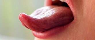
The inflammatory process affects the mucous membrane and causes severe symptoms:
- redness and swelling of the tongue;
- formation of white plaque;
- severe pain;
- the formation of many small blisters and ulcers.
Due to severe sensitivity and soreness of the tongue, the patient cannot eat food, fever, insomnia and irritability are possible.
Stomatitis under the tongue in adults, as in other parts of the oral cavity, is classified into two types.
- Aphthous. A form of inflammation in which the tongue becomes covered with papules and ulcers that turn into erosions (aphthae).
- Herpetic. The inflammatory process is manifested by swelling and redness of the tongue. Small bubbles are localized in groups.
Prosthetic stomatitis and ulcerative-necrotic form of pathology are extremely rare.
Without treatment, the signs of stomatitis completely disappear, but this does not mean that the patient is cured of the pathology. The inflammatory process becomes chronic and when the immune system is weakened or exposed to an irritant, a relapse occurs.
Tongue injuries
Another reason why the tongue hurts on the side lies in mechanical injuries to the organ. They occur when eating food with sharp parts (seeds, nuts, undercooked cereals), hitting, or biting. Damage can occur due to a seizure, sports, falls, or various accidents.
If you are absolutely sure that the soreness is the result of an injury, then you just need to give the tongue time to recover. Nevertheless, examination by a specialist to clarify the diagnosis and treatment will not be superfluous.
Causes of stomatitis
Stomatitis under the tongue in adults is a reaction of the immune system to certain types of irritants, which are any substances. The causes of stomatitis have not been clearly identified; presumably, the body exhibits an individual reaction to certain substances or microorganisms.
Possible causes of stomatitis on the tongue in adults:
- bacteria and infections of the oral cavity (caries, gingivitis, periodontitis, etc.);
- diseases of the gastrointestinal tract of an infectious or viral type;
- respiratory diseases (ARVI, influenza, etc.);
- frequent injury to the tongue by orthopedic or orthodontic structures;
- damage to the tongue due to a chipped tooth;
- fungal infection;
- bad habits (smoking or alcoholism);
- burning the tongue or eating “spicy” foods.
In addition to the listed causes of stomatitis on the tongue in adults, inflammation of the mucous membrane can be caused by the herpes virus (herpetic stomatitis).
Differentiation of plaque in gastritis from other diseases of the gastrointestinal tract
A thick, grayish coating is characteristic of dysentery.
It is important to suspect the onset of a dangerous disease in time. To take action and prevent complications from occurring. Differential diagnosis by tongue of gastritis from other diseases and conditions:
- A thick, grayish coating is characteristic of dysentery. In this case, the tongue looks cracked, and less saliva is produced than usual.
- Desquamative glossitis - this type of inflammation of the tongue is characterized by such symptoms as red spots of complete absence of epithelium or several altered taste buds on the tongue, covered with a white coating.
- Galvanic stomatitis is a form of inflammation of the tongue that arises as a result of a reaction to metal prostheses, manifested by spots in the form of pimples, and subsequently by the appearance of erosions against a background of white plaque.
- Infectious diseases - sore throat, scarlet fever, diphtheria, HIV infection can cause the appearance of a white coating on the tongue, but almost all of these infections are accompanied by high fever and skin rashes.
- Diseases of the heart and blood vessels - plaque is located on the anterior third of the tongue.
- Kidney disease - plaque on the tongue is localized at the back along the edges.
- Endocrine disorders - under the plaques of white plaque there are ulcers and erosions.
- Anemia is not a coating on the tongue, but blanching of the entire surface of the organ. Diseases of the respiratory system are often indicated by the localization of white plaque on the front and along the edges of the tongue.
- Diseases of the salivary glands - the appearance of a white coating is accompanied by the appearance of an unpleasant odor.
- Diseases of the liver and gall bladder - the color of the plaque is not white, but has a yellowish or brown tint. A white coating on the tongue can be caused by the consumption of dairy products, as well as the proliferation of bacteria and fungi in those who abuse sweets. Unlike plaque during gastritis, such layers are easily removed and do not form further.
Treatment Basics
If an adult has a swollen tongue, the surface is covered with plaque, or has severe hypothermia, you should consult a dentist. Even in the absence of neoplasms in the oral cavity in the form of papules, ulcers or erosions, a specialist will be able to determine the development of the inflammatory process. With timely treatment, the risk of pathology becoming chronic is minimal.
Treatment of stomatitis on the tongue in adults is carried out exclusively conservatively. Depending on the causes of the disease and the clinical picture, the doctor will determine a treatment regimen and prescribe effective drugs of systemic and local action.
The dentist’s task is not only to eliminate symptoms and inflammation, but also to identify the cause. If the provoking factor has a constant impact, then stomatitis under the tongue will constantly recur.
Treatment of stomatitis on the tongue in adults begins with the relief of concomitant diseases - the root causes.
Glossitis - symptoms and treatment
Folded or scrotal tongue. The term “scrotal tongue” comes from the external similarity of the affected tongue to the skin of the scrotum (from the Latin scrotum - scrotum). With this form of glossitis, a deep central groove runs along the back of the tongue with adjacent cracks and folds, which can be located longitudinally or randomly. The tongue is slightly enlarged [5]. Due to atrophy of the filiform papillae, it may look “smoothed out”, but this does not affect the taste sensation [16].
Diamond-shaped or median glossitis. This is a chronic disease of the mucous membrane of the tongue with partial or complete absence of filiform papillae closer to the posterior third of the tongue. The lesion has a diamond shape, a dense texture and a red varnished color. Usually it has clear contours and does not protrude above the mucous membrane. The lesion does not cause pain, but some patients perceive it as an aesthetic defect.
Other forms of rhomboid glossitis are less common - with tuberous outgrowths and papillomatous growths. The patient then feels the presence of a foreign body on the tongue [5][16].
Desquamative or “geographical” language. With this form of glossitis, the keratinization of the tongue is partially disrupted with signs of desquamation of the epithelium - desquamation (Latin desquamo - remove scales). The papillae of a significant part of the surface of the back of the tongue are changed. The lesions take on the appearance of red spots with a diameter of 0.5 cm and desquamated whitish epithelium along the edges. The spots can merge with each other and create a pattern similar to a geographical map.
Patients often complain of pain and burning when eating food or drinks that irritate the oral mucosa, and some of them note a short-term loss of taste.
There are two forms of desquamative glossitis: migrating and fixed [16].
The fixed form is more common with moderate damage to the stomach by the bacterium Helicobacter pylori. The disease is usually accompanied by persistent atrophy and smoothness of the filiform papillae of the tongue. The size and location of the lesions does not change.
The migratory form occurs when the stomach is heavily colonized with H. pylori. Foci of desquamation are up to 2 cm in diameter, their location is constantly changing.
Also, with desquamative glossitis, the tongue may increase in volume due to inflammation of the fungiform papillae, which are located at its root. They reach 0.8 cm in diameter and are covered with a white or light gray dense coating [14].
Black "hairy" tongue. This is a rare lesion in which the keratinized epithelium of the filiform papillae located in the posterior and middle third of the dorsum of the tongue does not slough off. Because of this, the papillae acquire a brown or black color, become stiffer, grow up to 2-3 cm, and are covered with a coating ranging from green and light brown to black [16].
The tongue takes on an unaesthetic appearance, patients complain of the sensation of a foreign body in the mouth and the appearance of a gag reflex [4].
Diseases of the gastrointestinal tract. A sign of damage to the gastrointestinal tract is a grayish-white coating on the back surface of the back of the tongue. There are no pain or any other subjective sensations.
Another manifestation of gastrointestinal tract damage is swelling of the tongue with tooth marks on its lateral surfaces without visible relief disturbances. At the same time, patients may be bothered by constant biting and burning when eating hot and spicy foods. The fungiform papillae located at the root of the tongue may also enlarge. They become bright red and rise above the surrounding tissue. The listed symptoms may accompany gastritis, gastric and duodenal ulcers[5].
With hepatitis A, areas of desquamation appear on the back of the tongue. Due to atrophy of the filiform papillae, the tongue becomes smooth and red. Patients often complain that the taste of sweet and bitter foods has become less pronounced [5].
Liver cirrhosis is manifested by swelling of the sublingual veins, atrophy of the oral mucosa and deepening of the natural folds of the tongue.
Lesions of the pancreas. In acute pancreatitis, the tongue becomes covered with a yellow-white coating, and the filiform papillae increase in size. Patients are often concerned about its dryness and disturbances in taste sensitivity.
In chronic pancreatitis, a secondary deficiency of B vitamins occurs, which is manifested by pain in the tongue and protruding bright red fungiform papillae. The tongue increases in size, becomes smooth and shiny.
Cardiovascular pathology. With hypertension, blisters with bloody contents are often present on the tongue, the so-called “vesical” symptom. They can suddenly appear and burst on their own, damaging the epithelium.
Diseases of the endocrine system. With diabetes, the body is dehydrated, so dryness appears, and the vessels of the tongue become overfilled with blood. A burning sensation occurs and long-healing ulcers form. Very often, symptomatic candidiasis is found on the tongue, especially if the patient does not regulate blood glucose levels.
Insufficient function of the thyroid gland, or hypothyroidism, leads to enlargement of the entire tongue or part of it and swelling, which does not form a dimple when pressed. Due to high cholesterol levels, the tongue is slightly yellowish in color.
Diseases of the blood and hematopoietic organs. With iron deficiency anemia, there is a need to eat unusual substances, such as chalk, clay, as well as a burning sensation and pain in the tongue. It is often pale in color and slightly increased in size.
With Addison-Beermer anemia, which occurs due to impaired absorption of vitamin B12, the filiform muscles weaken. The tongue becomes smooth and as if “polished”. This condition was called “Gunter's glossitis.” Painful red inflammatory streaks and spots appear. The tongue or just its tip turns bright red, and a tingling and burning sensation occurs [5].
Acute leukemia is manifested by ulcers of irregular shapes without clear boundaries with a necrotic coating on the tongue, without rims of hyperemia (overflow of blood vessels) along the edges. Pinpoint and small blood spots and hematomas may also appear on the mucous membrane of the tongue. One of the main symptoms of this condition is heavy bleeding.
Chronic leukemia occurs much less frequently than acute leukemia and lasts longer. Characteristic ulcers and leukemic infiltrates (fluid accumulation) appear on the tongue. At the acute and chronic stages of leukemia, they lead to the formation of ulcerative-necrotic lesions, which are difficult to treat.
Agranulocytosis is characterized by a significant decrease in the blood of granulocytes - a type of leukocyte, “protective” blood cells. The disease manifests itself as non-demarcated ulcers on the tongue and, unlike acute leukemia, an inflammatory reaction around them. Due to decreased immunity and the resulting increased susceptibility of the body to fungal diseases, candidiasis may develop.
Changes in the tongue due to hypo- and avitaminosis:
- hypovitaminosis A - dryness of the mucous membrane of the tongue and a tendency to increased keratinization of the skin (hyperkeratosis);
- hypovitaminosis C - swelling of the tongue with teeth marks on it; ulcerative necrotic lesions, usually covering a significant surface area of the tongue mucosa;
- hypovitaminosis PP causes a disease called “pellagra”, in which peeling of the surface layers of the epithelium occurs and a burning sensation of the tongue, it becomes smooth, shiny and crimson - the so-called “cardinal tongue”;
- hypovitaminosis B1 is characterized by pain in the tongue and the appearance of blisters;
- hypovitaminosis B2 is manifested by burning of the tongue, atrophy of the papillae and angular cheilitis - cracks in the corners of the mouth [4].
When infected with herpes simplex virus type 1 (HSV-1), patients complain of burning pain in the tongue. Upon examination, a deep crack is visible along its midline with several branches. In their depths there are painful blistering rashes, which over time burst with the formation of erosions.
Burning mouth syndrome. In the area of the tongue there is a feeling of a burn, paroxysmal burning pain, which intensifies with overwork and stress.
Galvanism. Patients experience a metallic taste in the mouth and rawness. Upon external examination, the tongue is swollen, its vessels are filled with blood, “bald spots” of the papillae and bright red erosions appear on its sides [5].
How is the treatment carried out?
Treatment for stomatitis is comprehensive.
- Anesthesia. To eliminate pain, the dentist may prescribe topical medications containing lidocaine or analgesics.
- Antiviral therapy. If the disease is caused by a herpes virus, the doctor prescribes antiviral drugs of the appropriate group of effects.
- Antibacterial therapy. In case of bacterial stomatitis, local treatment of the oral cavity with antiseptic solutions and dental ointments with antibacterial action is mandatory.
- Antifungal therapy. Stomatitis can be the result of fungal activity in the mouth. This form is more common in children, but is not excluded in adults. The specialist prescribes the patient a strict regimen of antifungal drugs.
- Anti-inflammatory therapy. It is carried out in cases of severe inflammation. The dentist recommends taking anti-inflammatory systemic drugs that have a targeted effect depending on the pathogen or cause of tongue stomatitis.
- Diet. Patients with stomatitis are prescribed a diet that excludes foods that can aggravate irritation of the mucous membrane and tongue. So dentists recommend avoiding hot, spicy, sour and salty foods. Smoking and drinking alcohol should be avoided.
Together with the treatment of stomatitis, treatment of diseases of the respiratory system, gastrointestinal tract, inflammation of the gums, caries, etc. can be carried out.
Catarrhal glossitis
If the tongue hurts on the side and a white coating covers its surface, then this is most likely catarrhal glossitis - a special case of this disease. It can be considered as a symptom of other pathologies. In particular, it is called:
- caries;
- stomatitis;
- gastrointestinal diseases;
- various infections (measles, diphtheria, etc.).
In addition to pain and plaque, symptoms of catarrhal glossitis include swelling of the tongue and a burning sensation, which is especially worse after eating or talking. The disease is treated by eliminating its causes and rinsing the mouth with antiseptic solutions.
General recommendations
For stomatitis in adults, treatment is carried out on an outpatient basis. The dentist can perform initial treatment of the oral cavity, then the patient will need to perform all the manipulations independently at home.
Antiseptic treatment of the entire oral cavity is a prerequisite for the successful treatment of stomatitis and the rapid recovery of the affected mucosa. For “disinfection”, solutions containing chlorhexidine, furatsilin or metronidazole are used. Dentists also recommend rinsing with a soda solution every 2-3 hours.
Locally in the affected areas, it is necessary to remove heavy plaque using gauze and apply anti-inflammatory and regenerating gels or ointments to the areas where ulcers accumulate. The procedure is unpleasant, but significantly speeds up recovery.
Treatment of stomatitis on the tongue in adults, subject to all prescriptions and recommendations of the dentist, takes no more than 10 days. Symptoms of the disease disappear after 3–5 days; a few more days are required to restore the affected tissues.