The main risk factors for the development of candidiasis include:
- improper oral hygiene;
- wearing orthopedic structures or removable dentures;
- long-term inhalation therapy;
- decreased salivation due to certain pathologies;
- hypothermia;
- stress;
- taking antibiotics;
- changes in hormonal levels;
- decreased immunity, including due to chronic diseases;
- endocrine pathologies, including obesity, diabetes;
- smoking;
- carrying out chemotherapy or radiation therapy.
Oral candidiasis occurs much more often in children than in adults. Newborns can become infected even in utero if the mother has thrush - infection occurs during natural childbirth. After birth, the infection can be transmitted to the child through the skin of the mother's nipples and hands, and from her oral mucosa. The source of infection is unsterile pacifiers and oilcloths, and medical personnel who do not follow the rules of asepsis and antisepsis during invasive procedures. In an older child, oral candidiasis is often caused by a weakened immune system or improper dental care.
Antifungal and anti-inflammatory drugs
07.03.2018
Salicylic ointment is a medicine indicated for chronic eczema , psoriasis , dyskeratosis, ichthyosis, oily seborrhea, acne , warts, calluses . It is applied to the affected surface 2-3 times a day.
The drug is contraindicated in case of hypersensitivity to salicylic acid and in childhood. Side effects are rare. These are local reactions in the form of skin rash, itching, burning, as well as allergic reactions .
Amphotericin B is an antifungal antibiotic intended for the treatment of candidiasis of the skin and mucous membranes, as well as candidiasis of the gastrointestinal tract and internal organs, chronic granulomatous and disseminated forms of candidiasis , coccidioidosis, histoplasmosis, chromomycosis, mold mycoses. The ointment is applied to the affected areas of the skin in a thin layer 2-4 times a day. The course of treatment for candidiasis of skin folds is 1-3 weeks, for diaper rash in children - 1-2 weeks, for lesions of the interdigital spaces and paronychia - 2-4 weeks.
liver and kidney dysfunction , diseases of the hematopoietic system, diabetes mellitus , and hypersensitivity to its components. When used externally, a burning sensation, tingling sensation, skin flushing, and rarely dry skin are possible.
Nystatin is an antifungal drug intended for the prevention and treatment of diseases caused by yeast-like fungi of the genus Candida, candidiasis of the skin , mucous membranes ( mouth , vagina ) and internal organs (gastrointestinal tract, lungs , kidneys ). For prophylactic purposes, it is prescribed to prevent the development of candidiasis during long-term treatment with penicillin and antibiotics of other groups, especially with oral use of tetracycline antibiotics, chloramphenicol, neomycin and others, as well as in weakened and depleted patients. When treating fungal infections of the mucous membranes and skin, nystatin ointment can be used. It can be combined with oral nystatin. For candidiasis of the lower intestines , colpitis and vulvovaginitis, rectal suppositories and vaginal globules containing 250,000 and 500,000 units each are used. They are administered 2 times a day, respectively, into the rectum or vagina ; The average course duration is 10–14 days.
Contraindications and side effects are the same as when taking amphotericin B.
Fluconazole is a broad-spectrum antifungal agent that has a powerful antifungal effect against the causative agents of candidiasis , thrush , some forms of lichen , dandruff (Candida, Microsporum, Cryptococcus neoformans, Trichophyton). Fluconazole is used for skin mycoses (including mycoses of the feet, groin area), pityriasis versicolor , onychomycosis, deep endemic mycoses, systemic cadidosis, cryptococcosis, for the prevention of fungal infections in patients with reduced immunity .
For candidiasis , mycoses of the feet, body, and groin area, 150 mg is prescribed once a week, treatment duration is 2–4 weeks. For cryptococcal infections, candidemia, disseminated candidiasis , and other invasive candidiasis infections, 400 mg is prescribed on 1 day, then 200–400 mg 1 time per day.
For deep endemic mycoses - 200-400 mg per day for a long time up to 2 years. The duration of therapy is determined individually; it can be 11–24 months for coccidioidomycosis, 2–17 months for paracoccidioidomycosis, 1–16 months for sporotrichosis, and 3–17 months for histoplasmosis. For children over one year of age, fluconazole is administered once a day at the rate of 1–3 mg/kg (for candidiasis of the mucous membranes) and 3–12 mg/kg (for systemic candidiasis or cryptococcosis) depending on the severity of the disease.
Fluconazole is contraindicated in case of hypersensitivity to its components, pregnancy , or in children under 1 year of age.
When using the drug, side effects are possible: nausea , headache, discomfort in the abdominal cavity, increased activity of liver enzymes, severe liver , headache, dizziness ; rarely - convulsions, agranulocytosis, leukopenia, thrombocytopenia; very rarely - skin rash, anaphylactic reactions.
Pimecrolimus is an anti-inflammatory drug indicated for atopic dermatitis ( eczema ). It is recommended for both short-term and long-term use in adults, adolescents and children (from 3 months). Prescribed externally 2 times a day.
The drug is contraindicated in case of hypersensitivity to its components.
When taking pimicrolimus, side effects are possible: burning at the site of application of the cream, local reactions (irritation, itching and redness of the skin, rash, pain, paresthesia, peeling, dryness, swelling, skin papillomas, boils), skin infections (folliculitis), suppuration, deterioration course of the disease, herpes simplex, dermatitis caused by the herpes simplex virus (eczema herpes ), molluscum contagiosum. In rare cases, alcohol intolerance appears (immediately after drinking alcohol, facial , rash, burning, itching or swelling were usually observed).
Dioxomethyltetrahydropyrimidine + sulfadimethoxine - trimecaine + chloramphenicol - a combined drug with antimicrobial, anti-inflammatory and local anesthetic effects, accelerating wound healing. Indicated for the treatment of purulent wounds in the first phase of the wound process. It is applied topically in the form of applications. Sterile gauze pads are soaked in the ointment and loosely filled into the wound. Dressings are performed daily until the wound is completely cleansed of purulent-necrotic masses. The ointment can be injected into purulent cavities through a drainage tube using a syringe.
The drug is contraindicated in case of hypersensitivity to its components.
When used, local allergic reactions in the form of skin rash are possible.
Published in Medicines Premium Clinic
Oral candidiasis is divided into 3 types:
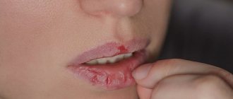
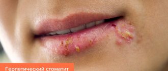
- Pseudomembranous. This is a classic thrush in the form of a superficial white film or white spots. Accompanied by a feeling of slight discomfort. In mild forms of the disease there may be only a few such plaques, and they can be easily removed. In severe cases, many large spots appear, they merge and gradually affect the entire mucous membrane, and can penetrate lower, affecting first the throat, and then the esophagus, up to the gastrointestinal tract. The spots may thicken, making them difficult to remove. Most often, ordinary acute candidiasis occurs in adults who have taken antibiotics, immunosuppressants or corticosteroids, as well as in infants.
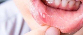
- Atrophic. It can be acute or chronic. In the first case, there may be no plaque, but the mucous membrane becomes inflamed - it becomes bright red, the patient complains of a strong burning sensation in the mouth, increased dryness, and sometimes an unpleasant taste with a hint of bitterness or metal. Most often, acute candidiasis is a consequence of drug therapy. The chronic form usually occurs due to wearing orthodontic structures or dentures. The symptoms of the disease are more subtle.
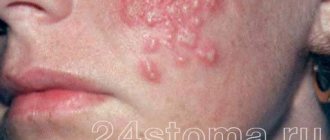
- Hyperplastic. Candidiasis in the mouth affects the lips, corners of the mouth, cheeks, soft palate, and back of the tongue. White spots are usually large, merge with each other, and may acquire a yellow tint. Gradually they become rough and cannot be scraped off. The disease is chronic and is diagnosed only in adults.
Fungal diseases of the pharynx
At the turn of the XIX-XX centuries. Almost all the main human mycoses and their causative agents have already been described. The names of Virchow, Hrubi, Remarque, Schönlein, and then Sabouraud are closely connected with the history of the development of medical mycology. Due to the widespread use of antibiotics, the second half of the 20th century. was accompanied by a significant increase in the incidence of mycoses, which currently affect from 5 to 20% of the adult population (A. Yu. Sergeev, Yu. V. Sergeev, 2003). Among all mycotic lesions of the human body, in second place after onychomycosis is candidiasis of the mucous membranes, up to 40% of cases of which, in turn, are oropharyngeal mycosis (V. Ya. Kunelskaya, 1989).
Among mycoses of the pharynx (pharyngomycosis), the most common (in 93% of cases, according to V. Ya. Kunelskaya (1989), and in 90%, according to T. N. Burkutbaeva (2002)) is candidiasis caused by the yeast fungus Candida, which combines 20 species (A. Yu. Sergeev, Yu. V. Sergeev, 2003). In patients with pharyngomycosis, as a rule, eight different types of pathogens are identified, among which four main ones “lead”: C. albicans, C. tropicalis, C. parapsilosis and C. glabrata. The first place is occupied by the disease caused by C. albicans. This species is found in the mouth and throat of 60% of healthy adults, more often in women and men who smoke. Other Candida species are significantly inferior to C. albicans in the number of discharges from healthy individuals, accounting for 10 to 20% of all cases of oropharyngeal candidiasis. In second place after C. albicans is usually C. glabrata, especially in elderly patients, less often - C. tropicalis, C. parapsilosis (in the latter case - in almost 50% of candidiasis-carrying children). With oropharyngeal candidiasis in HIV-infected people, rare species of Candida—C. sare, C. dubliniensis, C. famata, C. lipolytica and C. guilliermondii—more often appear among the pathogens. It has also been reported that C. rugosa has been isolated from diabetic patients, C. inconspicua and other yeasts from cancer patients. When treated with systemic antimycotics and antibiotics, the proportion of resistant species, C. glabrata and C. krusei, as well as C. kefyr, including those isolated simultaneously with resistant strains of C. albicans, may increase (MD Richardson, DW Warnock, 1997).
Much less often - in 5-6% of cases - mycosis of the pharynx is observed, caused by the bacterial microorganism Leptotrix buccalis and the fungi Aspergillus, Penicillium, Mucor, etc. In some cases, damage to the pharynx is caused by two or more types of fungi - Pharingomycosis mixta.
Almost all of these types of fungi are saprophytes, opportunistic microflora; they are activated and become pathogens when the body’s reactivity is impaired (I. V. Chumicheva, 2003).
The development of pharyngomycosis is provoked by diabetes mellitus, systemic diseases of the blood and gastrointestinal tract, especially intestinal dysbiosis. Deficiency of bifidobacteria and other lactic acid bacteria leads to disruption of the synthesis of B vitamins and to the unhindered colonization of fungi not only in the intestines, but also in other body cavities in contact with the external environment (nasal cavity, mouth, ear). In addition, malignant neoplasms have a negative impact, in which the balance of vitamins, carbohydrate and protein metabolism is disrupted, and the body’s general, including antimycotic, resistance suffers. Mycoses are especially common in AIDS patients, 10% of whom die from fungal infections. Often, pharyngomycosis of the pharynx and oral cavity develops with prolonged and improper use of topical glucocorticosteroids for bronchial asthma.
In the formation of pathogenetic mechanisms of fungal infection of the pharynx, the first place is taken by the restructuring of immune reactivity - the accumulation and circulation of fungal antibodies in the blood, which cause immediate and delayed reactions. Changes in cellular immunity are also important. An important link in the pathogenesis of pharyngomycosis is specific and nonspecific sensitization and allergy. Traumatic damage to the mucous membrane of the pharynx also plays a certain role here - as a factor predisposing to the development of a fungal process.
Mycotic foci in the pharynx are usually localized on the palatine tonsils, arches, velum, posterior wall, and tongue. Tissue reactions are expressed by atypical hyperplasia of the epithelium, the formation of granulomas, sometimes having a pseudotuberculous character, as well as necrotic changes of varying severity. Epithelial hyperplasia in the form of hyperkeratosis is especially pronounced in the crypts of the tonsils against the background of an inflammatory process in the stroma and can take the form of papillomatosis. In the tissues of the mucous membrane of the pharynx, tongue and stroma of the tonsils, fungal elements located subepithelially are detected.
Microscopy of a native preparation is the simplest and most reliable method for diagnosing mycoses, allowing not only to determine the presence of a fungus in the test material, but also to distinguish saprophyty from a fungal disease. With saprophyty, only single non-budding cells are found in the preparations, and with mycosis, all elements of fungi are constantly determined in each preparation: both mycelium and blastospores.
Material is collected from the pharynx for testing for fungi using ear tweezers, Hartmann ear forceps or a Volkmann ear spoon. It is unacceptable to use a cotton swab for this purpose, since the bulk of the sticky pathological content remains on the swab, and therefore a false negative result is possible during microscopic examination or culture. The collected material is carefully applied to a sterile glass slide. The material should not be rubbed on glass, as this may damage the delicate elements of the fungus, which also reduces the reliability of microscopy results. Microscopy of the unstained and Romanovsky-Giemsa-stained native preparation is performed, inoculation of the pathological discharge obtained by scraping from the tonsils or the posterior wall of the pharynx on Sabouraud's selective nutrient medium, followed by subculture of fungal cultures on Chanek's medium to identify the pathogen. When isolating a Candida culture, quantification is often required. In the case of actinomycosis, microscopy reveals a significant proliferation of granulation tissue with actinomycetes.
Immunological diagnosis is based on the detection of pathogen antigens in the blood. Serological diagnostics is a type of immunological diagnostics that makes it possible to detect antibodies to the components of the pathogen cell in the blood. A feature of immunological diagnostic methods is that serial sets of antibodies or antigenic diagnostics are developed only for a few of the most common pathogens (candidiasis, cryptococcosis, aspergillosis; diform fungi). The disadvantages of immunodiagnostic methods include insufficient sensitivity and specificity. However, with the help of immunodiagnostics it is possible, in particular, to monitor the effectiveness of treatment by reducing titers (A. Yu. Sergeev, Yu. V. Sergeev, 2003).
Mycotic lesions of the pharynx can be acute, in which case it is usually accompanied by widespread lesions of the oral mucosa and pharynx, and chronic, in which the lesion is often limited to the palatine tonsils (tonsillomycosis) or the posterior wall of the pharynx (pharyngomycosis); a distinction is made between recurrent and persistent forms of chronic pharyngomycosis. According to the clinical and morphological picture, pharyngomycosis is divided into pseudomembranous, which occurs most often in this localization; erythematous (atrophic), hyperplastic (hypertrophic, or candidal leukoplakia); erosive-ulcerative are rare (A. Yu. Sergeev, Yu. V. Sergeev, 2003).
The clinical picture of pharyngomycosis is varied and is determined not so much by the type of fungus that caused the disease, but by the general and antimycotic activity of the body. The clinical picture of candidiasis, penicilliosis and aspergillosis is almost identical (M. R. Bogomilsky, V. R. Chistyakova, 2001). The disease is characterized by sore throat, fever, hyperemia of the mucous membrane, the appearance of small white plaques at first, and then extensive necrosis of the epithelium, which occurs under the influence of its infection by fungi, which persist when treated with traditional conservative methods. On the mucous membrane, multiple grayish-white plaques of irregular shape are visible, located on the tonsils, arches, palate, as well as in other places and are difficult to remove; after their removal, an eroded surface remains. In the chronic form, typical complaints appear with a characteristic cyclicity, after two to three weeks. With superficial mycosis, a mildly expressed hyperemia of the mucous membrane is determined with small areas of translucent or dense peak-shaped plaques of a grayish or white color, often having a cheesy, lumpy character. The plaque is easily removed, revealing a smooth, hyperemic mucous membrane. In some cases, plaque can merge and become denser. After their removal, the mucous membrane is exposed, bleeding at the slightest damage, which is very reminiscent of damage to the pharynx due to diphtheria. With ulcerative-membranous lesions, the process is usually localized on infiltrated, tuberous tonsils and spreads to adjacent formations. Ulcers with uneven edges are covered with a white coating that can be easily removed. Regional lymph nodes (za- and submandibular) enlarge slightly and are slightly painful (I. V. Chumicheva, 2003).
Often such lesions are found not only in the pharynx, but also in other parts of the respiratory and digestive tracts, which significantly worsens the patient’s condition. Infection of internal organs by fungi is characterized by an exceptionally severe course, similar in symptoms to sepsis (V. Ya. Kunelskaya, 1989).
Pharyngomycosis is often combined with candidal lesions of the corners of the mouth - “jams”, candidal cheilitis, glossitis. The red border of the lips is sharply hyperemic, infiltrated, has transverse striations, and is covered with a thin grayish coating. In the corners of the mouth there are cracks covered with curdled crusts, painful when opening the mouth. The disease lasts 7-12 days and often has an undulating course. Recurrence of the process is noted in 20-22% of cases (I. V. Chumicheva, 2003).
Leptotrichosis of the pharynx is expressed by the formation of spiny, pointed, very dense outgrowths (stalked films in the form of spikes) of gray or yellowish-gray color on the surface of the unchanged mucous membrane of the palatine, lingual, pharyngeal tonsils, lateral ridges and granules of the posterior wall of the pharynx, as well as on the palatine arches. These outgrowths are a consequence of keratinization of the squamous epithelium, come off with difficulty, in parts and contain Leptotrix buccalis cells. The disease is often asymptomatic and long-lasting; sometimes patients complain of a sensation of a foreign body in the throat. There are no inflammatory changes in the mucous membrane and no fever. The course of the disease is persistent (V. Ya. Kunelskaya, 1989).
Actinomycosis is characterized by the formation of dense, lumpy infiltrates of a dark red color (specific infectious granuloma), slowly growing (sometimes the process manifests itself in the form of acute phlegmon), located most often in the area of the mouth and neck, less often in the area of the tongue, tonsils, nose and larynx. Over time, the tumor suppurates and abscesses form. The surrounding tissues of the chin and cheek are involved in the process, then the tumor can open on its own to form a fistula. In the contents you can find yellow-green druses of the fungus. Due to inflammatory swelling of the masticatory muscles, their trismus appears; regional lymphadenopathy is uncharacteristic (M. R. Bogomilsky, V. R. Chistyakova, 2001).
Candidomycosis of the pharynx should be distinguished from diphtheria, fusospirochetosis and damage to the pharynx due to blood diseases (V. Ya. Kunelskaya, 1989).
A comprehensive method of treating tonsillo- and pharyngomycosis includes three fundamental principles.
- General and local use of modern antifungal drugs (previously used antibiotics are canceled).
- Restoration of disturbed intestinal microbiocenosis. In correcting changes in intestinal microbiocenosis, in turn, three factors are important: normalization of chemical processes in the intestine and the fight against opportunistic flora with the help of diet (products with a bactericidal effect), antibacterial agents, intestopan, mexaform;
- taking bacterial preparations containing live cultures (bifidumbacterin, colibacterin, lactobacterin);
- increasing nonspecific defense reactions of the body, promoting the formation of healthy microflora; For this purpose, eubiotics (atsipol, bactisubtil, hilak-forte, linex) are used in complex therapy of candidiasis from three weeks to three months.
In most cases, treatment of pharyngomycosis begins with the prescription of local therapy, and then, if necessary, systemic therapy with azole derivatives is added: fluconazole (Diflucan, Mycosist, Flucostat, Flumicon) 100 mg/day, ketoconazole (Nizoral) 200 mg/day, and also itraconazole 100 mg/day, etc. In this case, systemic drugs are taken for one to three weeks from one to three courses (LL Patton et al., 2001). The antimycotic effect of these drugs is based on disruption of the synthesis of ergosterol (mycostatic) or its binding (mycocidal) to ergosterol, the main component of fungal cell membranes.
Drugs for local etiotropic therapy are divided into antiseptics and antimycotics. The duration of treatment of acute forms with local antimycotics is usually two to three weeks, with antiseptics - somewhat longer. Treatment is carried out until clinical manifestations completely disappear, and then continues for another week (H.-J. Tietz, 2002).
Antiseptics with antifungal effects are usually prescribed in the form of lubricants and rinses. Lubrication is carried out with 1-2% aqueous solutions of brilliant green or methylene blue, applying them to the previously dried surface of the mucosa. These drugs are widespread and available, but they are inferior in effectiveness to antimycotics, resistance to them quickly develops, and their continuous use leads to irritation of the mucous membrane. Lugol's solution diluted two to three times, or a 10-15% solution of borax in glycerin, are more effective. Rinsing with solutions of potassium permanganate (1:5000), 1% boric acid is carried out two to three times a day or after each meal (M. R. Bogomilsky, V. R. Chistyakova, 2001). It is recommended to alternate local antiseptics every week. Modern antiseptics are more effective - 0.12% chlorhexidine bigluconate or 0.1% hexetidine solution, which is also available in the form of an aerosol. For rinsing, you need 10-15 ml of any of these solutions; the procedure is performed for 30-60 seconds after meals, twice a day. The aerosol is sprayed for 1-2 s. Unlike rinses with antifungals, antiseptic solutions should not be swallowed.
Antimycotics - polyene antibiotics (nystatin, levorin, amphotericin, natamycin, etc.) and imidazole derivatives (fluconazole, ketoconazole, clotrimazole) - are prescribed in the form of solutions, aerosols, drops, regular and chewable tablets. It is necessary to explain to the patient that any drug for local treatment should remain in the oral cavity as long as possible. Nystatin tablets should be chewed and the pulp should be kept in the mouth for a long time. When quickly swallowed, they have no effect on the mycotic process in the pharynx, since they are not absorbed through the intestinal wall.
There is a preliminary report of the effectiveness of the topical non-steroidal benzydamine, available as a spray and rinse. It has anti-inflammatory and local anesthetic effects, promotes epithelization. Irrigation with benzydamine is carried out three to six times a day, an hour before or an hour after meals (I.P. Petrova, A.I. Mashchenko, 2002). However, the status of this study does not allow us to make a final conclusion about the effectiveness of the drug.
In case of widespread candidiasis of the pharynx, it is not recommended to carry out surgical interventions on the lymphatic pharyngeal ring, washing the lacunae of the tonsils, UHF and microwave therapy, steam inhalations, compresses on the neck, as well as the use of penicillin and tetracycline antibiotics (M. R. Bogomilsky, V. R. Chistyakova, 2001). In case of leptotrichosis, removal, cryo- or laser destruction of pathological structures is performed only on the palatine tonsils. In the case of actinomycosis, complex treatment is prescribed: antibacterial and antifungal therapy, iodine preparations orally, wide opening, drainage and washing of inflammatory infiltrates with antiseptics, immunotherapy (actinolysate injections). In severe cases, radiotherapy is performed.
The following recommendations have been developed for the prevention of oropharyngeal candidiasis.
The duration of courses of antibiotic therapy should be sufficient to eradicate the bacterial pathogen, but no more. Antibiotics should not be prescribed prophylactically, particularly for respiratory viral infections. When prescribing repeated courses of antibiotic therapy, mandatory concomitant antifungal therapy is indicated.
It is necessary to monitor the condition of the oropharyngeal mucosa during treatment with local and systemic corticosteroids; Nebulizers are used in the treatment of bronchial asthma.
After each meal, you should thoroughly rinse your mouth with boiled water, a 2-3% solution of baking soda, a weak solution of potassium permanganate, and also brush your teeth with pastes containing antimicrobial additives.
It is necessary to promptly treat caries, periodontitis, chronic tonsillitis and other diseases of the oral cavity and pharynx (A. Yu. Sergeev, Yu. V. Sergeev, 2003).
For questions about literature, please contact the editor
I. I. Akulich, Candidate of Medical Sciences A. S. Lopatin, Doctor of Medical Sciences, Professor of the Central Clinical Hospital of the Medical Center for the Administration of the President of the Russian Federation, Moscow
Treatment of candidiasis in the mouth
There are several types of candidiasis of the oral mucosa, but treatment tactics do not differ significantly.
The most commonly used drugs for oral candidiasis are local antimycotics in the form of aerosols, solutions, gels, for example, fenticonazole, miconazole or amphotericin B.
Additionally, antiseptics with antifungal activity are used in the form of solutions for rinsing or lubricating lesions. For rinsing, preparations based on hexetidine and chlorhexidine are often used. A good medicine for candidiasis in the mouth is Lugol's solution, it is used to lubricate plaques.
To strengthen the immune system, it is recommended to take vitamins, especially C, P and group B.
Oral and even infusional antifungal antibiotics for oral candidiasis (sertaconazole, miconazole, isoconazole) are required for severe or chronic disease.
It is important that during the therapy period patients follow a diet that excludes fast carbohydrates (all types of sweets), since they are an excellent breeding ground for candida. Patients should refrain from eating sour and spicy foods.
When treating oral candidiasis in children, it is recommended to carry out “alkalinization” - treating pacifiers, bottles and toys that the baby may put in the mouth with a baking soda solution.
Oral candidiasis
Treatment of oral candidiasis
Treatment of oral candidiasis in adults can be local or general. General treatment focuses on medications that affect the entire body. Taking antifungal drugs in this case allows you to destroy Candida fungi throughout the body.
Taking antifungal drugs
Polyene antibiotics (levorin and nystatin) are considered effective antifungal drugs, which are recommended to be taken for two weeks. A few days after starting to take these tablets, the patient’s well-being returns to normal, erosions heal and the white plaque disappears. Imidazoles such as econazole, clotrimazole, and miconazole have also shown their effectiveness in the treatment of candidiasis. The duration of their use and dosage are prescribed depending on the severity of the disease and the age of the patient.
Restoration of protective functions
Since oral candidiasis often occurs against the background of suppressed immunity, drugs to restore the body’s protective functions occupy a special place in the treatment of the disease. For this purpose, vitamins B, C, and PP are usually prescribed. To restore iron metabolism, which is significantly impaired due to the disease, it is advisable for the patient to take Ferroplex or Conferon. Despite the effectiveness of general treatment, it can also have a negative effect on the human body, as it has side effects.
Local treatment
Local treatment is safer than general treatment for the reason that it allows you to act directly on the source of inflammation. In addition, doctors prescribe drugs that are not absorbed into the blood, which eliminates possible complications after using antibiotics. Local preparations can quickly and effectively stop the proliferation of fungi, relieve symptoms of the disease and eliminate damage caused by candida.
For local treatment, doctors usually prescribe various aniline dyes: methylene blue, brilliant green, fucorcin solution. Iodine is used for application. Drugs such as Lyzac and lysozyme have a bactericidal effect. To eliminate white plaque, which is localized in the corners of the mouth, levorin and nystatin ointments are prescribed. During treatment of the disease, it is extremely important to pay special attention to eliminating all possible inflammatory processes in the oral cavity.
For more effective treatment of the disease, rinsing the mouth with special solutions of borax, boric acid, and baking soda is recommended. This procedure will help cleanse the oral mucosa of white plaque, eliminate inflammation, remove fungal colonies and speed up the healing of erosion. You should rinse your mouth with solutions three hours after eating.
Special diet
Diet is of great importance in the treatment of the disease. Excessive consumption of foods that contain yeast, as well as confectionery products, leads to the creation of good conditions for the proliferation of candida. Spicy and sour foods, which irritate the oral mucosa, also have a bad effect on the body.
During treatment, it is worth limiting the consumption of sweet fruits, coffee, tea, alcohol, carbonated drinks, ketchup, mayonnaise, mushrooms, fatty meats, smoked meats, and confectionery. The diet for candidiasis provides for the predominance of cereals, lean meat, herbs and vegetables, herbal teas, natural juices, coconut, flaxseed and olive oils, seeds, nuts, and fermented milk products in the patient’s diet. After recovery, the list of products can be expanded. However, it is advisable not to consume prohibited foods for a year in order to avoid a recurrence of candidiasis.
Prevention
To prevent the development of the disease, you should carefully monitor the condition of the oral cavity. Every person is recommended to undergo a preventive examination at the dentist once a year and have their teeth treated on time. People who wear dentures should do so more often. Smoking is also one of the causes of thrush, so it is advisable to give up this bad habit.
During the period of antibiotic therapy, it is necessary to take eubiotics to correct the intestinal microflora.
Effective protection of the oral mucosa
About the article
8490
0
Regular issues of "RMZh" No. 1 dated 01/04/2000 p. 53
Category: General articles
For quotation:
Effective protection of the oral mucosa. RMJ. 2000;1:53.
The oral mucosa, populated by a variety of microorganisms, is a place of delicate balance between the local bacterial flora and the body's defenses. When the body's defenses are weakened due to excessive proliferation of bacteria or when general and especially local immune defenses are reduced, the balance is disturbed, which contributes to the development of a focus of infection in the oral mucosa. The mucous membrane of the mouth is extremely intensively supplied with blood, has a relatively large surface, so it forms an entrance gate for infections to enter the body and serves as a place for colonization and infection by potentially pathogenic microorganisms in the event of overstrain of the body's natural defenses. The body's defenses include general and local factors. Local protection is provided by the integrity of the oral mucosa, the composition of saliva and lymphoid tissue. The integrity of the oral mucosa is the best guarantee of a good physiological barrier to infection. Due to the high content of immunoglobulins of the IgG, IgM and IgA classes, delivered along with the bloodstream or formed locally, the mucous membrane is involved in the creation of specific humoral immunity of the oral cavity. The protective factors of saliva are determined not only by its mechanical properties, but also depend on the biological compounds dissolved in it that can cause cell lysis. These substances include lysozyme, which has a bactericidal effect. In addition, saliva contains polymorphonuclear neutrophils, which have high bactericidal activity against oral microflora. Finally, secretory IgA contained in saliva is a powerful factor of local protection. Lymphoid tissue of the oral cavity includes: • palatine, lingual and nasopharyngeal tonsils; • lymphocytes and plasma cells of the salivary glands, taking part in the synthesis of secretory IgA; • accumulations of lymphoid tissue on the gums; • lymphoid cells of the lamina propria of the mucous membrane. Major inflammatory diseases of the oral cavity Gingivitis, periodontitis and stomatitis are among the most common diseases of the oral cavity. Gingivitis is an inflammatory disease of the gums, characterized by hyperemia, swelling and bleeding of the gums with minimal trauma. The main cause of the disease is poor oral hygiene, which results in the formation of dental plaque (colonies of microorganisms closely associated with the surface of the tooth). Local factors are also of considerable importance: improperly applied fillings and dentures, mouth breathing, food debris, tartar. Gingivitis often occurs in systemic diseases, diabetes mellitus and other endocrine disorders, in adolescents and pregnant women. Without treatment, gingivitis often progresses to periodontitis. Periodontitis is an inflammatory disease of the tissues surrounding and supporting the teeth, progressing to the destruction of the tissue of the interdental septa. Periodontitis develops under the influence of the same local and general factors as gingivitis. Late stages of the disease are characterized by tooth loss, and periodontitis is thought to be the most common cause of tooth loss in adults. Stomatitis is an inflammatory disease of the oral mucosa. Stomatitis is often a sign of a systemic disease. Possible causes of stomatitis are infection, trauma, irritating and toxic substances, allergic and autoimmune diseases, vitamin deficiency, leukemia and agranulocytosis. The main symptoms of the disease include hyperemia and swelling, itching, burning and dryness of the oral mucosa. Ulcerative stomatitis may be accompanied by halitosis and saliva mixed with blood. The persistence and recurrent nature of such inflammatory lesions requires not only the usual hygienic measures to care for the oral cavity and teeth, but also appropriate, reasonable therapy aimed at stimulating the defenses of the oral mucosa. Therapy of inflammatory diseases of the oral mucosa In light of the above pathophysiological data, the immunological way of solving the problem allows us to offer treatment that has two main effects - treatment of active inflammatory disease of the oral cavity and prevention of its relapses. One of the most effective drugs for the treatment of inflammatory lesions of the oral mucosa is the immunostimulant of biological origin Imudon. The drug is based on a polyvalent antigenic complex, which includes bacterial lizats of microorganisms that most often cause infectious processes in the oral cavity: Lactobacillus acidophilus, Lactobacillus fermentatum, Lactobacillus Helveticus, Lactobacillus lacTis, Streptocotis, Streptoc CUS Pyogenes, Streptococcus Sanguis, Enterococcus Faecalis, Staphylococcus aureus, Klebsiella pneumoniae, Corynebacterium pseudodiphteriticum, Fusiformis fusiformis, Candida albicans. Mechanisms of action and clinical effectiveness of Imudon The main mechanisms of action of Imudon are: • increased phagocytic activity due to qualitative and quantitative improvement of phagocytosis; • increased content of lysozyme in saliva; • stimulation and increase in the number of immunocompetent cells responsible for the production of antibodies; • stimulation and increase in the amount of secretory immunoglobulins IgA; • slowing down the oxidative metabolism of polymorphonuclear cells. Studies conducted by G. Jeanniard at the Laennec clinic (Paris) showed a significant increase in the content of lysozyme and immunoglobulins in saliva during treatment with Imudon (Table 1). In a recent study at the Department of Pediatric Therapeutic Dentistry of the Moscow State Medical University (head of the department - Prof. V.M. Elizarova), the effectiveness of Imudon therapy for acute herpetic stomatitis in 80 children aged 1 to 4.5 years was studied. Along with antiviral therapy, patients were prescribed Imudon 6-8 tablets for 5-7 days for mild disease, 8-10 days for moderate disease, 15 days for severe disease. Imudon reduced clinical symptoms such as pain and bleeding gums; in addition, positive dynamics of lysozyme and secretory IgA in saliva were observed (Table 2). The addition of Imudon to standard therapy shortened the time for epithelization of herpetic elements and recovery (Table 2). Thanks to the pleasant mint taste, the children took it with pleasure; there were no complications or side effects of the drug. Thus, Imudon, due to its anti-inflammatory effect and correction of local immunity, is highly effective in the treatment of acute herpetic stomatitis in children. At the Institute of Allergology and Clinical Immunology (Moscow), Imudon was studied in 88 patients with various diseases of the oral cavity (see Table 4). The clinical effect manifested itself on the 3-4th day of taking the drug in the form of a decrease in inflammation and soreness of the oral mucosa. Clinical recovery occurred in 24% of patients, significant improvement and improvement in 71%, the best results were obtained with stomatitis. Repeated courses of therapy with Imudon for chronic diseases (recurrent oropharyngeal candidiasis, gingivitis) extended the period of remission and reduced the number of relapses. The main indications for the use of Imudon: • periodontitis; • periodontal disease; • gingivitis; • stomatitis; • glossitis; • ulcerations caused by dentures; • infections after tooth extraction, implantation of artificial dental roots; • pharyngitis, laryngitis; • chronic tonsillitis. Dosage and method of administration of Imudon For acute inflammatory diseases of the oral cavity, Imudon is taken up to 8 tablets per day. The duration of the course of therapy is up to 10 days. For chronic diseases, the drug is prescribed 6 tablets for 20 days. It is recommended to carry out course therapy 2-3 times a year. The drug should be completely absorbed in the oral cavity; you must refrain from rinsing your mouth for 1 hour. Side effects and precautions when prescribing Imudon Cases of overdose and side effects when prescribing Imudon in recommended doses have not been described. Imudon does not interact with other pharmacological drugs. The drug can be used in women during pregnancy and lactation. When prescribing the drug to some patients with congestive heart failure, liver cirrhosis, etc., it should be taken into account that one tablet of Imudon contains 15 mg of sodium. Conclusion Imudon is a highly effective and safe drug for the treatment of periodontal diseases and inflammatory diseases of the oral cavity. Imudon restores local immunity of the mucous membranes, alleviates the condition of patients and has a therapeutic and preventive effect.
Literature
1. G.Jeanniard. Imudon. Therapeutique Actuelle. Jan. 1993.
2. Elizarova V.M., Drobotko L.N., Strakhova S.Yu. Prevention and treatment of diseases of the oral mucosa. // Rus. Honey. Magazine. – 1999. – vol.7. – P.919-20.
3. Sergeev Yu.V., Karaulov A.V., Sergeev A.Yu. Imudon in the treatment and prevention of inflammatory and infectious lesions of the oral cavity. // Attending doctor. – vol.2.
A. Seregin
Immunostimulant of biological origin –
Imudon (trade name)
(Solvay Pharma GmbH)
| Applications to the article |
Content is licensed under a Creative Commons Attribution 4.0 International License.
Share the article on social networks
Recommend the article to your colleagues
Popular questions about candidiasis
How to determine candidiasis?
The disease has a specific symptom - white plaque in different parts of the oral cavity.
Which doctor treats oral candidiasis?
Dentist-therapist.
How is candidiasis treated?
Doctors usually prescribe the following medications to treat oral thrush: chlorhexidine or hexetidine solution, as well as nystatin, amphotericin B, or miconazole. They are available in different dosage forms (in the form of a gel, solution, aerosol), everyone will choose the one that is convenient for themselves.
Treatment of dysbiosis
Remember, a disorder of the oral microflora is a complex, multifaceted and very individual disease that cannot be cured solely with probiotics. Such substances are used only as adjuvant therapy.
Dysbacteriosis should be treated only by a dentist based on smears from the mucous membrane. Therapy should be selected taking into account the general condition of the body and associated pathologies.
As a rule, the treatment of dysbiosis involves a whole range of measures and includes:
- rinsing the mouth with disinfectant solutions;
- use of special medicated toothpastes;
- the use of drugs aimed at restoring normal microflora with bifidobacteria and lactobacilli;
- taking lozenges, tablets and lozenges to eliminate pathogenic microorganisms and simultaneously restore normal microflora;
- changing the diet - including vitamin supplements in order to activate the body’s protective properties;
- taking immunomodulatory drugs to stop the development of pathogenic microflora and increase immunity;
In rare cases, with dysbiosis, dentists may prescribe antibiotics.
An assistant in restoring the microflora of the oral cavity is the probiotic complex ASEPTA PARODONTAL. This is an excellent source of lactobacilli for restoring the microflora of the oral cavity - it not only normalizes the bacterial microflora in the mouth, but also prevents the formation of biofilms of pathogenic microorganisms, reducing the formation of dental plaque - one of the factors in the occurrence of periodontal inflammation and caries.
So now you know a lot about probiotics and oral lactobacilli. We hope that you will not need treatment with such supplements, and the state of your microflora will always be excellent.


![Table 2. NSAIDs used in the treatment of back pain (Shtulman D.R., Levin O.S.) [9]](https://maestrostomat.ru/wp-content/uploads/tablica-2-npvs-primenyaemye-pri-lechenii-boli-v-spine-shtulman-330x140.jpg)