Surgical interventions in the complex treatment of periodontitis
The surgical stage is mandatory in the complex treatment of many inflammatory forms of periodontal disease. The main goal of periodontal surgery is to eliminate the etiotropic factor, create optimal conditions for improving oral hygiene, and eliminate periodontal pockets while stimulating the regeneration of periodontal tissue.
Treatment begins with a thorough examination of the patient, including general and local status. An important point is the assessment of the quality of oral hygiene at the preoperative stage of treatment, which is also a decisive factor in deciding whether to perform surgery.
At the initial stage of the oral cavity sanitation scheme, unpromising teeth are removed. When determining indications for tooth extraction, not only the degree of destruction of the marginal periodontium and pathological mobility are taken into account, but also the condition of the periapical tissues, the possibility of using the tooth to fix the prosthesis, position in the dental arch, continuity of the dentition, the form of destruction of the alveolar process, as well as the general health of the patient .
Indications for tooth extraction are the following clinical situations:
- degree III tooth mobility with bone loss of more than 70% in interalveolar height;
- damage to the bifurcation with loss of bone tissue in the interradicular space (grade III);
- destruction of bone tissue in single- and multi-rooted teeth below the apex;
- generalized loss of alveolar bone with retention of less than 3 mm at the apex;
- loss of bone tissue (more than 50%) in combination with damage to the dental pulp;
- the periapical focus of inflammation is connected along its length with the bone periodontal pocket;
- The periodontal focus of inflammation maintains a chronioseptic state in the patient and is the cause of focally caused diseases and autoallergic conditions (the focus of chroniosepsis).
- single standing movable teeth;
- preserved roots of decayed teeth that cannot be used in orthopedic structures;
- ineffective endodontic treatment (maintenance or increase in symptoms of periodontitis);
- the presence of foci of destruction at the root apex that do not tend to reverse development (based on x-ray).
Tooth extraction for inflammatory periodontal diseases is usually not difficult. After extraction, the wound heals by secondary intention. When the effect of the anesthetic wears off, slight pain appears in the wound, the severity of which depends on the traumatic nature of the procedure. From the socket of an extracted tooth, in addition to the primary one, secondary bleeding is possible, associated with chronic inflammation in the tissues, dilation of blood vessels and increased permeability of their walls.
Sometimes (from 2.7 to 10% of cases) alveolitis develops after tooth extraction. It can be caused by pathogenic microflora in the periodontal tissues and in the oral cavity, as well as the unsatisfactory general health of the patient.
For the treatment and prevention of alveolar bleeding and alveolitis, it is proposed to use various anti-inflammatory drugs: gelevit, oxycelodex, honsurid, catalugem, etc.
In particular, the use of a soft dressing “Solcoseryl dental adhesive paste” prevents bleeding from the socket and significantly reduces inflammation after tooth extraction in patients with periodontal diseases. The analgesic effect occurs immediately after applying the paste to the mucous membrane and lasts for 2-6 hours. The paste base consists of gelatin, pectin and carboxymethyl cellulose. After absorption of saliva or wound secretion, it forms a stable film.
Severe bleeding from the socket 15-20 minutes after tooth extraction is a contraindication for applying a dental dressing and requires careful hemostasis.
In some cases, tooth extraction due to periodontitis leads to complications associated with degenerative changes in hard and soft tissues. Microorganisms contained in periodontal pockets initiate the inflammatory process in the socket, since they can be pushed deep into the tissue during the application of forceps. Subgingival dental deposits remaining on the walls of the socket cause mechanical irritation and, in combination with existing pathological granulations, prevent wound healing. Gum tissues that have lost contact with the alveolar bone due to its destruction are often torn during tooth extraction, which maintains bleeding. Healing of the socket slows down because the marginal epithelium prevents the formation of granulations. In addition, a wide strip of marginal gingiva, when healing, forms a soft ridge along the alveolar edge, subsequently making it difficult to manufacture and fit orthopedic structures.
As a rule, supra- and subgingival deposits and granulations are not removed before tooth extraction. This is prevented by the mobility of teeth, which is actually the main reason for their removal.
In accordance with the facts presented, we have proposed a tooth extraction operation with preliminary excision of the epithelial edge of the gum (the instructions “Method of tooth extraction” are approved by the Ministry of Health of the Republic of Belarus).
Indications for surgery are the following factors:
- degree III-IV mobility of 2 or more teeth with uneven alveolar bone resorption;
- mobility of teeth III-IV degrees with mild gum recession and the presence of bone pockets;
- discrepancy between the clinical and radiological picture: degree II tooth mobility, gum recession of no more than 1/3 of the root; The radiograph reveals bone pockets up to the root apex.
Operation description
Pain relief is carried out using injection anesthesia, indicated for a specific area of surgical intervention.
Using an ophthalmic scalpel, incisions are made along the marginal gum from the vestibular and oral surfaces so that subsequently one flap is higher in height than the other. Their location depends on the location of the bone pockets and the level of alveolar bone resorption. From the side of the bone pockets, the incision passes below the marginal part of the gum by 1-2 mm; From the side of the preserved alveolar bone, the incision is made as close as possible to the edge of the gum. As a result, the last flap will be larger in height. The length of the incision has the shape of a straight line with the ends bent towards the crowns of the teeth that are not subject to removal (which subsequently close the dentition defect). An arch is formed at the end of the linear incision to prevent gum recession in remaining teeth.
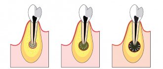
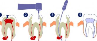
Deep into the tissue, the incision is made at an angle from the edge of the gum to the level of the bottom of the periodontal pocket and extends all the way to the cementum of the root. This direction allows excision of the epithelium along the edge of the gum, pathological granulations in the pocket and subgingival dental plaque. Therefore, the angle of inclination of the spatula depends on the depth of the pocket and the thickness of the alveolar wall (Fig. 1).
Rice. 1. Scheme of excision of the epithelium along the gum edge.
Hemostasis is carried out, and the operation to remove mobile teeth (usually 2-3) is immediately performed. When applying forceps, tissues excised from the oral and lingual sides of the tooth are captured. The hole is filled with a preparation containing hydroxyapatite. The material must be chemically stable in the physiological environment of the body, exhibit resistance to oxidation, prevent the accumulation of harmful reaction products, and not cause galvanic-electric phenomena.
These requirements are largely satisfied by materials containing hydroxyapatite [Ca10(PO4)6(OH)2] with a Ca/P ratio of 1.67, which is an analogue of the inorganic component of the bone and dental tissues of the body.
Calcium reduces the permeability of cell membranes and the vascular wall, preventing the development of inflammatory reactions. Hydroxyapatite gel helps accelerate the healing of bone wounds, the formation of a functionally and structurally complete osteoregenerate according to the type of primary healing. The use of the drug allows you to reduce the duration of pain in the postoperative period and promotes the rapid elimination of edema in all categories of patients.
The wound is sutured in such a way that the more movable flap covers the tooth socket. As a result, the suture passes not in the center, but along the edge of the alveolar ridge, which improves the conditions for wound healing. The number of stitches depends on the length of the incision: one stitch is placed at a distance of about 1 cm from the other.
The incisions made in this way ensure the extraction of the tooth(s) from the socket simultaneously with the inner epithelial lining of the marginal gum, granulations and subgingival deposits. As a result, the risk of microbial contamination of the tooth socket is reduced compared to the classic removal of mobile teeth. Wound healing is accelerated because it occurs by primary intention. In addition, partial excision of the marginal gingiva prevents the formation of a mobile marginal ridge along the alveolar bone. Filling the socket with a material containing hydroxyapatite reduces the risk of further atrophy of bone structures and a decrease in the level of the alveolar margin.
Depending on the clinical picture of periodontitis, other surgical interventions on the tissues of the oral cavity may be performed. For painless treatment of soft tissues, teeth and bones, local anesthesia is usually used: in the upper jaw - infiltration, in the lower jaw - often conduction. Depending on the goals, surgical interventions are divided into 3 groups: operations within the free and attached gums (curettage, gingivotomy, gingivectomy); operations within the free, attached gums and alveolar mucosa (frenulotomy, lip and tongue frenuloplasty, vestibuloplasty, closure of gum recession); operations on the gums, alveolar mucosa, bone tissue of the alveolar process (operations according to Widman; operations according to Neumann; modified operations according to Kirland, Ramfiord, Nissle; operations of apical displacement of the flap).
The purpose of the operations of the first group is the elimination (revision) of pockets (true and false), which are places where microbes and their metabolic products accumulate. A necessary condition for these interventions is the normal width of the attached gum and the absence of pathology of the architectonics of the vestibule of the oral cavity.
Closed curettage is indicated for shallow (up to 4-6 mm) gum pockets; as a palliative method of surgical intervention, it can also be performed for deeper pockets. Contraindications include fibrous changes in the pocket wall, thinned gums, and abscess formation. It is carried out with a sharp excavator and curettes after preliminary opening. In this case, subgingival dental deposits are removed and the inner walls of the pocket are scraped out. It is advisable to remove tartar using ultrasonic devices. This achieves greater surface cleanliness and increases the antimicrobial effect when using chlorhexidine solution as an irrigation liquid. The disadvantage is the lack of visual control of the walls and bottom of the pocket.
Open curettage was proposed to eliminate the shortcomings of closed curettage. A scalloped incision is made along the gingival margin, beveled inward to the base of the pocket. The flaps are peeled off, the remains of tartar and granulation are removed, the flaps are de-epithelialized, placed in place and fixed with sutures in the interdental space.
Gingivotomy is indicated for deep and narrow periodontal pockets. In this case, the pocket is dissected along its entire length, then the edges of the flap are separated, dental plaque is removed, the inner surface of the pocket is de-epithelialized, and the wound surface is treated with an antiseptic. The flaps are placed in place and secured with sutures.
If there is an abscess, the incision is made horizontally, the wound is washed and drained using a rubber strip. Gingivectomy is indicated in the presence of periodontal pockets. One of the conditions is a sufficient width (at least 2 mm) of the attached gum zone, which should remain after excision of the pocket to its full depth. The operation is not performed when bone pockets form.
Osteomucogingival operations (flap according to Widman, Neumann) are performed in the presence of bone pockets. Two vertical incisions are made, usually involving 4 teeth. A horizontal incision is made, slightly retreating from the gingival margin, on the vestibular and lingual sides. Mucoperiosteal flaps are peeled off. Granulation tissue and remaining dental plaque are removed, and bone pockets are eliminated. If necessary, the relief of the outer surface of the bone of the alveolar ridge is modeled, which ensures the maximum possibility of wound healing by primary application. The flaps are placed in place and fixed in the interdental spaces with interrupted sutures.
Modifications of flap operations are numerous, and all of them are aimed at increasing the effect by minimizing the disadvantages inherent in known interventions. The most typical is postoperative tissue retraction. In this regard, osteoplastic materials are used in combination with the technique of directed regeneration of bone structures.
Flap surgery with cementotomy and dental immobilization
The indication for surgical intervention is localized periodontitis, involving 3-4 teeth with exposed roots, grade I-II mobility, and the presence of bone pockets.
Anesthesia is administered to the corresponding area of the jaw. The operation begins with dissection of the soft tissues in the interdental spaces with a sickle-shaped scalpel. The direction of the incision in the interdental space is parallel to the base of the papilla between the proximal sides of the teeth. In this case, the cutting instrument, sliding along the vestibular surface of the tooth with its plane, enters the periodontal pocket and is directed to the middle of the proximal surface of the adjacent tooth until it stops. Following this, the cutting part of the instrument moves in the direction from the root to the crown of the tooth. Dissection of interdental soft tissues is carried out in a section of the dentition that exceeds the area of the intended intervention by 1-2 teeth on each side (modification of the incision by V. I. Lukyanenko, 1973). This makes it possible, while maintaining the area of the surgical field, to do without vertical incisions, which increase gum recession in the postoperative period. Vertical incisions are made in exceptional cases, when the surgical field is located on an area in the projection of no more than 3 teeth, especially in the lower jaw. Then, alternately from each side, sliding along the surface of the tooth with a rasp, they enter the periodontal pocket and, moving the instrument to the apical part, with fan-shaped movements, peel off the flaps on both sides of the alveolar process throughout the entire incision and to the depth of the lesion. Then the flaps are retracted from the bone to a distance that allows free manipulation in the surgical field.
Removal of granulations and vegetations of the epithelium begins from the apical parts of the surface of the flaps, and a scalpel can be used, which, during manipulation, is at an angle of 30° to 90° with respect to the surface of the flap and moves from the base of the flap to the periphery, scraping off granulations and vegetations . During the removal of granulations and vegetation from the flap, the outer surface of the flap is placed on the index finger. This ensures its reliable fixation and control of the depth of immersion of the cutting tool into soft tissue. Next, they begin to remove granulations adjacent to the tooth and alveolar bone. At this stage, scalers and small curettage spoons are used. If there are sharp bony protrusions on the alveolar process, they are ground off with a diamond head.
Treatment of the roots of non-pulp teeth is carried out by scraping with sharp instruments (excavator, sharp hooks of various shapes) surface deposits of tartar along with granulations and necrotic tissue on the surface of the cement. The roots of pulpless teeth are subjected to cementotomy. In this case, the root surface is periodically stained with a 2% aqueous solution of methylene blue or other neutral dyes. Cementomy is completed with an elongated bur with a blunted and polished end part, holding it at different angles relative to the surface of the tooth root (from 20° to 90°). Preparation is carried out until the stained areas of the tooth root disappear. After this, the treated surfaces are polished with polishers. The wound is thoroughly washed with antiseptic solutions (furacillin, 1% hydrogen peroxide solution, etc.) under pressure from a syringe with a blunt needle. Before placing the flaps in place, if necessary, the periosteum and submucosa are dissected along the entire length, creating conditions for displacing the flaps in the coronal direction. Due to the fact that during this operation the edge of the flap largely retains its original shape, it is possible to completely restore the anatomical configuration of the dental papillae. Sutures are made of catgut with an extended resorption period, the nodes are left at the level of the interdental spaces 1-2 mm below the base of the papilla.
When the interalveolar septa are resorption by more than half, the flaps are sutured with U-shaped sutures, creating so-called cuffs around the teeth.
Splinting teeth for periodontitis
To restore the chewing activity of mobile teeth, reduce the inflammatory process in the periodontium and prevent complications, splinting and/or the production of permanent dentures are used.
A splint is a type of denture that connects adjacent teeth into a single system, evenly distributing the chewing force and eliminating traumatic occlusion. In accordance with this, the splint must meet a number of requirements: firmly fix and not displace teeth without introducing excessive load, leave interdental spaces free, and not interfere with brushing and self-cleaning of teeth.
Indications for temporary splinting:
- tooth mobility I-III degrees;
- bone resorption of more than 1/2 of the root length;
- preparation for periodontal surgery;
- creating conditions for professional oral hygiene;
- replacement of minor dental defects;
- preparatory stage for permanent splinting;
- stabilization of the results of orthopedic and orthodontic treatment.
The simplest temporary structure is a wire splint that is placed tightly over the teeth just below the equator. In most cases, 6-8 teeth are connected with a block in such a way that the splinted teeth individually are not mobile.
Removable splints rest on the crown of the teeth and consist of hook clasps surrounding the teeth. In cases of low tooth mobility, these splints provide satisfactory support, but their fixing effect is significantly less than that of permanent splints.
When making removable splints, you need to ensure that they do not interfere with the closure of the dentition, so that at rest the splint fits exactly to the teeth without tension and maintains its position when chewing. The advantage of the splint is good fixation of the teeth in relation to horizontally acting forces, ease of manufacture: the teeth do not need to be ground, and the pulp does not need to be removed. The negative side of this splint is its low aesthetics due to the clasp located on the cheek side.
When making permanent (fixed) splints, artificial crowns are put on individual teeth, which are connected to each other, and thus a stable system is formed.
A fixed splint can be applied to teeth with intact pulp or root filling. The structure must be made in such a way that chewing pressure is evenly distributed over all supporting teeth, eliminating overload of individual teeth, and ensuring protection of the gums and periodontium.
Indications for adhesive prosthetics:
- splinting of the dentition for periodontal diseases complicated by partial edentia - included defects of the 3rd and 4th Kennedy classes of small extent (1-2 teeth in the anterior part of the dentition, 1 tooth in the lateral part);
- splinting a group of teeth after orthodontic treatment;
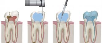
- splinting a group of mobile teeth in order to redistribute the load and ensure the stability of these teeth (Fig. 2);
Rice. 2. Splinting of teeth before surgical treatment.
- splinting the frontal area of the dentition if it is necessary to preserve aesthetics and it is impossible to manufacture traditional aesthetic structures;
- splinting of the dentition with a well-preserved anatomical shape of the coronal part of the tooth and with bone resorption of no more than 1/2 of the root length;
- splinting of the frontal area of the teeth with the possibility of preserving their vitality.
A correctly made splint secures the teeth well and leaves the interdental spaces free, thereby ensuring the ability to maintain oral hygiene, slows down or stops the development of the pathological process during the complex treatment of periodontitis.
Features and types of procedure
Surgical methods for the treatment of periodontal diseases are a set of measures aimed at eliminating the consequences and symptoms of severe forms of pathologies resulting from mechanical injuries or the development of infection. There are two groups of techniques, the main criterion for differentiation of which is the focus of the procedures performed.
Elimination of bone pockets
A bone pocket is a gap between the tooth and the gum, formed as a result of the growth of tartar, which destroys the periodontal ligamentous apparatus. The consequence of the development of this defect is the occurrence of complex pathologies that can cause the loss of elements of the dentition. Surgical treatment of periodontal diseases aimed at eliminating bone pockets includes:
- Open curettage – mechanical cleaning of the pocket from infected tissue;
- Gingivotomy – vertical excision of the gum for subsequent cleaning;
- Gingivectomy – removal of a pocket in a horizontal plane along the contour of the gum;
- Gingivoplasty – plastic modeling of the natural anatomy of the gum edge;
- Flap operations – excision, correction and fixation of the shape of the gingival margin;
- Bone grafting – increasing the required volume of bone tissue.
These operations are carried out both traditionally and using an electric coagulator, which allows to minimize the likelihood of bleeding and qualitatively align the contours of the gingival margin.
Correction of soft and mucous tissue defects
Plastic modeling of the frenulum and mucosa is often recommended for young patients, and helps avoid the development of serious pathologies, such as periodontal inflammation or gum loss.
To deepen the vestibule of the oral cavity, a vestibuloplasty technique is used to expand the area between the lips and the dentition. The procedure is determined by the presence of periodontal indications.
Closing gum recession eliminates the effects of gum reduction surgery that exposes the root area.
Indications and contraindications
The need for intervention is determined by the severity of the changes that the periodontium has undergone during the development of pathology. The list of indications includes the following situations:
- The depth of the bone pockets exceeds 5 mm;
- Significant growth of granulations was diagnosed;
- Interdental tissue areas are subject to deformation;
- There are abscesses or purulent inflammations;
- Pathologies in the formation and position of tissues were identified;
- Maintaining the gap between the gum and tooth after cleaning;
- Conservative treatment methods do not give the desired effect.
Contraindications that limit the possibility of treatment are differentiated into the following categories:
- Absolute – exclude the possibility of performing an operation. Such factors include cancer and severe chronic diseases;
- Relative - viral pathologies, inflammatory processes and local defects of the jaw row, after elimination of which surgical intervention is allowed.
Surgical treatment of periodontal diseases is carried out only after diagnosis, drawing up a clinical picture and assessing the general health of the patient.
Preparation and stages of treatment
Outpatient operations require compliance with antiseptic standards and involve the use of local anesthesia. Duration is no more than two hours. The operations are quite well tolerated by the patient's body, allowing for the possibility of short-term mild discomfort in the first hours after completion of the procedure.
The list of previous events includes:
- Sanitation and hygienic cleaning of the oral cavity;
- The use of medications that localize inflammation;
- Installation of splints or orthopedic structures that fix movable units;
- The use of antibiotics that enhance the immune system.
For good gum regeneration, a soft bandage is applied. In the postoperative period, if necessary, a course of antibiotics and antiseptic drugs is also prescribed to promote recovery.
Classification
Periodontitis is classified according to location, nature of the course, and severity.
According to the nature of the flow
There are acute and chronic forms. In the first situation, the signs of periodontitis are pronounced, the gums are loose, bleed, and hurt. If the patient does not consult a doctor or ineffective treatment is prescribed, the disease takes a chronic course. In the chronic form, the symptoms are smoothed out, there are no acute signs of inflammation, but the consequences are more severe. Chronic pathology is characterized by periodic exacerbations, when the symptoms become pronounced.
By localization
According to the distribution of the pathological process, periodontitis is divided into:
- Localized
(focal) - occurs in a small area, in the area of 1 or several teeth. - Generalized
periodontitis is inflammation of all or most teeth in a row.
The symptoms of localized and generalized forms of the disease are no different, the only difference is in the number of teeth around which the pathological process has spread.
Degrees of periodontitis
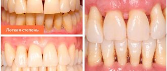
- Mild
– fiber disintegration of the compact plate, reduction of the interalveolar septum to 1/3 of the length of the tooth root, signs of osteoporosis, slight tooth mobility, periodontal pockets of 2.5-3.5 mm. - Medium
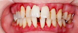
– the depth of periodontal pockets increases to 5 mm, moderate tooth mobility is observed (grades I-II), bone atrophy around the teeth reaches 1/2 of the roots. Exposure of the necks and roots of the teeth or inflammatory growth of the gums occurs, abscesses (abscesses in the gums) can form. The teeth begin to “diverge” under chewing pressure, and the gingival margin becomes deformed. This is especially noticeable in the area of the front incisors. The gums bleed when chewing or brushing teeth, the necks of the teeth are exposed, and react painfully to temperature stimuli. - Severe
– symptoms increase, exacerbations of chronic periodontitis in adults are accompanied by the formation of abscesses, severe swelling of the gums, and pain. The depth of periodontal pockets reaches 6 mm, right up to the root apex. The interdental spaces increase, there is strong tooth mobility (II-III degrees), bone resorption, up to complete resorption of the alveolar septum.
Depending on the individual characteristics of the body, the disease progresses differently. Aggressive forms of periodontitis are characterized by rapid, almost rapid destruction of periodontal tissue, loosening of teeth, and their painless loss. In another part of patients, the disease progresses slowly, with occasional exacerbations, and long remission.
The most severe form is necrotizing periodontitis.
, in which the gum tissue stops receiving nutrition, blood circulation in them stops, and necrosis of the periodontium, periodontal ligaments, and alveolar bone occurs. This form of the disease occurs in patients with severe immunodeficiency conditions (AIDS, DiGeorge syndrome, SCID, etc.).