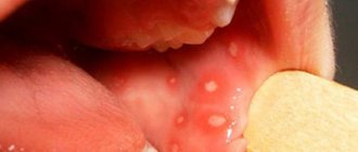
Herpetic gingivostomatitis
This form of the disease develops due to the herpes virus type I.
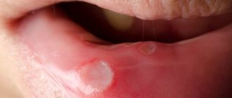
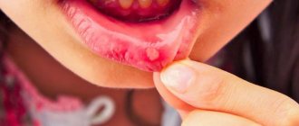
Its symptoms include round ulcers with a white coating, red at the edges, no larger than 0.5 mm in size, covering the mucous membrane of the mouth and gums. Acute gingivostomatitis is characterized by high (up to 38-39 degrees) temperature, bleeding and swollen gums, and inflamed lymph nodes. The number of ulcers sometimes reaches 15-20 pieces. Weakness occurs, problems with eating due to pain, appetite disappears, sleep is disturbed. These symptoms may be accompanied by signs of acute respiratory viral infection or catarrhal tonsillitis.
Other types of gingivostomatitis - allergic, plasmacytic and atypical - are much less common.
Causes of gingivostomatitis
For the development of ulcerative gingivostomatitis, a number of factors are necessary, the main one of which is a decrease in the body’s protective reaction.
The disease usually occurs when one or more of the following factors are present:
— An organism weakened due to illnesses; — Hypothermia, colds: — Chronic infections and diseases; — Inflammatory processes in the mouth, tartar deposits; — Chronic injury to the oral mucosa (for example, due to dentures); — Diseases of the immune system (including HIV); — Lack of vitamins, iron deficiency anemia; — Lack of oral hygiene measures for a long time; - An allergic reaction to toothpaste, medications or any food.
Acute herpetic gingivostomatitis
The incubation period most often lasts from 2 to 6 days, but can last up to 17 days. Acute herpetic gingivostomatitis, as a rule, affects children of toddler and preschool age, most often from 1 year to 3 years. In recent years, cases of illness in children 6–10 months of age, artificially fed from the first months of life, have become more frequent.
A generalized form of herpes is possible in a child born to a mother who does not have antibodies to the herpes simplex virus. Infection of such a child, who has not received passive immunity from the mother, leads to the development of a severe septic disease with damage to the serous membranes of the brain and internal organs. Extensive necrosis occurs in the oral cavity; most children die.
Herpetic gingivostomatitis is contagious. It occurs acutely with a pronounced disturbance of the general condition and local symptoms, depending on which mild, moderate and severe forms of the disease are distinguished.
Mild form of acute herpetic gingivostomatitis in children In mild form of acute herpetic gingivostomatitis, the general condition of the child is slightly disturbed, the body temperature is subfebrile, less often normal, the prodromal period is not always pronounced. The first clinical sign is pain when eating. Upon examination, the doctor detects hyperemia and swelling of the oral mucosa and individual, usually non-confluent, round-shaped erosions with a diameter of 1 - 5 mm, covered with fibrinous plaque. The rash is usually one-time, no new elements appear in the following days, the duration of the disease is 4-5 days.
Moderate and severe forms of acute herpetic gingivostomatitis in children Moderate and even severe forms of acute herpetic gingivostomatitis are more often diagnosed in children. The onset of the disease is acute, as in most other infectious diseases, body temperature is above 38°C, and in severe cases can reach 40°C, intoxication is pronounced: the child is lethargic, capricious, sleeps poorly, complains of a headache, appetite is significantly reduced even before the onset of the disease. erosions in the mouth. Some children experience nausea, vomiting, and stool disorders. Then catarrhal symptoms often appear: runny nose, cough, conjunctivitis. Usually during this period the child is examined by a pediatrician and in most cases diagnosed with an acute respiratory disease. However, with a careful examination of the maxillofacial area, the pediatrician may detect enlarged, painful submandibular, chin, cervical lymph nodes and initial signs of catarrhal gingivitis; the gingival margin is brighter in color than the rest of the oral mucosa, the apices of the interdental gingival papillae are not sharp, as is normal, but rounded.
On the 2nd, 3rd or (less often) 4th day of the disease, rashes of individual and grouped blisters with a diameter of 1 - 3 mm appear on the mucous membrane of the mouth, and often on the red border of the lips and facial skin. On the skin and red border of the lips, the vesicle stage is easily determined; at first the vesicles have transparent contents, after 1–3 days their contents become cloudy, then shrink into a crust. If the lining of the vesicle is damaged, skin erosion occurs. On the oral mucosa, intraepithelial vesicles quickly open and the doctor sees round-shaped erosions - aphthae. Aphthae are sharply painful, they are localized on the tongue, mucous membrane of the lips, cheeks, and less often on the palate, arches, and gums. With a massive rash, aphthae merge with each other and form extensive erosions of various shapes. The oral mucosa, free from erosion, is swollen, hyperemic, and the tongue is coated. The gingival margin is also swollen, hyperemic, and erosions often form along the edges of the gums. Salivation increases, but the saliva is viscous with an unpleasant odor.
The period of rash lasts 2–4 days, the child’s condition remains serious, some children refuse not only food, but also drink, which increases intoxication. Of great importance in the pathogenesis of the disease is secondary infection of erosions with endogenous mixed, primarily coccal, microflora of the oral cavity, which becomes pathogenic in a child weakened by a viral infection. Some children with severe forms of the disease develop deep ulcerative-necrotic lesions of the oral mucosa.
The duration of the disease depends on its severity and the effectiveness of treatment and lasts 7–15 days, aphthae heal without scarring, and the symptoms of gingivitis persist longer. The disease does not recur if stable immunity is developed. In recent years, many children have experienced relapses of the disease.
Acute herpetic gingivostomatitis should be distinguished from drug-induced stomatitis, erythema multiforme and similar syndromes, from diphtheria and other stomatitis in acute infectious diseases.
Severe form of acute herpetic gingivostomatitis in children (Pospischilla aphtoid)
Pospishill described a severe form of strictly herpetic gingostomatitis in children debilitated by infectious diseases. With this form of erosion, erosions occurred not only in the oral cavity and on the skin of the face, but also on the skin of the fingers, including near the nail plates, as well as on the genitals. Abroad, this disease is called Pospischilla aphtoid.
Gingivostomatitis in children
Most often in childhood, gingivostomatitis occurs, caused by the herpes virus.
The disease occurs in an acute form, usually of moderate or severe severity. Gingivostomatitis in a child begins with a sharp rise in temperature, lethargy, and headache. All these symptoms in children appear even before ulcers appear in the mouth, and they can easily be confused with signs of a cold or flu: there is a cough, runny nose, and sometimes diarrhea.
Upon examination, enlarged lymph nodes under the jaw and on the neck and reddened gums are detected. For 2-3 days, the oral mucosa becomes covered with ulcers, which are very painful. The illness lasts one to two weeks.
Treatment of gingivostomatitis consists of aseptic treatment and anesthesia of the oral cavity, as well as antiviral therapy and strengthening the immune system.
Feline gingivostomatitis. New in treatment
Feline gingivostomatitis is an inflammation of the oral cavity (gums, corners of the mouth) with the formation of wounds or ulcers around the teeth and on the mucous membrane. Also, the process of inflammation can spread to the tongue, hard and soft palate, the mucous membrane of the inner surface of the lips and cheeks, to the parotid and submandibular lymph nodes.
The presence of dental calculus leads to the development of gingivostomatitis. They consist of deposits of microbes and organic substances held together by an inorganic matrix of hydroxyapatite, calcium and phosphorus (from saliva). Certain bacteria found in the folds of the gums produce toxins (hyaluronidase and lysosomal enzymes) which, when combined with a large influx of inflammatory cells, irritate the gums and release inflammatory cellular elements that lead to swelling, redness and loosening of the gums. Common bacteria that cause oral lesions in cats: Pasteurella spp., Fusobasterium nucleatum, Actinobacillus spp., Staphylococcus spp., Streptococcus spp., Pseudomonas spp.
Other causes of gingivostomatitis include:
- a diet containing a high percentage of starch and vitamin D;
- viruses (of all infectious factors, Feline calicivirus (FCV) is most often associated with gingivitis and lymphocytic stomatitis; FHV, FIV and FeLV are less common);
- anodontia/oligodontia - reduced number or complete absence of teeth;
- eosophilic granuloma complex (usually perioral lesions (eg, basal cell carcinoma)) and other skin lesions;
- oncological formations in the oral cavity.
Genetics also have an influence (juvenile gingivitis in cats of Abyssinian, Persian and Maine Coon breeds) and the structure of the oral cavity (in Persian breeds, other breeds with brachycephalic syndrome).
Clinical signs of gingivostomatitis. Diagnostics
An attentive owner can easily identify the signs of gingivostomatitis in a pet. The animal becomes lethargic and apathetic, sometimes showing aggression and irritability. A visual examination of the oral cavity shows red, swollen gums.
Cats with gingivostomatitis often stop eating. They experience polydipsia (frequent drinking of water) and anorexia. Excessive salivation (hypersalivation), sometimes mixed with blood, is common. If the cat does try to eat, it may become awkward, even causing pieces of food to fall out of its mouth. Sometimes a hungry animal swallows large pieces of food without chewing, which can lead to stomach upsets. The cat washes itself more often, carefully rubbing the area of its mouth, as if something is bothering it there.
Gingivostomatitis is easily diagnosed visually - the veterinarian will see red, inflamed gums and oral mucosa. Next, you need to determine the type of disease to determine the causes of inflammation and prescribe the optimal course of treatment. Therefore, diagnostics may include additional procedures:
- mucosal inoculation for various types of pathogenic microflora;
- blood and urine tests;
- virological tests.
The most common catarrhal stomatitis is the beginning of all complicated stomatitis and the development of gingivostomatitis. Classic signs are intense redness, swelling, sore gums, increased salivation, and unpleasant odor. The saliva is viscous and smells unpleasant.
In addition to catarrhal, stomatitis is classified as follows:
- fibrinous (white discharge);
- ulcerative (weeping ulcers on the mucous membrane);
- gangrenous (a complication of ulcerative disease, a strong foul odor is added to the symptoms);
- hemorrhagic (pink discharge mixed with blood);
- papillomatous;
- uremic syndrome;
- autoimmune stomatitis and others.
Gingivostomatitis can develop into a chronic form that is extremely difficult to treat.
Gingivostomatitis can develop into a chronic form that is extremely difficult to treat
Treatment
Depending on the type of infection detected, the doctor prescribes a course of treatment, which may include:
Antiseptics:
- Lugol's solution - shows excellent disinfecting properties: to treat ulcers directly - smear or spray (important: long-term use is avoided, as it can provoke the proliferation of Pseudomonas aeruginosa, which is not affected by iodine);
- Chlorhexidine 0.05% - for washing the mouth or treating wounds and ulcers directly;
- Dentavedin/Metrogil Denta - used up to 3 times a day, applied in a thin layer to sore gums or placed directly into the sockets in case of tooth extraction;
- Stomatidin - irrigate the mouth during inflammation or cauterize ulcers, wounds or sites where papillomas are removed.
Anti-inflammatory medications - to reduce redness and/or soreness. There are two main types used:
- corticosteroids - can be given as tablets or injections (These drugs are good for reducing inflammation; they also have the positive side effect of stimulating appetite, which is very important for this problem. However, long-term use of steroids can cause unwanted side effects in the body);
- non-steroidal anti-inflammatory drugs (NSAIDs) - are considered safe and are widely used in these cases (Usually in the form of injections or drops that are administered into food or directly into the mouth. NSAIDs are recommended for use in combination with gastroprotectors (due to the negative effect on the gastrointestinal tract), the course should last no more than 5 days (exception: the drug Metacam, a course of up to 14 days is possible)).
Antibiotic therapy:
- Lincomycin 10% - a course of 3 to 7 days at a dose of 2 ml/10 kg for intramuscular administration and 1 ml/10 kg for intravenous administration;
- Amoxicillin 15% - single injection in a dose of 1 ml/10 kg under the skin or into the muscle; If necessary, you can re-inject after 48 hours.
Immunostimulants - to stimulate the immune system and enhance the body's response to the inflammatory process. Apply:
- Stimulus - 0.5 ml/kg once a day, up to 10 days;
- L-cin - 0.5-1.0 ml/animal, depending on weight, once a day, course - 5 days.
Dental care . Any tartar will aggravate the inflammation, even if it is not the main cause. In more severe cases, it is possible to remove all molar teeth. If conservative treatment is not possible, surgical removal of the affected mucosal tissue that has undergone changes is performed.
Diet - complete refusal of milk (the milk environment is beneficial for the development of bacteria), transferring the cat to food that is easier to accept (usually canned soft food). Food should be at room temperature.
In some cats, stress also plays a role in initiating outbreaks of gingivostomatitis. Removing stressors or giving the cat a "safe" place can help in these cases.
Treatment of gingivostomatitis most often takes place at home with a visit to the veterinarian on a set schedule. In this case, the owner’s main task is to strictly follow the recommendations.
In complex advanced forms, the veterinarian may recommend inpatient treatment with round-the-clock monitoring, administration of medications and supportive drugs through a drip. In some cases, the animal may require surgical intervention (removal of teeth or parts of the mucous membrane).
You should also keep in mind that not all treatments are suitable for cats. There are cases when an animal does not respond completely to treatment, regardless of the procedures performed. Therefore, some animals require repeated treatment or constant monitoring and therapy throughout their lives.
Experience with the use of the drug Ciprocoline
Based on market needs, the Ukrainian created a combination drug Ciprocolin containing two antibiotics: ciprofloxacin and colistin sulfate. As a result of the synergy of these active ingredients, it was possible to create a formula whose activity is higher than that of its individual components.
When administered orally, ciprofloxacin irrigates the inflamed areas in the oral cavity and begins to be absorbed directly into the oral cavity. Entering the blood, it penetrates all organs and tissues of the body, reaching maximum concentration after 1-2 hours. Therapeutic concentration lasts up to 24 hours. The mechanism of action of ciprofloxacin lies in the inhibition of bacterial DNA gyrase, which prevents the process of DNA replication, thereby disrupting the integrity of the bacterial cell membrane and its death. Ciprofloxacin hydrochloride quickly eliminates R-plasmids, which prevents the development of resistance of microorganisms to the drug.
Ciprofloxacin is effective against gram-positive and gram-negative microorganisms, namely: against E.coli, Staphylococcus spp., Streptococcus spp., Pseudomonas spp., Salmonella spp., Shigella spp., Enterobacter spp., Klebsiella spp., Proteus spp., Campylobacter spp. ., Brucella spp., as well as Mycoplasma spp. and Chlamydia spp.
Colistin sulfate is an antibiotic that is synthesized by the aerobic spore-forming bacillus Bacillus polymyxa. Colistin sulfate has a bactericidal effect on gram-negative bacteria (E. Coli, Salmonella spp., Pasteurella spp., etc.). Colistin sulfate binds to phospholipids of the cytoplasmic membrane, increasing its permeability to components, which leads to destruction of the bacterial cell.
Cyprocoline is a powder that, after dissolving in plain water, turns into a white suspension. It has a sweetish taste and smell of bacon, which is attractive to animals. Used to treat dogs, cats and poultry for all types of stomatitis and gingivitis; mixed and secondary infections in diseases caused by microorganisms sensitive to ciprofloxacin and colistin.
Cyprocoline is administered orally. It is set with a dosing syringe at the rate of 1 ml of the finished suspension per 1 kg of animal weight.
Watering is carried out once a day. The course of treatment is 7-10 days, depending on the clinical signs and severity of the disease. After drinking the drug for use in dentistry, it is advisable not to feed or water the animal for 2 hours to ensure longer contact of the drug with inflammation in the oral cavity.
Clinical case
The owners of a Thai breed cat named Solomon contacted the WSW CLINIC of Dr. Velichko (Kyiv). Age 8 years. Weight 5 kg. Lives in an apartment. Vaccinated. Oral hygiene is carried out periodically, removing plaque from teeth using cotton pads with chlorhexidine. Recently, the owners have noticed that the cat has become slower and more careful in eating and, while washing its face with its paw, rubs its mouth area more often.
Upon careful examination, the veterinarian discovered intense redness, swelling, soreness of the gums, and deposits of tartar on the molars and premolars of the upper and lower jaws.
A diagnosis was made of gingivostomatitis in the initial stage. It is proposed to remove supragingival tartar and plaque using ultrasound. The manipulation was carried out under sedation with the drug Relax.
View of the animal's teeth during the initial examination at the clinic
Home treatment prescribed:
- Chlorhexidine 0.05% - for sanitation of the mouth after each meal, 30 days:
- Cyprocoline - drinking 5.0 ml of the prepared suspension once a day, course 10 days;
- Metrogyl Denta - treat inflamed areas of the gums with a thin layer once a day at night, 30 days;
Visit the doctor again after 60 days.
According to the owners, after 3 days from the start of treatment, the cat showed positive dynamics. The animal's condition has improved, it actively consumes food while eating, and there is no pain syndrome.
There is also a significant improvement in gum health in the mouth. The redness and inflammation that was caused by the presence of tartar has disappeared.
The course of treatment was continued according to the doctor’s recommendations.
Thus, the drug Ciprocoline in the complex treatment of gingivostomatitis showed positive dynamics.
Recommended by veterinarians in practice for the treatment of dental problems.
Drinking the drug Ciprocoline at home
The full text of the article was published in the journal VET TOPic (Veterinary Topic), No. 4/2021.
Publications in the media
Acute herpetic stomatitis is a viral infection of the oral mucosa.
Etiology and pathogenesis. The disease is caused by HSV and is one of the clinical forms of manifestation of primary herpetic infection. Transmission of infection occurs by contact and airborne droplets. The incidence of children from 6 months to 3 years is explained by the disappearance of AT received from the mother.
Clinical manifestations. A viral disease has five periods of development: incubation, prodromal, period of disease development, extinction and clinical recovery. Depending on the severity of general intoxication and local manifestations in the oral cavity (area of damage), the disease can occur in mild, moderate and severe form.
• The disease begins acutely with general malaise, weakness, headaches, nausea, lack of appetite; is accompanied by a rise in body temperature, reaching 41 °C or more in severe forms. Enlargement and tenderness of the submandibular and, in severe cases, cervical lymph nodes are determined.
• When the body temperature rises, against the background of hyperemia and swelling of the oral mucosa, lesions appear on the lips, cheeks and tongue (from 2–3 to several dozen, depending on the severity of the disease).
• In severe forms of the disease, the lesions are localized not only in the oral cavity, but also on the skin of the face, earlobes and eyelids.
• At the initial stage, gingivitis is detected, accompanied by severe bleeding of the gums.
• With the addition of fusospirillary microflora, gingivitis becomes ulcerative-necrotic in nature.
• Hypersalivation is noted, although the saliva becomes viscous and viscous. Bad breath develops. Lips are dry, cracked, covered with crusts, there is maceration in the corners of the mouth.
• The severe form is accompanied by profuse salivation and damage to the skin with the appearance of aphthae-like elements. Young children may develop delusional states.
• Violation of the general condition is confirmed by laboratory data.
• Depending on the severity and intensity of treatment, recovery occurs within 1–3 weeks. Aphthae heal without scarring.
Diagnostics. Changes characteristic of an acute inflammatory process are detected in the blood. The content of lysozyme in saliva decreases, IFN is absent. The pH of saliva changes: first to the acidic side, then to the alkaline side. The cytological picture is nonspecific. In the first days, the virus is detected in the contents of the vesicles. Subsequently, the FN titer is determined.
Differential diagnosis. With other viral diseases (foot and mouth disease, etc.) and allergic lesions.
TREATMENT
Regime and diet. It is necessary to follow a diet: predominantly liquid or semi-liquid food and plenty of liquid (tea, fruit juices, fruit infusions). Before feeding, the oral mucosa is anesthetized with a 5% anesthetic emulsion. After eating, be sure to wash or rinse your mouth.
Drug treatment. Determined by the stage and severity of the process. It is recommended to treat the child’s oral cavity with a solution of proteolytic enzymes (trypsin, chymopsin, pancreatin, etc.), which help dissolve plaque, and IFN. Applications are made with antiviral ointments (oxalic, bonaftone, etc.). Later keratoplasty is used. General treatment is required, including antiviral drugs and vitamins.
Forecast. Usually favorable. An unstable, non-sterile immunity occurs. The herpes virus usually remains in the body for life. Transition to a chronic form is possible.
Prevention. It is necessary to take precautions due to the highly contagious nature of the disease.
ICD-10 • B00.2 Herpetic gingivostomatitis and pharyngotonsillitis
Herpetic stomatitis - symptoms and treatment
Treatment of patients with mild and moderate forms of the disease is carried out on an outpatient basis, i.e. at home, but with regular visits to the doctor. Patients with severe forms of stomatitis, complications and generalized forms of the disease - when the virus affects the entire body - are subject to hospitalization. Treatment includes adherence to the regimen, diet, local and general therapy [5].
All patients require gentle treatment . If the child is lying down, he needs to be provided with fresh air and regular changes of linen. If he plays and refuses to go to bed, there is no need to put him to bed by force - it is better to occupy him with quiet games, reading, etc. You should not go with a sick child to kindergarten, school, shops, shopping centers, cinemas, playgrounds. It must be remembered that the disease is very contagious, and other children can become infected. But during the recovery period, walks in the fresh air separately from other children are possible.
The room where the child is located must be ventilated 3-4 times a day, daily wet cleaning is necessary. The child also needs more sleep. But if he sleeps almost all day and has difficulty waking up or, on the contrary, hardly sleeps, then an additional examination by a doctor is required.
But refusing to eat is not so bad. Eating becomes very painful, so you should not insist, much less force-feed the child. It is worth offering liquid food, porridge, jelly, pureed soups.
It is important to prevent dehydration . Small febrile children become dehydrated very quickly, this increases intoxication and aggravates the condition. Therefore, it is necessary to give the child plenty of water. It is worth offering any liquids: water, compote, tea, diluted juices without sugar. The amount of liquid you drink should be at least 1.5-2 liters. If there is vomiting or the child refuses to drink, you need to give him a teaspoon every 5-7 minutes.
Pain relief also plays an important role . At the height of the disease, patients experience severe headaches and mouth pain. For pain relief, paracetamol and ibuprofen are most often used. The dose is calculated depending on body weight, drugs can be alternated.
Fever above 38.5℃ in young children, in children with a history of febrile seizures or with poor tolerance should be brought down. Fever above 39.5℃ should be reduced in all patients. If, when taking an antipyretic drug, the temperature drops by at least a degree, this is good, but if it does not decrease or rises even higher, then an additional examination by a doctor is required.
With mild severity, it is quite possible to get by with local treatment . This requires careful oral hygiene, rinsing or treatment with antiseptic solutions that do not contain alcohol: furatsilin 1:5000, 2% aqueous solution of chlorhexidine, etc. Acyclovir ointment is used to treat aphthae - 3% for mucous membranes, 5% for skin . From about the third day of illness, healing agents based on an oil solution of vitamin A, sea buckthorn oil, aekol, and solcoseryl are added. Local treatment is carried out 5-6 times a day until the lesions are completely healed.
IMPORTANT : the use of local anesthetics based on lidocaine is not recommended for children under 12 years of age [5]. The use of local aerosol antiseptics for acute herpetic stomatitis is also undesirable.
For moderate forms, local treatment alone is not enough. General antiviral treatment is added . For this purpose, acyclovir and valacyclovir (Valtrex) are used in children. The first drug is used in an age-specific dosage 5 times a day - this is important . Valtrex is used 2 times a day, which is more convenient.
IMPORTANT : interferon preparations (viferon, genferon, cycloferon), as well as homeopathic antiviral agents (anaferon, aflubin) and immunomodulators (imudon, thymogen, lykopid) are not indicated, since their effectiveness and safety in the treatment of infections caused by herpes viruses has not been proven .
Recurrent forms require additional examination by a doctor and the appointment of a prolonged course of antiviral treatment.
Severe and complicated forms of the disease are treated in a specialized hospital, since outpatient treatment will be ineffective [1][2]. It involves taking antiviral drugs in the form of tablets and injections, detoxification therapy, replenishment of lost fluid and electrolytes, and intravenous supply of necessary solutions [11].
It is worth remembering that acute herpetic stomatitis is a disease caused by a virus. Therefore, in routine cases, antibiotics are not required , even if the fever lasts more than three days - this is a typical condition for the disease.