The disease is considered quite rare, but can occur in people of all ages, although the main risk group is men 40+. Women get sick less often than men.
Palate cancer is often not recognized as a primary pathology. It is believed that it becomes a consequence of metastasis of tumors that arise in the head and neck area.
If we take into account the location of the neoplasm, then there are two types:
- Cancer of the hard palate - in which the process of tumor development affects bone structures;
- Cancer of the soft palate - affects the muscular and mucous parts of the fornix.
Often, if we take into account the source of tumor growth, a different classification of the disease is given:
- Cylinder. The source of pathogenic cells is glandular tissue. This form of neoplasm grows rapidly and quickly metastasizes.
- Adenocarcinoma. A tumor of epithelial origin, found in oncology of any part of the palate.
- Squamous cell carcinoma. The most common source of cancer cells is mucous tissue.
Also, to determine the degree of infection, the condition of the lymph nodes, and the presence/absence of metastases, the international TNM classification is used.
Classification of oncological pathology of the palate
In half of all cases, malignant tumor of the hard palate is squamous cell carcinoma.
Nonsquamous cells, including minor salivary gland cancer, sarcoma, and melanoma, account for the other half of hard palate neoplasms. In 80% of cases, a tumor of the soft palate is squamous cell carcinoma. The remaining 20% are benign neoplasms.
The histological distribution of malignant neoplasms of the hard palate is as follows:
- squamous cell carcinoma - 53%;
- adenoid cystic carcinoma - 15%;
- mucoepidermoid carcinoma - 10%;
- adenocarcinoma - 4%;
- anaplastic carcinoma - 4%;
- other - 14%.
The histological types and frequency of neoplasms of the minor salivary glands of the palate are as follows:
- Benign - 26%
- Malignant - 74% overall Adenoid cystic carcinoma - 30%
- Mucoepidermoid carcinoma - 16%
- Adenocarcinoma - 18%
- Malignant mixed tumor - 8%
- Other - 2%
Causes of palate cancer
The causes of this disease have not been established, but it is known that certain factors may increase the risk of developing malignant tumors of the palate.
Smoking and chewing tobacco
Active smoking, especially unfiltered cigarettes and cigars, at least doubles the risk of developing malignant neoplasia of the oral cavity. Their development is also facilitated by the use of chewing tobacco mixtures - betel nut, gutka, snus, etc.
Alcohol
Strong alcohol is a recognized risk factor for palate cancer. This is mainly associated with the direct damaging effect of ethyl alcohol on the oral mucosa.
Human papillomavirus
HPV infection has been significantly associated with an increased risk of developing oral squamous cell carcinoma in recent studies.
Chronic inflammatory diseases of the oral cavity
Sluggish inflammatory processes, such as chronic gingivitis or periostitis, increase the likelihood of the formation of palate tumors.
Precancerous diseases
In the presence of pathologies of the mucous membrane such as leukoplakia or erythroplakia, the likelihood of developing cancer becomes extremely high - up to 90%.
Reasons and factors
The cause of the disease can be any damage to the oral mucosa.
This may occur if exposed to:
- Nicotine;
- Alcohol;
- Narcotic drugs.
Damage and inflammation of healthy cells due to prolonged contact with carcinogenic substances is a path to the appearance and development of palate cancer.
In addition, factors contributing to the transformation of healthy cells into cancer cells may include:
- Hereditary predisposition;
- Eating excessively hot food;
- Eating sour and spicy foods;
- Lack of vitamin A in the body;
- Mechanical injuries.
Of course, precancerous diseases should not escape attention, primarily leukoplakia and papillomatosis.
Symptoms of the disease
Squamous cell carcinoma appears as ulcerative superficial lesions. However, there are often no signs of palate cancer in the initial stages. The first manifestations appear, as a rule, already in the advanced stages of the disease.
Palate cancer - symptoms and first signs:
- bleeding;
- difficulty swallowing;
- speech disorders;
- bad breath;
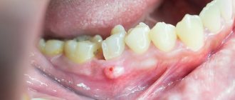
- wounds in the mouth that do not heal for a long time;
- loosening of the teeth of the upper jaw;
- pain when swallowing;
- weight loss;
- ear pain;
- swelling of the neck;
- white spots in the mouth that do not disappear for a long time.
A swelling on the roof of the mouth, bleeding, foul odor, or loose teeth may be symptoms in patients with hard palate cancer. Patients with advanced soft palate cancer may have difficulty swallowing, altered speech, trismus, or neck swelling.
Because the area is easily visualized, tumors are often discovered in the early stages by chance by the patient or physician.
Diagnosis of diseases accompanied by the formation of lumps in the mouth
Tumors have appeared in the mouth, and the question is, which doctor should I contact? This symptom is a prerequisite for visiting the dentist. To diagnose the disease, the doctor will first conduct a visual examination and palpation, and collect an anamnesis. A puncture is taken from the resulting lump and a bacteriological examination is carried out, which makes it possible to determine the cause of its occurrence.
Based on the results obtained, the doctor determines the need to use other diagnostic methods:
- general tests;
- X-ray;
- Ultrasound;
- biopsy.
If the cause of the formation of a lump on the upper palate is beyond the scope of the dentist’s competence, the patient is prescribed consultations with other specialists (pediatrician, therapist, gastroenterologist, endocrinologist, hepatologist). Only after making a diagnosis will the doctor determine how to treat the disease.
Stages of development of palate cancer
The stages of malignant neoplasms of the palate are determined based on the international TNM system. With its help, the characteristics of the tumor are described according to three main criteria.
Primary tumor stage (T)
- TX—primary tumor not evaluable
- T0 - no evidence of a primary tumor (T - carcinoma in situ.)
- T1—tumor 2 cm or less in greatest dimension
- T2 - tumor larger than 2 cm but not larger than 4 cm in greatest dimension
- T3 - tumor larger than 4 cm in greatest dimension
- T4 - Tumor invades adjacent structures (eg, cortical bone, soft tissue of the neck, maxillary sinuses)
Regional lymph node involvement (N)
- NX—regional lymph nodes not evaluable
- N0 - Absence of regional metastasis in the lymph node
- N1 - Metastasis in one ipsilateral lymph node, 3 cm or less in greatest dimension
- N2 - metastasis in one ipsilateral lymph node, more than 3 cm, but not more than 6 cm in greatest dimension; in several ipsilateral lymph nodes, not more than 6 cm in greatest dimension; or in bilateral or contralateral lymph nodes, not more than 6 cm in greatest dimension
- N3 - Metastasis in a lymph node greater than 6 cm in greatest dimension
Distant metastasis (M)
- MX - The presence of distant metastases cannot be assessed
- M0 - no distant metastases
- M1 - distant metastases
Based on the TNM characteristics, the so-called clinical stage is determined.
| Stage | Characteristics |
| 0 | Tis N0 M0 |
| I | T1 N0 M0 |
| II | T2 N0 M0 |
| III | T3 N0 M0 |
| (T1, T2, T3) N1 M0 | |
| IVA | T4a ( N0 or N1) M0 |
| (T1, T2, T3 or T4a) N2 M0 | |
| IVB | Any T N3 M0 |
| T4b Any N M0 | |
| IVC | Any T Any N M1 |
Diagnosis of palate oncology
In most cases, detection of oncological pathologies of the palate occurs during a routine examination by a dentist. The diagnosis is then confirmed by biopsy findings.
However, to assess the extent of the tumor in the structures of the upper jaw, pharynx, etc. Additional diagnostic tools are required:
- MRI (magnetic resonance imaging) - to detect tumor growth in the sinuses of the upper jaw or the nasal cavity.
- PET (positron emission tomography) - to detect metastasis in regional lymph nodes and separated metastasis.
Symptoms of cleft palate in a child
The larger the cleft in the palate, the more noticeable the presence of pathology. With an incomplete cleft, the child chokes when sucking, eats poorly, and milk may leak from the nose. If the cleft is through, complete, the baby has difficulty breathing, and in principle he cannot suck. Often during natural childbirth, amniotic fluid enters the respiratory tract of such children, so they require emergency assistance.
When examining the oral cavity and pharynx, a hole is noticeable in the place where a solid soft palate is normally located. If the cleft also affects the lip, then the division of the upper lip into two or more halves is outwardly noticeable.
Treatment of the disease
Radiation therapy
Radiation therapy to treat palate cancer can be used in several ways.
- As the primary treatment for small, early-stage tumors.
- As an adjunct to surgery or ongoing chemotherapy. In this case, it is possible to use adjuvant radiation therapy as after surgery to destroy tumor remnants. So is the use of radiation before surgery to shrink the tumor and make it easier to remove—neoadjuvant radiation therapy.
- Radiation therapy can also be used to relieve symptoms of advanced, inoperable cancer, such as pain, bleeding, problems swallowing, and problems caused by bone metastases.
Modern radiotherapy for palate cancer in Belgium
Radiation in cancer centers in Belgium is carried out using techniques that help doctors focus the radiation more precisely.
There are two main such methods.
- Three-dimensional conformal radiation therapy (3D-CRT);
- Intensity modulated radiation therapy (IMRT).
These methods rely on the results of imaging tests, such as MRI, and special computer programs to pinpoint the location of the tumor. The beams are then shaped and directed at the tumor from multiple directions, reducing damage to healthy nearby tissue.
Another modern way of delivering radiation is to place radioactive materials directly into the tumor. This is called brachytherapy.
To do this, a small grain of radioactive material is placed into the tumor. In this case, the radiation spreads only over a very short distance and is limited to the tumor.
Surgery
The type of operation is selected depending on the location and extent of the tumor.
For small to medium-sized tumors up to T3, an oral approach is often used without trauma to the mandible. For most tumors that do not have regional spread, surgery to remove the hard or soft palate is used - palatectomy.
For tumors that have grown into the paranasal sinuses or nasal cavity, a unilateral or bilateral maxillectomy (removal of the upper jaw) may be required.
Chemotherapy
Treatment of palate cancer with chemotherapy is considered as an additional method in combination with radiation therapy and surgery.
The chemotherapy drugs most often used for cancer of the oral cavity and oropharynx are:
- Cisplatin;
- Carboplatin;
- 5-fluorouracil (5-FU);
- Paclitaxel (Taxol);
- Docetaxel (Taxotere).
Targeted and immunotherapy
As a rule, targeted and immunological drugs for this disease are used for metastatic inoperable forms, or for relapses.
Cetuximab (Erbitux®) is a monoclonal antibody that targets the epidermal growth factor receptor (EGFR), which helps cells grow and divide. Palate cancer cells often have higher than normal amounts of EGFR. By blocking EGFR, cetuximab can help slow or stop cell growth.
Pembrolizumab (Keytruda®) and nivolumab (Opdivo®) are drugs that target the PD-1 checkpoint. It is a protein on the membranes of T cells that normally helps keep these cells from attacking other cells in the body.
By blocking PD-1, these drugs boost the immune response against cancer cells, which can shrink some tumors or slow their growth.
The nature of the pain and the need to contact a specialist
If the roof of your mouth hurts, the nature of the course of this symptom usually depends on the cause that led to the inflammatory process. To understand what exactly caused this reaction, you will have to seek medical help. If this is simple damage from rough food, the wound may swell for several days, but the sensations will be tolerable, and the discomfort should gradually subside.
To speed up the healing process, experts recommend being careful when eating and rinsing your mouth with a light antiseptic solution or a soothing herbal infusion. You should consult your doctor personally about suitable medications for symptomatic therapy.
If the pain does not go away within 3-4 days and only gets worse, you should definitely visit a dentist. If the situation is ambiguous, the specialist will suggest undergoing a diagnostic examination. To determine the cause of the pathological process, you may have to undergo x-ray diagnostics, as well as visit other specialized doctors, for example, an ENT specialist, an infectious disease specialist, a therapist or an oncologist.
Rehabilitation after treatment
Treatment of malignant tumors of the palate is always very traumatic. The consequence of the operation is, at a minimum, a violation of the process of swallowing and speech. In multidisciplinary clinics in Belgium, rehabilitation doctors and speech therapists begin working with the patient immediately after the operation - this greatly shortens the rehabilitation period and increases its effectiveness
Reconstructive surgery - palate surgery in Belgium
Palatectomy and maxillectomy operations require subsequent prosthetics.
Small defects of the hard palate can be covered with local flaps from the rest of the hard palate or from the buccal mucosa. Larger defects are adequately and effectively treated with obturators. Because the hard palate is not dynamic, obturators are very effective and well tolerated.
The soft palate is a dynamic structure and its optimal functioning requires muscular action to lift and contract the palate during swallowing and relax it during nasal breathing. Therefore, any reconstruction of the soft palate using flaps and prosthetics does not reproduce its functionality - it will be limited even with the most successful outcome.
Complications
Complications of surgical resection of the soft palate may include:
- velopharyngeal insufficiency (most often);
- hypernasal speech;
- dysphagia (impaired swallowing);
- effusion in the middle ear due to scarring at the opening of the eustachian tube.
The extent and likelihood of these complications depend on the extent of the resection, the size of the defect, and the method of reconstruction. The larger the resection and defect, the greater the likelihood of these complications occurring.
With soft tissue resection preserving only the bony palate and soft palate, the resulting defect heals with granulation and epithelialization, and no complications are expected. If patients have previously had radiation therapy in this area, healing may be delayed.
Palate cancer and its treatment can sometimes cause problems such as loss or changes in taste, dry mouth, or even tooth loss. This can make it difficult to eat, leading to weight loss and weakness due to poor nutrition.
Radiation therapy may also cause long-term or permanent side effects.
- Damage to the salivary glands. It may cause dry mouth. This leads to problems with eating and swallowing. Lack of saliva can also lead to tooth decay (cavities).
- Damage to the jaw bone. This problem, known as osteoradionecrosis of the jaw, can be a serious side effect of radiation therapy. This condition is difficult to treat. Osteoradionecrosis is manifested by pain in the jaw, and sometimes by fractures, which require surgical treatment.
Newer radiation treatments such as IMRT may help reduce this side effect. The drug amifostine (Ethyol®) may also help reduce this side effect by limiting radiation damage to normal tissue. It is injected into a vein over 15 minutes before each radiation treatment.
Malignant tumors of the lower jaw
Before treating malignant tumors of the lower jaw, it is necessary to sanitize the oral cavity. If the patient is to be exposed to radiation, the metal prostheses should be removed, or better yet, isolated with a plastic mouth guard.
The choice of treatment method depends on the type, location, extent of the tumor, age and general condition of the patient.
Cancer of the lower jaw is treated mainly with a combined method, which is also indicated for Ewing sarcomas, reticulosarcoma and hemangioendothelioma. But it should be borne in mind that with Ewing’s sarcoma, even after combined treatment, the prognosis is poor. Treatment of osteogenic and chondrosarcomas, which are radioresistant, is surgical.
In a combined treatment regimen for malignant tumors of the lower jaw, a preoperative course of remote gamma therapy is first carried out. Sessions are daily, the number of radiation fields depends on the size of the lesion and the presence of regional metastases. The total focal dose per course is 40-50 Gy (4000-5000 rad).
The surgical stage is performed 3 weeks after completion of radiation therapy. During this time, radiation reactions occur on the skin and mucous membrane of the oral cavity.
Before surgery, taking into account clinical, radiological and morphological data, you should consider its volume, the method of fixation of the remaining jaw fragment, and the possibility of performing primary bone grafting of the jaw defect. It is also necessary to take into account the presence or absence of regional metastases. We must remember that the primary task of the surgeon is radical removal of the tumor. Therefore, if one-stage plastic surgery is impossible without compromising the radicality of the operation, it should be postponed. Paches A.I. In general, he believes that bone grafting of a defect after resection of the lower jaw for a malignant tumor should be performed no earlier than 2 years after treatment in the absence of relapse and metastases.
Surgeries for malignant tumors of the lower jaw (resections) can be of several types:
- resections with disruption of the continuity of the lower jaw (segmental);
- resections without breaking the continuity of the lower jaw (segmental);
- segmental resection of the lower jaw with disarticulation;
- half resection of the lower jaw with disarticulation;
- resection of the lower jaw (one of the options) with soft tissues.
When choosing the type of surgical intervention, you should remember the possibility of a malignant tumor growing into surrounding organs and tissues: the floor of the mouth, tongue, palatine arches and tonsils, lower lip, skin of the chin, submandibular salivary glands, parotid salivary glands. In this case, the scope of the operation must be expanded to include a block of tissue removed, organs affected by the tumor. In addition, primary malignant tumors of the mandible can spread along the neurovascular bundle in the mandibular canal. Experience shows that even with superficial damage to the alveolar process or the lower edge of the mandible (if regional metastasis spreads to the bone), it is not worth performing resection while maintaining the continuity of the organ, because this is fraught with relapse.
If the neurovascular bundle is involved in the tumor process, resection with disarticulation is indicated.
If the tumor is localized in the chin area, resection is performed from angle to angle of the jaw.
If the tumor is localized in the body area, resection is performed from the middle of the chin to the mandibular foramen.
If the angle of the mandible is affected, half resection with disarticulation is indicated.
According to Kabakov B.D. et al., 1978, resection of the lower jaw while maintaining continuity is very controversial and can be indicated for secondary cancer, when the mucous membrane is affected and there are only initial bone lesions. It must be remembered that the desire to simultaneously preserve the function and appearance of patients with any location of a malignant tumor is fraught with its relapse.
Resection of the lower jaw is performed under endotracheal anesthesia. Resection of the jaw without disarticulation is technically easier.
Technique of half resection of the lower jaw
The patient lies on the operating table with his head turned in the direction opposite to the affected one. An incision is made in the skin and subcutaneous tissue from the middle of the chin to the mastoid process 1.5-2.0 cm below the level of the lower edge of the lower jaw. For a better view, you can additionally make a midline incision in the lower lip. Then the mucous membrane of the vestibule of the oral cavity is dissected to the branch, 2 cm away from the tumor infiltrate. The buccal flap is separated within the incision with a scalpel and scissors. An incision is made in the mucous membrane along the alveolar process from the lingual side from the frenulum of the tongue (at the point of attachment to the lower jaw) to the angle. The soft tissues are carefully separated so as not to damage the tumor. The central incisor on the affected side is removed. At the level of the socket of this tooth, a Jig-li saw is carried out and, protecting the soft tissues with Farabeuf hooks, the lower jaw is sawed in the frontal region. The assistant uses a hook to move the jaw down.
The next stage of the operation is the separation of soft tissues in the area of the jaw branch. To do this, the masticatory and internal pterygoid muscles are cut off from the jaw, and the neurovascular bundle is crossed between two ligatures above the mandibular foramen. An osteotomy of the branch is performed using a Gigli saw. When performing an osteotomy of the lower jaw, the doctor should be guided by the radiograph and retreat from the boundaries of the lesion visible in the image by 3-4 cm.
The removed tumor should be sent for histological examination. The wound is carefully examined, washed with antiseptics and, after hemostasis, sutured in several rows with catgut and lesa.
At this stage, it is important to take into account reliable separation of the wound from the oral cavity, otherwise the wound may become infected with the contents of the oral cavity, which leads to failure of the sutures and the formation of a large defect. To prevent the cutting of sutures in the mouth, immediately after osteotomy of the lower jaw, its stump must be smoothed with a milling cutter, and during suturing, carefully covered with soft tissues.
Resection of the lower jaw with disarticulation
All stages of the operation are performed as described above. After lowering the lower jaw and cutting off the neurovascular bundle, the tendon of the temporal muscle and the external pterygoid muscle in the area of the condylar process are cut off from the coronoid process. After this, they begin careful dislocation movements, avoiding fracture of the jaw and damage to the tumor, since this leads to contamination of the wound with tumor cells. Suturing the wound begins with the oral mucosa.
If surgery on the regional lymphatic system is indicated, then lymphadenectomy is first performed, and at the final stage the lower jaw is included in the block of tissue removed. This guarantees the ablastic nature of the intervention.
It is very important in the postoperative period to keep the jaw fragments in the correct position. For this purpose, Tigerstedt and Weber splints with one or two inclined planes are used, less often - Vankevich or Stepanov splints, and the Rudko apparatus.
When resection of the frontal fragment of the lower jaw with tissues of the floor of the mouth, the operation often begins with the application of a tracheostomy. When part of the tongue is included in the block of tissue to be removed, tube feeding is indicated
Rehabilitation of patients
Restoring the anatomical integrity of the lower jaw, and thereby function and appearance, is a complex and controversial issue. Plastic surgery of a bone defect is a complex operation, which in a cancer patient weakened by previous treatment may end in failure, since the regenerative abilities of the tissues are sharply reduced. With malignant tumors, even after radical treatment, relapses are possible.
Some surgeons resort to primary bone grafting only after resection of small tumors that do not extend beyond the bone, because otherwise it is impossible to create a good receptive bed for the graft.
To hold jaw fragments in the correct position, implants are recommended to use various designs that approximate the shape of the missing area of the jaw. In this case, materials indifferent to body tissues are used: plastic, stainless steel, tantalum, Vitaly. It should be taken into account that these devices do not always provide rigid fixation of jaw fragments and can cut through soft tissue. Then they need to be removed and replaced with a tire.
Secondary plastic surgery of the defect after resection of the lower jaw, according to the recommendation of A.I. Paches, should be performed no earlier than 2 years later in the absence of relapse and metastases. It is carried out using both a preserved graft (which is preferable, since it eliminates the operation of taking an autograft from a weakened patient) and an autograft.
When planning reconstructive operations in this group of patients, the doctor must carefully consider the method of fixation of the lower jaw.
Prognosis for a malignant tumor of the lower jaw
The results of treatment of malignant tumors of the lower jaw are unsatisfactory. A 5-year cure after combined and isolated surgical treatment is observed only in 20-30% of patients. The results obtained after removal of sarcomas are even worse, and a 5-year cure occurs in less than 20% of patients.
Relapses of malignant tumors of the lower jaw usually occur in the first 1 to 2 years after treatment. Tumors of this location are insensitive to chemotherapy. The main reason for the high mortality rate of patients with this tumor location is late diagnosis and untimely initiation of treatment.
If patients are cured, their ability to work, as a rule, decreases, but some patients get the opportunity to return to their previous profession. Such patients themselves raise the question of plastic surgery within a few months after discharge from the hospital.
Prevention
Limit smoking and alcohol
Tobacco and alcohol are the most important risk factors for these cancers. Not starting to smoke is the best way to limit your risk of cancer. Quitting tobacco also significantly reduces the risk of developing these cancers, even after years of use. The same applies to alcohol - limiting its amount helps reduce the risk of developing palate cancer.
Preventing HPV infection
Vaccination against HPV reduces the risk of mouth and throat cancer. But vaccines are only effective if they are given before someone gets HPV, given at a young age, before they become sexually active.
Correctly selected dentures and proper oral hygiene
Avoiding sources of oral irritation (such as dentures that don't fit properly) can help reduce your risk of developing oral cancer.
Popular questions and answers
The pediatrician, the chief pediatrician, works to solve the problem of cleft palate along with surgeons, orthodontists and other specialists. The pediatrician makes sure that the child eats normally, helps reduce the risk of infections, and gives advice on caring for the baby. Pediatrician Daria Shchukina will tell you more about the treatment of children with cleft palate .
What complications can occur with cleft palate?
Such a child cannot eat normally without food being thrown into the nasal cavity, which provokes the development of chronic inflammation and infections of the ENT organs. These defects lead to psychological trauma and speech development disorders. Children with a cleft palate are more likely to suffer from acute respiratory viral infections and may be delayed in growth and development. They may also have associated developmental defects.
When to call a doctor at home for a cleft palate?
Diagnosis and treatment of cleft palate is planned; a doctor’s home visit is not required. If a child with a large cleft palate has difficulty breathing, signs of infection, or a high temperature, an ambulance is most likely required. How early can pathology be detected in a child? Can this be somehow influenced in the womb? The first trimester of pregnancy is the most dangerous in terms of the development of defects. It is believed that cleft lip and palate are formed as a result of the combination of hereditary characteristics and adverse environmental influences. Mother's age over 35 years is also a risk factor. It is impossible to influence this when the fetus has already formed. Most often, pathology is detected at the birth of a child. However, a pronounced defect may be visible on ultrasound. Fetoscopy and fetoamniotomy may also help. However, diagnostic success rates during pregnancy range around 30%.