- Causes of salivary gland cancer and risk groups
- Symptoms of the disease
- Diagnostic methods
- Classification: types of salivary gland cancer
- Stages of the disease
- Treatment
- Complications and relapses
- Life prognosis at different stages of cancer and prevention
The salivary glands produce saliva, a liquid found in the oral cavity that performs some important functions: wetting the mucous membrane, food, facilitating articulation, swallowing, protecting against pathogenic bacteria, etc. There are two groups of salivary glands:
- Large: sublingual, parotid, submandibular.
- Small ones have a microscopic structure, scattered throughout the oral cavity - there are several hundred of them in total.
Cancer can develop in all of these glands. Most often (in 7 out of 10 cases), benign and malignant tumors arise in the parotid salivary glands. Approximately 1–2 tumors out of ten occur in the submandibular salivary glands; in 50% of cases they are malignant. In rare cases, cancer develops in the sublingual or minor salivary glands.
Causes of salivary gland cancer and risk groups
A normal salivary gland cell becomes cancerous when a certain set of mutations occurs in it. The development of malignant tumors is caused by mutations in oncogenes (genes that activate cell reproduction) or tumor suppressor genes (suppress cell reproduction, “repair” damaged DNA, trigger programmed cell death - apoptosis). In each specific case, it is very difficult to judge the reasons for the mutations that occurred in cells. It is impossible to say why exactly they happened.
There are some risk factors that increase the likelihood of developing a malignant tumor:
- The older a person is, the more changes in his genes accumulate, the higher the likelihood of developing various types of cancer.
- Salivary gland cancer is more common in men than in women.
- Irradiation of the head. For example, this may be a previous course of radiation therapy, exposure to ionizing radiation in the workplace.
- There is evidence that the risk of salivary gland cancer is increased in people who have certain occupational hazards: contact with asbestos, nickel alloy dust, work in enterprises that produce rubber and woodworking.
The role of heredity is currently considered insignificant. Most patients do not have a family history (close relatives who have been diagnosed with the same type of cancer). The role of alcohol and tobacco has not been proven. These unhealthy habits are known to increase the risk of head and neck cancer in general, but no association has been found with salivary gland cancers.
Is a mobile phone dangerous? In one study, researchers found that heavy cell phone users were more likely to have parotid tumors (usually benign). But other studies have not found such a relationship.
Symptoms of the disease
Benign and malignant tumors of the salivary glands present with similar symptoms. One of the main differences is that cancer grows much faster and more often leads to a number of symptoms associated with growth into surrounding tissues and compression of nerves.
You need to visit a doctor if you are bothered by the following symptoms:
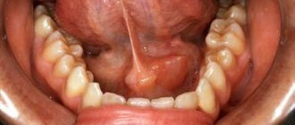
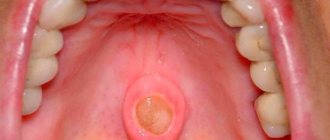
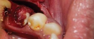
- A lump or swelling has appeared in the mouth, cheek, jaw, or neck.
- You have noticed that your face has become asymmetrical.
- I am worried about pain in the mouth, neck, ear, cheek, jaw. It doesn't last long.
- Numbness of part of the face.
- Weakness in the facial muscles, because of this, half of the face may be “lowered,” which becomes especially noticeable when baring teeth or frowning.
- I can't open my mouth wide.
- Difficulty swallowing.
Neoplasm of the parotid salivary gland - symptoms
A tumor of the parotid gland in the initial stages can be practically asymptomatic. The first witnesses of the disease may be causeless dry mouth or, on the contrary, excessive salivation. Further dynamics of the disease are often characterized by the following clinical manifestations:
- numbness of the face or part of it in the area of the salivary glands;
- swelling, painful lump in the neck, mouth or jaw;
- pain while swallowing;
- increased body temperature;
- dizziness;
- discomfort when opening the mouth;
- muscle pain or weakness (paresis) in a certain area of the face.
However, these symptoms may also indicate the occurrence of other benign neoplasms, for example, a salivary gland cyst. If you experience one or more of the above symptoms, you should consult a qualified physician for a diagnosis. Oncologists at the Yusupov Hospital, thanks to their professionalism and extensive experience working with patients of different ages, will competently prescribe treatment and all the necessary diagnostic measures.
Diagnostic methods
During the initial appointment, the oncologist talks with the patient, questions him, trying to find out risk factors, and conducts an examination. The doctor feels the tumor on the face and cervical lymph nodes, evaluates the sensitivity of the face and the work of facial muscles. After this, the patient can be referred for additional consultation to an ENT doctor.
Computed tomography and magnetic resonance imaging help assess the size, shape and location of the tumor, and detect lesions in the lymph nodes and other organs. The current gold standard for searching for distant metastases is PET scanning. If the tumor is located near the jaw, an x-ray is taken. A chest x-ray helps detect metastases in the lungs and evaluate the condition of the lungs and heart before surgical treatment.
The most accurate method for diagnosing salivary gland cancer is a biopsy. During this procedure, the doctor receives a fragment of pathologically altered tissue and sends it to the laboratory for cytological and histological examination.
The most common procedure is fine-needle aspiration biopsy. Tumor tissue is obtained using a hollow needle similar to the one used for injections. If the required amount of tissue cannot be obtained, the doctor performs an incisional biopsy: makes an incision and removes part of the tumor.
If tumor cells are found in the sample, the diagnosis of cancer is virtually certain.
Classification: types of salivary gland cancer
The salivary glands are made up of different types of cells, any of which can give rise to a malignant tumor. Therefore, there are different types of cancer:
- Mucoepidermoid carcinoma of the salivary gland is the most common type. Most often it is found in the parotid salivary glands, less often in the submandibular and small ones. These tumors are rarely aggressive.
- Adenoid cystic carcinoma is characterized by very slow growth and rare metastasis. However, this malignant tumor can be very difficult to get rid of: it can recur a long time after treatment.
- Adenocarcinoma is a malignant tumor that develops from glandular cells. Different types of adenocarcinomas can occur in the salivary glands: acinic cell carcinoma, low-grade polymorphic adenocarcinoma, basal cell adenocarcinoma, clear cell carcinoma, cystadenocarcinoma, etc.
- Rare types of malignant cancers of the salivary glands include: squamous cell carcinoma, epithelial-myoepithelial carcinoma, anaplastic small cell carcinoma, undifferentiated carcinomas.
Stages of the disease
Salivary gland cancer, like other malignant tumors, is classified into stages according to the generally accepted TNM system. The letter T in the abbreviation denotes the characteristics of the primary tumor: the size and degree of its growth into surrounding tissues, N - spread to the lymph nodes, M - the presence of distant metastases. Depending on these indicators, the following stages are distinguished during salivary gland cancer:
- Stage 0 is “cancer in situ” (carcinoma in situ). The tumor is located within the layer of cells that form the salivary gland and does not grow into neighboring tissues.
- Stage I is a tumor that is located within the salivary gland and measures no more than 2 cm.
- Stage II - the tumor reaches a size of more than 2 cm, but not more than 4 cm.
- Stage III - a tumor that reaches a size of more than 4 cm and/or spreads into surrounding tissues, or a tumor of any size that has grown into surrounding tissues, has spread to one cervical lymph node on the same side, and the focus in the lymph node is no more than 3 cm and does not extend beyond its borders.
- Stage IV includes substages IVA, IVB or IVC. The first two are characterized by varying degrees of spread of the malignant tumor to the anatomical structures of the head, neck, and lymph nodes. If stage IVC is diagnosed, it means that there are distant metastases.
In addition to stages, there are three degrees of malignancy of salivary gland cancer:
- Grade I - low degree of malignancy. Such tumors are called highly differentiated. Tumor tissue is as similar as possible to normal salivary gland tissue. It grows slowly, and the prognosis for such patients is most favorable.
- Grade II are moderately differentiated tumors. Tumor tissue differs more significantly from normal tissue. This cancer is more aggressive and has a poorer prognosis.
- III degree - poorly differentiated tumors. Cancer cells almost completely lose the features of normal ones. Such tumors behave the most aggressively.
Determining the degree of malignancy helps the doctor predict how the cancer will behave and plan treatment correctly.
Tumors of the salivary glands
SPECIALIZED ONCOLOGY CARE www.OncoClinic.com
Tumors of the salivary glands account for 1-5% of all neoplasms of the human body. Most often occur in the parotid salivary glands. The age of patients is mainly 40-60 years. 60-80% of tumors are benign. The most common histological form of benign tumors is pleomorphic adenoma (60%).
Malignant tumors often develop in the sublingual, submandibular and minor salivary glands (50-80%). The main histological forms are mucoepidermoid carcinoma and adenoid cystic carcinoma. Metastasis of carcinomas to regional lymph nodes is observed in 25-50% of cases. Adenoid cystic carcinoma (cylindroma) is characterized by distant hematogenous metastasis to the bones and lungs (40-45%).
International histological classification of salivary gland tumors
1. Adenomas: 1.1. Pleomorphic adenoma. 1.2. Myoepithelioma (myoepithelial adenoma). 1.3. Basal cell adenoma. 1.4. Adenolymphoma. 1.5. Oncocytoma. 1.6. Canalicular adenoma. 1.7. Sebaceous adenoma. 1.8. Ductal papilloma. 1.9. Cystadenoma.
2. Carcinomas: 2.1. Acin cell carcinoma. 2.2. Mucoepidermoid carcinoma. 2 3. Adenoid cystic cariinoma. 2.4. Polymorphic low-grade adenocarcinoma. 2.5. Epithelial-myoepithelial carcinoma. 2.6. Basal cell adenocarcinoma. 2.7. Sebaceous cell carcinoma. 2.8. Papillary cystadenocarcinoma. 2.9. Mucinous adenocarcinoma. 2.10. Oncocytic carcinoma. 2.11. Salivary duct carcinoma. 2.12. Adenocarcinoma. 2.13. Malignant myoepithelioma (myoepithelial carcinoma). 2.14. Carcinoma in pleomorphic adenoma (malignant mixed tumor). 2.15. Squamous cell carcinoma. 2.16. Small cell carcinoma. 2.17. Undifferentiated carcinoma. 2.18. Other carcinomas.
3. Nonepithelial tumors.
4. Malignant lymphomas.
5. Secondary tumors.
6. Unclassified tumors.
7.0 tumor-like processes.
International classification according to the TNM system
Applicable for cancer of the parotid, submandibular and sublingual salivary glands.
T - primary tumor: Tx - insufficient data to assess the primary tumor, T0 - the primary tumor is not determined, T1 - tumor up to 2 cm in the greatest dimension without spreading beyond the gland, T2 - tumor up to 4 cm in the greatest dimension without spreading beyond the gland , T3 - a tumor with spread beyond the parenchyma without affecting the VII nerve and/or from 4 to 6 cm in the greatest dimension without spreading beyond the gland, T4 - a tumor more than 6 cm in the greatest dimension and/or with damage to the base of the skull, the VII nerve. Note. All categories are divided into a - no local distribution and b - local distribution. Extension beyond the gland is indicated by clinical or macroscopic signs of skin, soft tissue, bone, or nerve invasion. Microscopic features alone do not indicate tumor extension beyond the parenchyma for classification purposes.
N/pN - regional lymph nodes: N/pNx - insufficient data to assess damage to regional lymph nodes, N/pN0 - no signs of metastatic damage to regional lymph nodes. pN0 - histological examination of material from a selected area of neck tissue includes 6 or more lymph nodes; histological examination of the material obtained using radical cervical lymphadenectomy includes 10 or more lymph nodes, N/pN 1 - metastases in one lymph node on the affected side, up to 3 cm or less in the greatest dimension, N/pN2 - metastases in one or more lymph nodes on the affected side, up to 6 cm in greatest dimension or metastases in the lymph nodes of the neck on both sides or the opposite side, up to 6 cm in greatest dimension: N/pN2a - metastases in one lymph node on the affected side, up to 6 cm in greatest dimension dimension, N/pN2b - metastases in several lymph nodes on the affected side, up to 6 cm in greatest dimension, N/pN2c - metastases in lymph nodes on both sides or on the opposite side, up to 6 cm in greatest dimension, N/pN3 - metastasis in a lymph node, more than 6 cm in greatest dimension.
M - distant metastases: Mx - the presence of distant metastases cannot be assessed, M0 - no distant metastases, M1 - distant metastases.
The requirements for determining the categories рТ and рМ correspond to the requirements for defining the categories ТМ.
Grouping by stages
Stage I Т1-2 N0М0 Stage II ТЗN0М0 Stage III Т1-2N1М0 Stage IV Т4N0М0 ТЗ-4N1М0 Any Т N2-3М0 Any Т Any NM1
Clinic. Benign tumors of the major salivary glands develop painlessly and slowly, sometimes over decades. The skin over the tumor does not ulcerate and remains mobile. The huge size of the tumor is not evidence of its malignancy. A tumor of the pharyngeal process of the parotid gland can cause the development of dysphagia, otalgia or trismus. From the side of the oral cavity, deformation of the pharyngeal wall and arches of the soft palate is detected.
The clinical course of malignant neoplasms in the early stages differs little from the course of benign tumors. Indirect signs of malignancy or malignancy are paralysis of the facial nerve, the appearance of pain, and a shorter history of the disease. As the malignant tumor grows, its displacement is limited, infiltration of the subcutaneous tissue (mucous) and redness of the skin appear, which subsequently ulcerate. Further spread of the tumor leads to the involvement of the masticatory muscles and skull bones in the process.
Diagnosis is based on clinical signs of malignancy (rapid growth, immobility of the tumor node, pain, paresis of the facial nerve, metastasis), results of ultrasound, x-ray (sialography), cytological and morphological studies. Differential diagnosis is carried out with inflammatory processes, cysts, tuberculous lesions. Tumors of the parotid salivary gland must also be differentiated from metastatic lesions of the periglandular and lymph nodes of the parotid salivary glands.
Treatment of benign tumors of the salivary glands is surgical (with intraoperative histological examination). When the size of the primary tumor is up to 2 cm, enucleation of the tumor or resection of the gland (for pleomorphic adenoma) is performed; in other cases, subtotal resection or parotidectomy with preservation of the branches of the facial nerve is performed. Relapses of pleomorphic adenoma are treated in combination.
Well-differentiated mucoepidermoid tumors are treated with surgery (parotidectomy). Preservation of the facial nerve is acceptable in the early stages (T1-2), if there is no clinical evidence of its damage. Combined treatment of adenocystic and poorly differentiated mucoepidermoid carcinoma, undifferentiated carcinoma and adenocarcinoma (radiation therapy + surgery). External beam radiation therapy is carried out in a total focal dose of 50-60 Gy to the entire gland. In case of regional metastases and poorly differentiated tumors, the irradiation field includes the cervical lymph nodes on the side of the primary lesion. After 2-3 weeks, parotidectomy is performed.
The presence of metastases in the cervical lymph nodes is an indication for fascial-sheath excision of the tissue or Crail's operation. In this case, the regional lymphatic apparatus is removed in a single block with the gland. Treatment of malignant tumors of the submandibular salivary gland is carried out according to the same principles with the obligatory performance of cervical lymphadenectomy on the affected side. When treating malignant tumors of the minor salivary glands, prophylactic cervical lymphadenectomy is not performed. For local-regional spread and distant metastases of poorly differentiated tumors, chemotherapy is used. For this purpose, regimens including cisplatin, methotrexate, doxorubicin, and 5-fluorouracil can be used. Palliative chemoradiotherapy (60-70 Gy) in some cases makes it possible to transform a locally advanced tumor process into a resectable state.
Forecast. The five-year survival rate in the treatment of high-grade mucoepidermoid carcinomas is 92%, low-grade - 68%, for the fibrous variant of ade. nocystic carcinoma - 85%, mixed - 50%, solid - 0% (Garry L. Claiman, 1997). For adenocarcinoma and other types of carcinomas, cure is observed in 20-25% of cases.
Share link:
Treatment
The patient is treated by a team of doctors, which may include: a clinical oncologist, an ENT doctor, an oncologist-surgeon, an oral and maxillofacial surgeon, a chemotherapist, a radiotherapist, etc. The treatment program is determined by the stage of cancer, the histological type of the tumor, its location (which gland is affected) , age, general condition and concomitant diseases of the patient.
Surgery
If the tumor has not grown much into the surrounding tissue, then it is resectable, that is, it can be removed surgically. The surgeon’s task is to excise the tumor while capturing the surrounding tissue so that there are no cancer cells left on the cut line, that is, to ensure a negative resection margin. If tumor cells have spread to the lymph nodes, or a biopsy reveals aggressive cancer, the lymph nodes are also removed.
For parotid salivary gland cancer, surgery presents certain difficulties, because the facial nerve passes through the gland, which controls the work of facial muscles. If the tumor affects only the superficial lobe of the gland, you can remove it separately - perform a superficial parotidectomy. There is no risk of damaging the facial nerve. In some cases, it is necessary to remove the entire gland, and if the tumor has grown into the facial nerve, then it too.
For cancer of the sublingual and submandibular gland, the surgeon removes the gland itself and some of the tissue located around it, including, possibly, bone tissue. In some cases, it is necessary to excise the nerves that control sensitivity, movements in the lower part of the face, in the tongue, and the sense of taste.
For cancer of small glands, the affected gland and part of the surrounding tissue are removed. The extent of the operation depends on the size and location of the tumor.
Radiation therapy
Indications for the use of radiation therapy for malignant tumors of the salivary glands:
- To combat malignant tumors that cannot be removed surgically due to their location or size. Sometimes radiation is supplemented with courses of chemotherapy.
- After surgical treatment. This type of radiation therapy is called adjuvant and is sometimes combined with chemotherapy. Radiation after surgery helps destroy remaining cancer cells and prevent recurrence.
- For advanced cancer. In this case, radiation therapy is aimed at combating pain, difficulty swallowing, bleeding and other symptoms.
Radiation is typically given five days a week for 6–7 weeks. If radiation therapy is used for palliative purposes, the course will be shorter.
Chemotherapy
Chemotherapy is used quite rarely for malignant neoplasms of the salivary glands. Anticancer drugs can reduce the size of the tumor, but are not able to completely destroy it. They are most often prescribed for advanced cancer as palliative treatment or in addition to radiation therapy.
Depending on the type and other characteristics of the cancer, the doctor may prescribe combinations of different chemotherapy drugs: carboplatin, cisplatin, 4-fluorouracil, doxorubicin, paclitaxel, cyclophosphamide, vinorelbine, docetaxel, methotrexate. Chemotherapy for cancer is always given in cycles. The patient is administered the drug, then takes a “break” for several days. The course of treatment may consist of several cycles.
Rehabilitation
After treatment, some problems associated with nerve damage may persist: dysfunction of the facial muscles, speech disorders, swallowing, and cosmetic defects. Some side effects of chemotherapy and radiation therapy go away after treatment is completed, while others persist for a long time. In such cases, rehabilitation courses are indicated. The doctor draws up a rehabilitation treatment program individually, depending on the severity and nature of the disorders.
CYLINDROME
CYLINDROME
(
cylindroma
; Latin cylindrus, from Greek kylindros cylinder + -oma; synonym:
adenoid cystic carcinoma, cystadenoid carcinoma, basal cell carcinoma with hyaline stroma
) is a malignant epithelial tumor characterized by the formation of caster-like structures and hyaline stroma.
Most researchers believe that the term “cylindroma” was first proposed in 1856 by T. Billroth. The basis for the introduction of this term was the presence in the tumor of the so-called. cylinders - rounded cylinder-like sections. Such structures are also found in other tumors (for example, mixed tumors of the salivary glands, benign skin tumors). Therefore, the term “cylindroma” has long been associated with a long-term benign course of the tumor process, and cylindroma was classified as a group of mixed tumors (see), which was greatly facilitated by the often observed peculiar long course of the process, as well as the features of the histological structure of cylindroma (lack of signs of cellular polymorphism and mitotic activity). Subsequently, it turned out that cylindromas of various localizations often recur and metastasize. Therefore, at present, the term “adenocystic cancer” has been adopted to designate tumors of this structure, which is included in the WHO International Histological Classification of Tumors.
Cylinder most often occurs in the salivary glands, but can also develop in the lacrimal glands, mucous glands of the upper respiratory tract, bronchi, esophagus, cervix, and mammary glands. A benign tumor of the skin appendages—eccrine cutaneous cylindroma—should be distinguished from the cylindromas of the listed localizations (see Skin, tumors).
Rice. 8-10. Microscopic specimens of salivary gland cylindroma. Rice. 8. Cribose variant of the structure of a cylindroma: bizarre growths of small monomorphic tumor cells (1) along the periphery of rounded spaces filled with mucus-like basophilic (2) or dense hyaline-like (3) substance; hematoxylin-eosin staining; x120. Rice. 9. Solid variant of the structure of a cylindroma: dense, solid accumulations of various sizes of monomorphic tumor cells (1) with single round spaces filled with a mucus-like basophilic substance (2); hematoxylin-eosin staining; x100. Rice. 10. Microscopic specimens of salivary gland cylindroma. Infiltrative (perineural) growth of cylindroma into surrounding tissues: a large nerve trunk (1) is surrounded by growths of tumor tissue (2): hematoxylin-eosin staining; x60.
Macroscopically, cylindroma is a dense node of whitish or yellowish color with a uniform consistency. In most cases, a clear boundary between the tumor and surrounding tissues is not determined. However, when the tumor is small, it sometimes has distinct contours. Upon microscopic examination, cylindroma is characterized by a monomorphic cellular composition: it consists of rather small cells with scant cytoplasm. The cell nuclei are large with marginal clumpy arrangement of chromatin. In some nuclei, nucleoli are detected. Mitoses are rare. The round spaces of varying sizes between the tumor cells are filled with a mucus-like basophilic or denser hyaline-like substance (tsvetn. Fig. 8), which gives the tumor a characteristic latticework (cribrotic) appearance. In some cases, the accumulation of an amorphous hyaline-like substance between groups of cells is accompanied by the formation of trabecular structures. In addition to the described most typical and frequently occurring cribriform variant of the cylindroma structure, there is a solid variant with a dense arrangement of tumor cells (tsvetn, Fig. 9). Regardless of the size of the tumor, microscopic examination, as a rule, reveals infiltrative growth of the tumor into the surrounding tissues, often perineural spread of tumor cells (tsvetn. Fig. 10).
Clinical signs of cylindroma are not very specific and usually correspond to the manifestations of other malignant tumors of similar localization. For example, with a cylindroma of the salivary gland in the form of a single node, relatively slow tumor growth is usually observed, pain is often noted, which is associated with the perineural spread of tumor cells typical for cylindroma.
Regardless of the location, cylindroma is characterized by a relatively long process, late, predominantly hematogenous metastasis, the possibility of long-term existence of metastases in the lungs, and a slow increase in the size of metastatic nodes. Lymph node metastases from cylindroma are rare.
When treating cylindroma of the salivary, lacrimal glands and mucous glands of the upper respiratory tract, a combined method is preferable - surgery in combination with radiation therapy. The prognosis depends on the location of the tumor, its size and the variant of histological structure.
The prognosis is less favorable when the tumor is localized in the minor salivary glands, large sizes of the primary tumor, spontaneous paralysis of the facial nerve (if localized in the parotid gland), or a solid variant of the structure of the tumor.
Treatment methods for cylindroma of the esophagus, cervix, and mammary gland are the same as for other malignant epithelial tumors of these organs (see Uterus, Breast, Esophagus). The prognosis in these cases is more favorable than with other types of cancer of these localizations.
Bibliography:
Pathological diagnosis of human tumors, ed. N. A. Kraevsky et al., M., 1982; Evans RW a. CruickshankA. N. Epithelial tumors of the salivary glands, Philadelphia, 1970; L ucas RB Pathology of tumors of the oral tissues, NY, 1976; Peters GN a. W o 1 ff M. Adenoid cystic carcinoma of the breast, Cancer, v. 52, p. 680, 1983.
T. A. Belous.
Complications and relapses
Even if the treatment is successful and the examination results show no signs of the presence of cancer cells in the patient’s body, a relapse may occur in the future. Therefore, you need to regularly see an oncologist, come for examinations, undergo various studies and take tests.
Typically, the doctor prescribes examinations once every few months for several years, then less frequently.
- If cancer recurs, treatment options may vary:
- If the tumor can be removed, surgery is performed followed by a course of radiation therapy.
- If the tumor cannot be removed surgically, the doctor prescribes radiation therapy in combination with chemotherapy.
- If there are distant metastases, chemotherapy becomes the main treatment method. Radiation therapy and surgery can be used to control some symptoms.
With advanced cancer with metastases, achieving remission becomes extremely unlikely. In this case, treatment will be aimed at slowing the progression of cancer, combating symptoms, and prolonging the patient’s life.
Euroonco doctors undertake cancer treatment at any stage. For us there are no hopeless patients. You can always help, and we know how to do it correctly, we have all the necessary technologies, the latest generation drugs.
What you can do at home
Treatment of inflammation of the salivary glands at home is acceptable, but only at the very initial stages of the disease or in combination with traditional methods of therapy. To avoid complications, you must consult a doctor .
To speed up recovery, you can drink and rinse your mouth with decoctions based on the following herbs:
chamomile; mint; raspberries; needles; eucalyptus; feverweed; sage; elder.
An excellent folk remedy for reducing pain and inflammation is aromatherapy with essential oils of fir, pine needles, eucalyptus and many other oils.