Each person goes through the stages of the eruption of the first teeth, the development of milk teeth and their subsequent replacement by permanent ones. Despite their similar appearance and function, temporary and permanent teeth have differences, which we will talk about, at the same time we will consider the timing of the appearance of the main teeth, possible problems with them in the process of their development.
The photo shows a diagram of the structure of human teeth
The structure of human teeth
Teeth are not only intended for mechanical processing of food, but are also necessary for the formation of speech, breathing, and influence facial features. To navigate what dentists advise, how to take care of your teeth, and what the risks of disease are, it is useful to know how they work.
Anatomical structure
3 parts that make up a tooth:
- Crown. The visible part of the tooth used for chewing. The outside is covered with durable enamel, which protects it from bacteria, chemicals contained in food, water, and saliva. The surfaces have their own names: Facial (vestibular) - in contact with the lip or cheek.
- Lingual (lingual) – the opposite of the facial, involved in the formation of speech.
- Occlusion – the upper surface in contact with the tooth of the opposing jaw.
- Contact (approximal) – contacts with adjacent teeth.
Milk teeth, having a largely similar structure, also have differences in anatomy:
- They are noticeably smaller in height than permanent ones.
- The crown is much wider than the root.
- Enamel is thinner and more fragile.
- The roots are more round.
- The wear of baby teeth, as well as their spontaneous loss, is a normal physiological process.
Histological structure
The structure has several layers:
- Enamel is the most durable fabric. When a tooth just erupts, a cuticle is located on it, which is gradually, under the influence of saliva, replaced by a pellicle.
- Dentin is a highly mineralized tissue that resembles bone, but has better mechanical strength. Instead of enamel, the root part of the dentin is covered with cement.
- The pulp, the central part of the tooth, is a soft connective tissue containing a large number of blood vessels. Caries and inflammatory processes “owe” pain to the pulp with its large number of nerve endings.
Milk teeth are distinguished by dentin with a lesser degree of mineralization, which weakens their protection against caries. The volume of pulp occupies most of the tooth, and small protective layers (enamel and dentin) provide less protection against the penetration of bacteria and the development of inflammatory processes.
Types of teeth
There are 4 groups:
- Incisors. 4 chisel-shaped cutters. The largest are a pair of upper central incisors, and the situation is opposite from below - the lateral incisors are slightly larger than the central ones.
- Fangs. 2 on the upper and the same number on the lower jaw. Their length is longer than the others, the front wall is convex.
- Premolars. There are 8 in total, prismatic in shape, the upper surface with two tubercles (buccal and lingual). Premolars have 2 roots. The second premolar has a larger buccal surface. There are no primary premolars.
- Molars. The first molar (molar) is the largest tooth in the upper jaw. The chewing surface has four tubercles, 3 roots. The cubic-shaped second molar is smaller, and the buccal tubercles are larger than the lingual ones. The third (“wisdom tooth”) is in many ways similar to the second, but not everyone has it.
Dental formula
In order to improve the convenience of describing each tooth, numbering them, and filling out cards, it is customary to record the order of the teeth using a special formula. There are several varieties of it.
Zsigmondy-Palmer system (quadratic-digital)
Arabic numerals are used, numbering starts from the central incisors in each direction:
- 1 and 2 – incisors.
- 3 – fang.
- 4, 5 – premolars.
- 6-8 – molars.
Milk teeth are designated differently - using Roman numerals:
- I and II – incisors.
- III – fang.
- IV and V – molars.
Two-digit Viola system
Teeth numbering uses 2 digits. The jaws are divided into 4 quadrants. The first digit shows its number.
For adults this is:
- 1 – upper jaw on the right.
- 2 – upper jaw on the left.
- 3 – lower jaw on the left.
- 4 – lower jaw reference.
For a similar description of baby teeth, numbers 5 to 8 are used.
So, there are 8 teeth in each quadrant, its number is shown by the second digit. Thus, the first molar of the lower jaw on the left is designated 35, and the child’s canine from the lower right is designated 43. Therefore, the phrase that “treatment of the 48th tooth is required,” or, for example, the 55th, does not indicate the doctor’s lack of qualifications or what - or pathology in your child, who suddenly acquired so many teeth.
Dental development
The differences between primary and molar teeth begin with their number - only 20 primary teeth, 8 incisors and molars, and 4 canines. This is explained by the fact that children simply have nowhere to fit more teeth. In this regard, there are no primary premolars. By the time the permanent teeth appear, the adolescent's jaws are already sufficiently developed for all teeth to appear.
The formation of tooth buds in humans begins at the 6th week of intrauterine development, and at the 14th week hard dental tissue appears. The crown develops first. The development of the rudiments of permanent teeth begins in the 5th month.
By the time a child is born, the formation of the rudiments of both milk and permanent teeth is almost complete. The process of development of permanent teeth, which have no analogues among milk teeth, begins a year after birth.
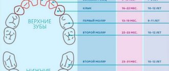
While the first teeth may appear at 4 months, and their eruption may be delayed for up to a year, permanent teeth erupt in everyone at approximately the same age. The sequence of their eruption is the same as in the case of milkweeds:
- 6-7 years. The central incisors appear from below.
- 7-8 years old. The central incisors on top and the lateral incisors on the bottom are replaced.
- 8-9 years old. The lateral incisors of the upper jaw appear.
- 9-12 years old. Canines and premolars are replaced.
- From the age of 12. From this age, molars begin to change, and from about 14 years of age, teeth appear, which were not among the milk teeth.
Interesting Facts
Enamel color
The natural color of enamel can be absolutely anything and it is extremely rare that it is snow-white. As a rule, the enamel is slightly yellowish or grayish. In children, teeth may even have a bluish tint, which indicates the presence of a large pulp and its close location to the enamel
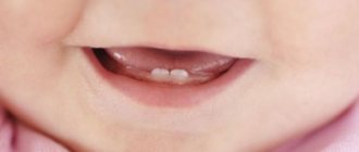
About a child's first teeth
Some babies may be born with one or even more teeth, which form in the womb. It also doesn’t matter the age when the baby’s first teeth appear – at three or ten months. The main thing is that, in general, the milk bite is formed completely and correctly.
About the important role of baby teeth
Milk teeth serve primarily as the basis for permanent ones - they literally pave the way for them. If the first teeth grow straight, it is not at all necessary that the subsequent ones will grow as well. However, the opposite situation is also possible, so the formation and growth of all teeth should be monitored under the supervision of the attending physician.
About wisdom teeth
Wisdom teeth may not erupt – this is a completely normal and fairly common occurrence. Whether to remove or leave grown ones depends on their condition: if they only bring discomfort, in the presence of caries or pulpitis it is extremely difficult to cure them, it is quite possible to sacrifice such teeth, since they do not bear any functional load
About oral hygiene
According to statistics, only 57% of women and 46% of men cleanse twice a day. The rest periodically skip such an important procedure, which allows you to effectively cope with plaque and thereby protect the enamel and gums from attack by harmful bacteria.
About using dental floss
According to research, today only 50% of the population flosses daily. Meanwhile, this is an excellent way to prevent many dental diseases: it is the floss that effectively cleans the interdental spaces from food debris and plaque. Today, the only alternative to thread can be an irrigator - a brush cannot cope with such narrow spaces
Signs of the imminent appearance of molars
You can determine when you should soon wait for the baby teeth to begin replacing with permanent teeth based on several signs:
- The gradual growth of the baby's jaws leads to increasing gaps between the teeth.
- The tooth begins to wobble. This is due to the fact that the already small root begins to gradually dissolve, causing the fixation of baby teeth to be significantly weakened.
- A fallen tooth indicates that the formed permanent one, which is about to appear, pushed it out.
- Swelling and redness may appear on the gums at the site of the eruption of a permanent tooth.
- Pain in the gums, where the permanent tooth erupts, increased temperature, and poor health of the child indicate problems have arisen, and it is necessary to see a doctor. The process of erupting molars should be painless.
The structure of tooth enamel -
The main structural unit of enamel is the so-called “enamel prisms”, between which there is an interprismatic substance that glues the prisms together. In this section we will analyze their structure, and also talk about the structure of apatite crystals, the structure of the interprismatic substance, as well as such formations as enamel plates and bundles, enamel spindles.
1) Enamel prisms and their structure -
Enamel prisms are formed from apatite crystals that are adsorbed onto an organic matrix. The latter has a fibrillar structure - in the form of a thin protein mesh, which evenly penetrates all the prisms and the interprismatic substance. The prisms themselves have the shape of thin elongated formations that pass through the entire thickness of the enamel - from the enamel-dentin border to the tooth surface (Fig. 4-5). The enamel of one tooth consists of a total of several million enamel prisms.
Longitudinal section of enamel prisms –
The thickness of the prisms ranges from 3 to 6 microns, and as they approach the enamel-dentin border to the surface of the tooth, their diameter increases by approximately 1.5-2 times. This is due to the fact that the area of the enamel-dentin junction (where the prisms begin) is significantly less than the surface area of the tooth enamel. The prisms have a radial direction and lie in relation to the enamel-dentin border - almost at a right angle. But, as for the enamel surface, in the area of occlusal surfaces they will lie parallel to the long axis of the tooth, and on the lateral surfaces of the crown - perpendicular to the tooth axis.
As for the length of the enamel prisms, it will depend on the thickness of the enamel layer on different surfaces of the tooth crown, and the length of each prism will in any case be greater than the thickness of the enamel layer. The latter becomes possible due to the fact that the bundles assembled from enamel prisms have wavy bends along their course (in the shape of the letter S). The appearance of such a radial structure with pronounced S-shaped bends in the enamel is associated with a functional adaptation that prevents the appearance of radial cracks under the influence of occlusal load (Fig. 6).
Cross section of enamel prisms –
On transverse sections of teeth, prisms can have oval, hexagonal, polygonal, but most often - the shape of arcades that resemble fish scales or a keyhole (Fig. 7). According to R. Frank, this form of prisms occurs due to the uneven mineralization of enamel prisms that occurs during their development. Thus, one side of the prisms mineralizes and becomes hard earlier than the other, which causes compression of the softer part of the prism. According to research by J. Saot and N. Symons, only 2% of prisms had a regular hexagonal shape, 57% had an arcade shape, another 31% of prisms were polygonal or oval, and another 10% had an irregular shape.
It is worth noting that the surface of each enamel prism is surrounded by a shell, which is called the “prism cortex”. However, such a shell is not considered as an independent formation, and is distinguished by the fact that it is less mineralized and contains significantly more enamel proteins than the rest of the prism. As a result, the shell is more resistant to acids (compared to the prism core). Below you can see an electron microscopy image showing enamel subjected to acid demineralization for 5 days (Fig. 8). As we see, only the outer shell of the prisms and the interprismatic substance have been preserved.
Enamel prisms after demineralization –
Prismatic enamel –
However, the innermost layer of enamel, adjacent to the enamel-dentin junction, does not contain enamel prisms. This layer is often called the term “initial enamel” (the thickness of this layer is only 5-10 microns). This layer of enamel consists exclusively of small crystals of hydroxyapatite, only 3-5 nm thick. The formation of prismless enamel is due to the fact that in the initial period of its formation, enameloblasts still lack Toms fibers (Toms processes). Only later do enameloblasts begin to form short protoplasmic processes, which give rise to enamel prisms.
The outer layer of tooth enamel is formed in a similar way (at the final stages of its development). During this period, Toms' processes in enameloblasts already disappear, and therefore the most superficial layer of enamel (terminal enamel) will also be devoid of enamel prisms. The layer of the so-called “final enamel” is more pronounced in permanent dentition teeth, but in primary teeth, electron microscopy shows a predominantly prismatic structure of the surface layer of tooth enamel.
2) Features of apatite crystals –
We have already said above that of the different types of crystalline apatites, enamel contains the most hydroxyapatite [Ca10(PO4)6(OH2)], the share of which is 75%. Hydroxyapatite crystals are covered with a hydration shell 1 nm thick. The microspaces between apatite crystals are filled with water, which is called enamel fluid. The water content in enamel is about 2-3%, and its function is the transfer of ions, which ensures the processes of mineralization/demineralization.
In turn, the hydroxyapatite crystals themselves have the form of hexagonal plates - with an average length of about 200 nm (but crystals with a size of 500-600 nm, and even up to 1000 nm can be found), as well as a width of 40-90 nm and a thickness of 25-40 nm . The direction of the crystal axis in relation to the long axis of the prism differs in its different sections. In the central part, the crystals will lie parallel to the long axis of the prism, and on the periphery, they move away from this axis, forming an increasingly larger angle with it. For example, with an arcade-shaped enamel prism, this angle will be about 40–65°.
3) Interprismatic substance –
We have already said above that enamel prisms are, as it were, cemented in a thin layer of interprismatic substance, the thickness of which is less than 1 micron (Fig. 10). By the way, it is worth noting that with the arcade configuration of enamel prisms, the latter are in such close contact with each other that the interprismatic substance between them is almost completely absent. The interprismatic substance also consists of apatite crystals, which are located at an angle to the enamel prisms (often even at an angle of 90°).
The interprismatic substance is less mineralized than the enamel prisms themselves, and therefore, in comparison with them, it has less strength. As a result, cracks that occur in the enamel usually pass through the interprismatic substance, without affecting the prisms themselves. Below you can see longitudinal and transverse sections of the enamel, on which crystals of the interprismatic substance are located between the enamel prisms (at an angle to the enamel prisms).
Tooth enamel (electron microscopy image) –
4) Enamel plates and enamel bundles –
Such formations are characteristic of mature enamel (Fig. 12-13). Both plates and bundles are low-mineralized areas of enamel prisms and interprismatic substance, differing from each other only in position and shape. Enamel plates are very thin “sheet-like” structures that pass through the entire thickness of the enamel (most of them are in the neck of the tooth). They contain enamel proteins as well as organic matter from the oral cavity.
On enamel sections, they look exactly the same as enamel cracks, but are only filled with organic matter. It should be noted that they can serve as entry points for the development of caries. In turn, enamel tufts are small cone-shaped formations (resembling the shape of “spearing tufts of grass”) that extend from the enamel-dentin border. The distance between the beams is approximately 30 to 100 μm.
5) Enamel spindles –
Enamel spindles are flask-shaped structures that extend from the enamel-dentin border at a right angle (Fig. 14). Their formation is due to the fact that during tooth development, some of the processes of odontoblasts penetrate the enamel-dentin junction, which is apparently necessary for communication between odontoblasts and secretory anameloblasts. Thus, enamel spindles are structurally dentinal tubules.
In addition to odontoblast processes, enamel spindles also contain tissue fluid and other organic components. According to most authors, enamel spindles play an important role in the mineralization of deep layers of enamel from the dental pulp side. Below you can see exactly what enamel spindles look like:
6) What are Gunter-Schräger stripes –
We have already said above that enamel prisms have a wave-like curvature along their course (in the shape of the letters S). This leads to the fact that on a longitudinal section of a tooth it is impossible to cut each enamel prism strictly longitudinally along its long axis and along its entire length. Therefore, it turns out that some sections of the prisms will in any case be ground in the longitudinal direction, and their extensions – in the transverse or oblique directions. The areas of the prisms that will be dissected longitudinally look light (parazones). Sections of prisms cut transversely will appear dark (diazons).
As a result, a correct alternation of transverse and longitudinal sections of bundles of enamel prisms appears on the tooth section. When studied in reflected light, they appear in the form of dark and light stripes, crossing the entire thickness of the enamel in an arc in the radial direction. They start from the enamel-dentin junction and end in the superficial layer of enamel. Such stripes are called Gunter-Schräger stripes, and they can be clearly distinguished even with low magnification (Fig. 15-16).
Possible problems
At the moment the molars appear, certain dental problems are possible. In order to take timely measures to eliminate them, parents must have an idea about them.
Molars do not erupt
A situation is possible in which baby teeth do not fall out in a timely manner, or they have fallen out, but molars have begun to appear in their place. The reason for this must be determined by the dentist, who must be visited without delay. A general x-ray is usually taken to show the degree of development of the molars.
Among the options for the lack of eruption of molars in due time can be indicated:
- Hereditary predisposition, which is the cause of a possible delay in the appearance of molars. If the x-ray shows that the process of forming the rudiments of teeth is underway, then you will just have to wait a little for their appearance.
- Adentia. Disturbances in the processes of formation of tooth germs during the intrauterine development of a child, inflammatory processes can lead to a similar pathology - the absence or death of tooth germs. The solution is prosthetics.
Pain
The first time after teething, the tooth is poorly protected from caries and the effects of various bacteria. This is explained by the low degree of enamel mineralization at the initial stage. Almost nothing interferes with the development of caries; tooth tissue is destroyed, pulpitis occurs, with the subsequent risk of its transition to periodontitis. Severe pain, changes in body temperature and deterioration in well-being may occur.
It is highly advisable not to let the situation get worse, not to cause severe pain, but to visit a dentist as soon as painful sensations appear. If a child is predisposed to caries, it is better to carry out preventive procedures, for example, fissure sealing. The folds on the chewing surface are covered with a composite material that protects such natural cavities from the accumulation of food debris in them, the development of bacteria, and inflammatory processes.
In the worst case, you can lose a tooth.
Teeth grow crooked
A common situation is when the molar has already begun to erupt, but the baby tooth does not want to fall out. The result is that the new tooth seeks alternative growth paths, which leads to its displacement and change in the direction of growth. Hence the malocclusion and the alignment of the dentition. Treatment by an orthodontist will be required.
If this situation occurs, you should not remove or loosen a baby tooth yourself; you should visit a doctor.
Loss of molars
An alarming symptom of the presence of diseases (caries, etc.) in the oral cavity, or there are problems with the entire body (connective tissue diseases, diabetes, etc.). A visit to the doctor is mandatory.
This is necessary to develop a strategy for restoring a lost tooth. This is necessary for the proper growth of the remaining teeth and the formation of the maxillofacial system. Considering that the jaw tissue is still in the process of growth, prosthetics are only possible temporary, which must be adjusted as the jaws develop. Permanent prosthetics will be available only after their formation is completed.
Injuries
The first few years after teething, teeth are at increased risk of injury from impact. Sports injuries, falls, and blows can lead to chipping of parts of the tooth and cracks. Be sure to contact a dentist who will restore the lost part with modern materials.
Organic and inorganic enamel matrices –
Tooth enamel is quite unique, and in many ways is absolutely unlike other hard tooth tissues. Firstly, enamel is the only dental tissue that comes from the ectoderm (the development of all others is associated with the mesoderm). Secondly, if the organic matrix of other dental tissues is formed mainly by collagen fibers, then the organic enamel matrix does not contain collagen and is formed by the so-called “enamel proteins”. Thirdly, hydroxyapatite crystals in tooth enamel are much larger than in other mineralized tooth tissues.
The last point is that other hard dental tissues continue to be synthesized by cells during the life of an individual (odontoblasts and cementoblasts, respectively), but in mature enamel there are no cellular elements, and therefore, after its maturation, no growth can occur. This is due to the resorption of enameloblast cells during the process of enamelogenesis.
1) Organic matrix of tooth enamel –
We have already said above that the organic matrix of tooth enamel consists of non-collagenous proteins (proteins), which are the product of secretion of enameloblasts, and they are called the term “enamel proteins”. The function of the organic matrix is to adsorb minerals, resulting in the formation of apatite crystals around the enamel proteins. However, as the enamel matures, the organic matrix is almost completely lost.
All enamel proteins are conventionally divided into four types - 1) enamelins and 2) amelogenins, 3) ameloblastins and 4) taftelins. Enamelins are acidic glycoproteins with a large molecular weight, which are characterized by a high content of glycine, serine, aspartic and gamma-carboxyglutamic acids. In turn, amelogenins are hydrophilic glycoproteins (2 times less molecular weight), enriched with proline, leucine, histidine and gamma-carboxyglutamic acid.
Ameloblastins and taftelins occur only during the period of amelogenesis (enamel formation). In addition to enamelins and amelogenins, the organic matrix of mature enamel also contains glycosaminoglycans, proteoglycans, as well as various classes of lipids. All these organic substances are one way or another involved in the processes of mineralization of the organic matrix (protein calcification).
2) Inorganic enamel matrix –
According to research by E.V. Borovsky, tooth enamel contains the following inorganic compounds (average values):
- hydroxyapatite [Ca10(PO4)6(OH)2] – 75.04%
- carbonate-apatite [Ca10(PO4)6(CaCO3)2] – 12.06%,
- chlorapatite [Ca10(PO4)6(Cl)2] – 4.39%,
- fluorapatite [Ca10(PO4)6(F)2] – 0.66%,
- calcium carbonate – 1.33%,
- magnesium carbonate – 1.62%.
These compounds contain about 37% calcium and about 17% phosphorus. Thus, the main mineral salt in the composition of enamel (as well as dentin and tooth root cement) is “calcium phosphate” in the form of apatite crystals, which will additionally contain either hydroxyl residues, or a carbonate group, or chlorine or fluorine. But in addition to these elements and compounds, lead, zinc, aluminum, copper, molybdenum, sodium, strontium, sulfur, tin and titanium are also included in enamel apatite crystals (in extremely small quantities).
In the superficial layers of enamel there are more apatite crystals containing fluorine, lead or zinc, but in the deep layers of enamel their content will be less. At the same time, on the contrary, there will be more apatite crystals containing sodium, magnesium or carbonates in the area of the enamel-dentin junction, and fewer in the surface layers of enamel (24stoma.ru). This "ion gradient" has a certain meaning. For example, apatites with inclusions of sodium, magnesium or carbonates are highly resistant to splitting along the enamel-dentin junction.
More superficially located apatites with inclusions of fluorine, lead and zinc - thanks to these elements, they acquire special strength and resistance to acids. Enamel containing such apatite crystals (such as fluorapatite) is significantly resistant to caries, because fluorapatite begins to degrade at a lower pH value compared to ordinary hydroxyapatite. For example, for ordinary hydroxyapatite the critical pH value will be 5.5, but for fluorapatite – pH 4.6.
In addition, it should be noted that enamel hydroxyapatite crystals (compared to dentin and cement) will have a significantly larger size. For example, in dentin, hydroxyapatite crystals are 20 nm long, 18-20 nm wide, and 3.5 nm thick, which indicates their small size and needle-shaped shape. In turn, in enamel, hydroxyapatite crystals look like hexagonal plates with a length of about 200 nm (but crystals with a size of 500 to 600 nm can also be found), a width of 40–90 nm and an average thickness of 25–40 nm.
Important: due to the absence of enameloblasts in mature enamel, enamel does not have the ability to regenerate (like tooth root cement or dentin). However, despite this, the inorganic enamel matrix is in the process of constant remodeling - thanks to the ongoing processes of mineralization / demineralization. Moreover, the entry of calcium, phosphorus, and fluorine ions into the enamel occurs not only from saliva, but also from the dentin and pulp of the tooth (thanks to the so-called “enamel spindles”).