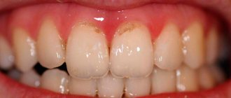
Cervical caries, which occurs at or under the gum, is considered one of the most dangerous forms of caries. In the cervical area, tooth enamel is more vulnerable to cariogenic microbes, so pathology develops very quickly. In the article we will consider the features of the course and methods of treatment of cervical caries.
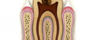
The structure of a tooth, when it comes to its structure, includes three main elements: the root, the neck and the crown. The first two are hidden by soft tissue. The enamel on them is thinner, and there are fewer minerals in it than on the visible part, so it is more vulnerable to cariogenic bacteria. Caries that develops in the area where the tooth comes into contact with the gums and under them is called cervical or root caries. It occurs mainly in adults over 30 years of age, and can appear on both the front and chewing teeth.
In this article
- Why does cervical caries occur?
- Stages of cervical caries
- How is cervical caries diagnosed?
- How to treat initial cervical caries
- How is advanced cervical caries treated?
- How to treat cervical caries on the front teeth
- What you can do at home
The problem with cervical caries is that it is almost asymptomatic until the third stage. The person does not complain of pain or discomfort, there is no visible destruction, and therefore treatment begins late. At the same time, basal caries progresses rapidly. The disease does not pose a serious danger if you visit the dentist every six months. People who rarely visit dental offices are at risk of losing a tooth. Let's talk about the causes and symptoms of cervical caries.
What type of caries is this?
Cervical caries is a clinical form of the carious process localized in the neck of the tooth - a small space between the root and crown, covered by the gum. It is also called basal caries. The disease develops regardless of age. Even an infant can get sick if he is fed haphazardly and often given sweetened water.
Based on the number of lesions, cervical dental caries is divided into:
- single – from 1 to 3;
- multiple – from 3 to 10;
- systemic – 10 or more.
The disease may be:
- primary – develops for the first time;
- secondary – develops again after treatment has already been carried out.
And:
- uncomplicated - if the destruction involves only hard tooth tissues - enamel and dentin;
- complicated - when infection penetrates into the dental cavity, root canals and periodontium with the development of inflammation in these tissues (pulpitis, periodontitis).
Cervical caries
Features of the disease
The cervical form of caries is particularly aggressive and inconspicuous in its early stages. Symptoms of this form of caries can appear when the tooth appears intact at first glance. According to statistics, more than 70% of cases of cervical caries are detected too late, when the root canals are damaged or the neck of the tooth is fractured.
Dentists call root caries the most aggressive form of the disease. This statement is based on three significant factors.
The first factor is specific localization
The neck of the tooth, connecting the root and crown, has the thinnest layer of enamel. In addition, this part is covered by the gingival margin, which is why more bacteria and substances that contribute to the destruction of the hard surface of the teeth accumulate on its surface.
Since the thickness of the enamel on the neck is not large enough, and the degree of its mineralization is lower than on the crown, destruction occurs faster. As a result, caries deepens into the dentin and pulp in a matter of weeks, that is, many times faster than when the crown is damaged.
The second factor is specific distribution
Cervical caries tends to spread the pathological process under the gingival margin. In this case, the lesion not only goes deeper inside the tooth, but also around its circumference. This leads to the breaking off of part of the crown at an early stage of the disease.
Important! When the carious lesion covers the entire surface of the cervix, we are talking about the transition of cervical caries to circular. In this case, it is almost impossible to save the tooth.
The third factor is a blow to aesthetics or the invisibility of caries
When located on the front teeth, cervical caries cannot be hidden: it is clearly visible when talking and especially when smiling. This has an extremely negative impact on the psychological state of the patient.
Cervical caries located on chewing teeth, on the contrary, is often ignored and left without treatment. Therefore, more than 80% of patients with this diagnosis turn to dentists when the disease is complicated by pulpitis and the spread of the inflammatory process to the periosteum.
Causes of the disease
The main cause of cervical caries is a bacterial infection that constantly lives in the oral cavity. If the crowns of the teeth are not completely cleared of food, then colonies of bacteria settle on them, forming soft and hard (tartar) plaque. Bacteria secrete enzymes that destroy tooth enamel and penetrate first deep into hard tissues and then into the dental cavity, affecting the pulp, root canals and moving onto the periodontium (the ligament that holds the tooth in the cell).
The localization of the carious process in the neck of the tooth determines the characteristics of its occurrence and course. This:
- the area with the thinnest layer of enamel - about 0.01 mm (average thickness in the crown area - 2 mm);
- the part of the tooth next to the gum is difficult to clean during daily hygiene procedures, so food debris accumulates in it, first soft and then hard plaque (tartar) is formed, destroying the gums, forming pockets between the gum and tooth, where even more food debris accumulates - it creates an ideal breeding ground for bacteria, and their waste products destroy the enamel;
- the neck is covered by the gum, so the onset of the carious process occurs unnoticed, the carious process is detected already at advanced stages, when the body signals it with pain;
- The pathological process proceeds very quickly with the development of complications.
Factors predisposing to the development of caries in the cervical region are:
- poor oral hygiene;
- frequent consumption of sweets without subsequent rinsing of the mouth;
- use of certain medications (glucocorticoids, antibiotics, etc.)
- smoking - poor circulation in the gums contributes to their inflammation and the formation of pockets between the gums and teeth where food gets stuck; smoking also destroys and stains enamel yellow;
- defects in the development of enamel - a thin layer or completely absent, slit-like depressions;
- immunity disorders;
- constant dry mouth, which develops for various reasons - saliva washes away bacteria;
- endocrine disorders of the thyroid and pancreas (diabetes mellitus);
- pregnancy - calcium is used to build fetal tissue.
- occupational hazards - persons at risk are those who work in industries with acids, heavy metals (lead, copper, brass), industrial dust and gases, sudden temperature changes, and infections.
- concomitant diseases - at risk are persons suffering from diabetes mellitus, thyroid diseases, dysfunction of the salivary glands, vitamin deficiency, as well as those with a family history (having close relatives with multiple or systemic caries).
Why does cervical caries occur?
The main cause of any type of caries is damage to the enamel by cariogenic microorganisms from the group of streptococci. They live in the oral cavity of almost all people. Under certain conditions, microbes begin to actively multiply and secrete acids that corrode the enamel, washing away minerals from it. Demineralization of the enamel surface causes exposure of hard tooth tissues. Bacteria penetrate microcracks in dentin and form a carious cavity. Without treatment, it spreads and leads to gradual tooth decay. However, microbes alone are not enough for the development of caries. They are dangerous only in the presence of other factors for the occurrence of carious lesions. Among them:
- Insufficient oral hygiene. If you brush your teeth twice a day, use dental floss and irrigators for cleaning, then the risk of caries is quite low. Otherwise, deposits and food debris will accumulate in the interdental spaces and gum pockets. They are a favorable environment for the spread of microbes.
- Poor nutrition. Cariogenic bacteria feed on carbohydrates. Immediately after eating sweet foods, which contain a lot of fast carbohydrates, active proliferation of microbes begins. This process is accompanied by a sour taste in the mouth. The more sweets a person eats, the more often he develops tooth decay.
- Nicotine addiction. Smoking contributes to the formation of plaque on the enamel. Over time, it mineralizes and hardens, leading to the formation of tartar, which contains many bacteria. They may also be covered with plaque, as a result of which caries begins to develop between the enamel and tartar.
- Disorders of the salivary glands. Saliva not only washes away food debris and microbes from the oral cavity, but also neutralizes the waste products of cariogenic bacteria. Some diseases are accompanied by insufficient saliva production, which can become a factor in the development of caries.
- Enamel abrasion. The enamel surface protects the hard tissues of the tooth from external factors. Some people's enamel is very thin and tends to wear away, resulting in more tooth decay.
These are the main factors in the development of caries. But there are many other reasons, including weak immunity, mechanical injuries to teeth, hormonal imbalances, endocrine pathologies, etc. If they are not eliminated, the disease will periodically recur.
Symptoms
A stain on a tooth is a sign of caries
First, if you look closely at the tooth, you can see a small white spot on its frontal surface near the gum. But very often it is covered by gums or tartar, so only a dentist can notice it (this is why it is so important to undergo medical examinations twice a year). The tooth doesn't hurt.
After this, thin enamel and dentin are destroyed quite quickly with infection penetrating into the pulp (soft tissue penetrated by nerve endings and blood vessels). This is a complicated carious process. A person experiences pain when eating cold, hot, sour and sweet foods. The pain is severe and does not go away immediately after rinsing.
The next thing that appears is severe throbbing pain, sometimes accompanied by an increase in body temperature - a sign of periodontitis. If it is not treated promptly, the infection can spread to the soft tissues and bones of the jaw.
Causes of caries in the gums
The main cause of the disease is cariogenic microbes that cause damage to enamel and dentin. They exist in everyone, but they begin to develop only when favorable conditions are created: consumption of large amounts of carbohydrate foods, poor oral hygiene and due to some other reasons. The development of caries at the base of the tooth is associated with its localization and is caused by the following factors:
- Due to the characteristic structure of the tooth, plaque constantly accumulates in the root area, which cannot always be removed even with a toothbrush. If you do not make an effort when cleaning, then gradually the neck of the tooth will begin to collapse under the influence of cariogenic bacteria contained in plaque.
- In the depressions near the gums, the so-called “pockets,” food debris accumulates. If oral hygiene is not sufficiently thorough, food decomposes and lactic acid is formed, which destroys tooth enamel.
A risk factor that provokes the development of cervical caries may be the consumption of large quantities of too acidic foods that contain quickly fermentable carbohydrates. As a result of their fermentation, organic acid is released, which corrodes tooth enamel and washes away calcium.
Stages
Cervical caries: medium, deep and complicated
Cervical caries occurs in stages. They replace each other faster than in the carious process of other localizations and cause complications more quickly.
Spot stage
The main symptom is a white spot on the enamel. The stain is matte - the enamel loses not only calcium, but also its shine. There are no complaints, only occasionally there is a feeling of a sore throat. If the patient does not visit the dentist for preventive purposes during this period, cervical caries may not be noticed.
Surface
The enamel continues to lose minerals and gradually deteriorates, its surface becoming slightly rough. The color may change from white to yellowish. The sore throat intensifies, and it hurts when eating very cold or hot food. At this stage, the patient may already suspect something is wrong if he carefully listens to the signals of his body. But this rarely happens, so this stage in most cases remains without treatment.
Average
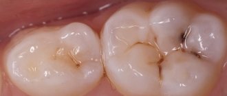
At this stage, there is already complete destruction of the enamel and deepening of the carious cavity to the middle of the dentin layer. The defect is black in color and visible in the form of a hole in the tooth. It can only be missed if the cavity is covered by gums. At this stage, it hurts from the effects of temperature and chemical (sour, sweet foods) irritants, but the pain goes away quite quickly. It is at this stage that most patients turn to the dentist.
Deep
The carious cavity increases in width and depth, reaching the dentin border. A thin layer of dentin separates it from the dental cavity with pulp. The pain intensifies, the tooth reacts to all types of irritants, including mechanical ones - pressure when chewing. The tooth can hurt for a long time and quite severely. Food gets stuck in the resulting cavity.
Circulatory
Circulatory caries in a child with a broken tooth
This caries of the cervical region has its own characteristic features. There is a circular damage to the enamel and dentin of the cervical area and the teeth simply break off. The front teeth are mainly affected, the disease proceeds unnoticed and very quickly, reaching deep stages in weeks and even in a matter of days.
It mainly affects children from infants to preschool age, which is why its second name is bottle disease. In children in the first months and years of life, it is associated with improper frequent feeding without subsequent cleansing of the oral cavity. This leads to the accumulation of food debris on the crowns, the proliferation of bacteria and the destruction of several teeth at once.
In adults, circular cervical caries also occurs; its causes are: impaired immunity, smoking and alcohol abuse, poor oral hygiene, crowded teeth, and malocclusion.
Stages of cervical caries
Cervical caries develops according to the same scenario as other types of this disease, but much faster. Let us describe the stages of development of root caries:
- Initial (white spot stage). It is asymptomatic. A demineralized area of enamel forms in or under the gum area. It looks like a white or chalky spot. Gradually it begins to fade, but this is difficult to notice with the naked eye, especially if the pathology is localized on the back surface of the tooth or on the molars and premolars. If it starts under the gum, then it can only be detected using hardware diagnostics.
- Superficial. The carious spot becomes larger and acquires a yellowish or brownish tint. Destructive processes begin on the tooth surface. At this stage, the pathological focus has not yet spread to hard tissue. The patient may experience discomfort when eating sweet foods. It disappears immediately after the stimulus is removed. Often people attribute this symptom to increased sensitivity of the gums.
- Average. Damage to hard tissues is observed. A person experiences severe discomfort when eating sweet, sour, cold or hot foods. Possible pain. These signs disappear as soon as you brush your teeth.
- Deep. The last stage is an advanced form of the disease. Deep damage to dentin occurs. The pathological process involves the tooth root and pulp. The tooth may split into several parts or break in the neck area. The patient almost constantly experiences pain, which painkillers do not help eliminate.
Treatment for caries can vary depending on the degree of damage to the tooth. The method is selected individually after diagnosis.
How dangerous is the disease?
If cervical caries is not treated, the following complications may quickly appear:
- Pulpitis is an inflammation of the soft tissue (pulp) located in the dental cavity. The infection enters the pulp when hard tissue is destroyed. Symptoms: severe excruciating pain in the tooth, appearing regardless of food intake, including at night.
- Periodontitis is an inflammation of the ligament that holds the tooth root in the cell (periodontal). The infection enters the periodontium through the root canals. The pain becomes unbearable, often pulsating, and intensifies with chewing (mechanical irritation). Fever and general malaise appear.
- An abscess at the root apex is a capsule filled with pus. Severe pain, severe fever, chills.
- Osteomyelitis is an inflammation of the jaw bone. The condition is serious and urgent hospitalization is required.
- Periodontitis – inflammation spreads to the periodontal tissues, the gums become inflamed and bleed. Pockets form between the gum and neck, into which food becomes trapped, maintaining the infection. It is painful. Treatment takes a long time and is not always successful; much depends on the patient’s immunity.
- Lateral spread of infection to the necks of adjacent teeth, development of multiple caries.
- Circular lesion of the neck with crown fracture.
If you suspect the presence of a carious process under the gum, you should immediately contact your dentist: complications with this form of the disease develop very quickly!
How to treat cervical caries on the front teeth
Cervical caries of the anterior teeth causes the most discomfort. Firstly, it develops and progresses faster than on molars and premolars. Secondly, the front teeth fall into the smile zone, so a cosmetic defect also occurs. Treatment is carried out according to the usual scheme, but the material for the filling is selected more carefully. It should not differ in color from the rest of the tooth, otherwise it will look unaesthetic. The cost of such a filling is significantly higher than the standard one.
How to distinguish between cervical caries and wedge-shaped defect
Wedge-shaped defect before and after treatment
These pathological processes are similar, but there are a number of significant differences between them. A wedge-shaped defect is a non-carious process. The main reason for its development is the incorrect distribution of mechanical load on the teeth due to occlusion pathology, periodontal disease and endocrine pathology. It is necessary to distinguish cervical caries from a wedge-shaped defect, since these diseases require different approaches to treatment.
Differences:
- wedge-shaped defect (CD) is localized more often on the lateral teeth (canines and molars), which have a high chewing load, and cervical caries (CC) - on the anterior ones;
- externally, the CD initially appears as a small yellowish area on the front surface of the neck of the lateral tooth under the gum; the enamel is not altered or rough; then a gradually deepening V-shaped defect appears; PC is first a white spot, then a black-brown carious cavity;
- CD lasts for years and decades, while cervical caries can reach a deep stage in a matter of weeks.
How is advanced cervical caries treated?
Deep caries is often accompanied by damage to the pulp, so depulpation is required. After this, root canal treatment and filling are performed. In some cases, nerve removal can be avoided. Then treatment is carried out according to the standard algorithm:
- Anesthesia.
- Removal of deposits and tartar.
- Selecting the shade of the future filling.
- Retraction of the gingival margin.
- Preparation of a carious cavity.
- Tooth isolation.
- Grinding.
- Filling.
- Polishing the filling.
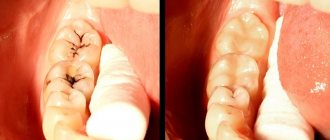
The procedure can be more complicated if the carious lesion is located under the gum. It has to be cut to open access to the neck and root. After the main treatment, the doctor applies sutures, which dissolve within a few days.
If the patient goes to the doctor late and the tooth is almost completely destroyed, it is removed. The question arises about installing an implant.
Diagnostics
The dentist begins by examining the patient's mouth using a dental mirror. If there is a defect in the crown, it is examined using a probe. After this, the doctor prescribes:
- X-ray diagnostics; if cervical caries of only one tooth has been detected, targeted radiovisiography is prescribed to determine its depth and volume; if a general X-ray of the jaw is necessary, an orthopantomogram is prescribed;
- electroodontodiagnostics (EDD) – determination of pulp viability and pain sensitivity of the affected tooth;
- laser fluorescence – illumination of pathological foci using a laser device;
- painting the stain with water-soluble paints (methylene blue or magenta); in places where the enamel is damaged, it becomes permeable to paint and the pathological focus acquires clear contours;
- X-raying the crown using a special lamp to detect any hidden cavities.
Diagnosis of the disease
Cervical caries can be diagnosed during a dental examination of the oral cavity. The specific location and rough structure of the carious spot make it possible to differentiate the pathology from a wedge-shaped defect and simple caries.
Additionally, instrumental diagnostic methods are used: dental radiography, radiovisiography, transillumination and others. They allow you to assess the degree of penetration of caries into the tooth cavity, see hidden lesions and predict the effectiveness of treatment.
Treatment
Features of the treatment of this form of the disease are that the hard substance (enamel and dentin) in the area of the tooth neck has minimal thickness, so treatment of the resulting cavity may result in penetration into the pulp chamber and be complicated by pulpitis. The proximity of the gums also creates difficulties in treating and filling carious cavities. Cervical caries on the front teeth also requires restoration of the aesthetics of the tooth.
But there is good news: after local anesthesia, the entire treatment is absolutely painless. You can find out how cervical caries is treated and treatment methods at different stages of the disease below.
Treatment at the spot stage
Cervical caries begins with a process of demineralization (the enamel just begins to lose minerals). It can be suspended without instrumental intervention. To restore the enamel, professional teeth cleaning is carried out, then a course of remineralization is prescribed: applications with special products containing calcium and fluoride. To combat relapses, after treatment, the doctor recommends daily use of toothpaste with fluoride and minimal abrasive properties, as well as rinsing with light antiseptics or solutions with fluoride. The course of treatment is 10 procedures plus oral calcium supplements, followed by the use of fluoride-containing oral hygiene products for 1 to 2 months.
Treatment at the superficial stage
Enamel destruction requires instrumental treatment. First, professional teeth cleaning is also carried out, and then the resulting roughness on the enamel is sanded off. The doctor decides individually how cervical caries is treated further. Sometimes a course of remineralization is enough, but in some cases the doctor prepares the cavity, then etches its walls with acid, applying a special gel to improve the adhesion (sticking) of the filling material. Filling should be done with a liquid composite followed by light curing. The filling is ground and polished. Anesthesia is not performed, since enamel treatment is painless. But if gum separation is required, topical anesthesia is sometimes used. All treatment is carried out within half an hour.
Treatment at the middle and deep stages
The treatment process is carried out under local anesthesia. Average caries is treated in one session (30 – 40 minutes). It is carried out: removing plaque, if necessary, pushing back the gums with a special tool, opening and preparing the carious cavity, treating its walls with an antiseptic, etching with acid, applying an adhesive. Installation of a filling for cervical caries at this stage is usually performed on the same day.
Deep caries in the cervical area is treated in two visits. First, plaque is removed, the gums are necessarily pushed back, preparation and medicinal treatment of the cavity are carried out. To combat infection and mineral loss, fillings have to be done in two stages. First, a therapeutic pad with antiseptic and remineralizing properties and a temporary filling are installed at the bottom of the cavity. Return visit after 1 – 2 weeks. If there is no pain during this time, an insulating gasket is placed at the bottom of the prepared cavity. Then a permanent filling is performed.
What can you do at home?
It is impossible to cure cervical caries on your own. Even at the earliest stage, the patient himself will not be able to carry out high-quality demineralization at home; the process will quickly progress with the development of complications. But you can fight this process by preventing its appearance or identifying spots at the earliest stage. At home, the main thing is the prevention of cervical caries.
How to treat initial cervical caries
At the white spot stage, caries can be treated using therapeutic procedures. It is necessary to restore the enamel structure by saturating it with minerals. First, the dentist will clean the affected tooth from plaque and tartar, and then apply a solution containing fluoride, magnesium, phosphorus and calcium to it. Under the influence of the active components, the enamel is remineralized and the pathological process is blocked. The patient is also prescribed mouth rinses with an antiseptic solution and toothpaste with fluoride.
How to treat superficial caries
If the carious area has managed to destroy the enamel, but has not penetrated into the hard tissues, remineralization is carried out, as in the first stage. The dentist grinds the affected area of the tooth, removes damaged tissue and applies a solution to the enamel to restore it. Several procedures may be required - 5-10 sessions. Throughout the treatment, the patient uses medicated toothpaste.
Treatment of cervical caries at the middle stage
As a rule, people consult a doctor when root caries has reached the third stage. At this stage, tooth filling is required. The doctor removes the affected tissue, carries out antiseptic treatment of the carious cavity and fills it with filling material. The procedure is performed under local anesthesia.
Prevention
To combat the development and recurrence of the carious process, you need:
- stop consuming large amounts of easily digestible carbohydrates - sweets, baked goods, sweet carbonated drinks;
- do not have frequent snacks;
- regularly include dairy products in the diet, which the body needs as a source of calcium and phosphorus;
- Brush your teeth twice a day with a soft toothbrush and use toothpaste recommended by your dentist (the choice of toothpaste depends on the condition of the teeth, heredity and the fluoride content of the water in the area);
- After each meal, rinse your mouth thoroughly and use dental floss or an irrigator to clean the interdental spaces;
- visit the dentist twice a year to identify and prevent the initial stages of cervical dental caries, and carry out professional cleaning.
If you regularly adhere to these requirements, you can maintain healthy teeth for a long time.
Traditional methods in the fight against cervical caries
In order to get rid of cervical caries, you can also use folk remedies. However, you need to understand that they can only help at the initial stage of progression of this disease, when a small stain has just appeared on the tooth.
What means are suitable for this?
- Decoction of onion peels: boil the peels, let them sit overnight and rinse at least after each meal;
- Propolis: rinse with a solution (four teaspoons of pharmaceutical solution per glass of warm water) three times a day;
- Sage: pour a glass of water over a spoonful of dried leaves, leave for an hour and rinse as often as possible.
- Melissa: pour four tablespoons of the herb with a glass of boiling water, leave for an hour and rinse after eating.
Before using any of these methods, it would be a good idea to undergo an examination by a dentist and consult with him about the most effective treatment method in this case.
What to do if a tooth hurts after treatment of cervical caries
If a slight pain after removal of cervical tooth caries lasts 2–3 days and gradually subsides, then this is normal. If there is severe, increasing pain, as well as an increase in body temperature or malaise, you should immediately consult a dentist. Possible reasons:
- insufficiently thorough treatment of the carious cavity before filling led to the spread of infection and relapse of the disease, including perforation of the dentin barrier, penetration into the pulp and the development of a complication - pulpitis; the dentist will remove the filling, open the tooth cavity and treat pulpitis; and only then can it be re-sealed;
- periodontitis (inflammation of the periodontal tissues) – the gums become swollen, painful, their edges move away from the crown, and bleeding may occur; this condition also requires dental treatment.
Free consultation on the cost of treatment in our dentistry
Leave a request and the clinic administrator will contact you within 15 minutes!
Treatment of medium and deep cervical caries
When treating medium and deep cervical caries, drilling the tooth is no longer necessary.
To rid a tooth of caries and then restore it with a filling, the dentist must remove all tissue affected by caries. A drill is used for this purpose. But there is no need to be afraid of pain - before starting caries treatment, the doctor will definitely give you an injection of an anesthetic, which will “freeze” the tissues and nerve endings in the area of the diseased tooth and eliminate discomfort. Treatment of medium and deep cervical caries is carried out in several stages:
1. First, the doctor will clean your teeth of plaque. This is necessary to improve the quality of caries treatment in general, to identify all carious lesions and to correctly select the color of the filling material that will be used to restore the tooth in the future.
2. Using a drill, the doctor will remove all tissue destroyed by cervical caries and form a cavity for installing a filling. Before using a drill to drill out a tooth and remove tissues damaged by caries, the patient will be given local anesthesia!
3. The finished cavity is washed with an antiseptic solution, and a rubber dam is placed on the tooth - a special insulating gasket that will protect the treatment area from saliva and moisture.
4. The cavity under the filling is treated with antiseptics and an adhesive composition, which will increase the adhesion strength of the filling and natural tooth tissues.
5. The dentist will install the filling and polish and grind it so that the restoration after caries treatment is invisible.
At this point, the treatment of cervical caries can be considered complete. Upon completion of all treatment procedures, the doctor will give you advice on dental care, which will prevent the recurrence of cervical caries in the future.
USEFUL TO KNOW: Advanced cervical caries requires emergency treatment! The neck of the tooth is located too close to the pulp of the tooth and its root part, and if measures are not taken to treat cervical caries in time, the tooth can be lost. If a complication such as periodontitis develops in a tooth with cervical caries, in 90% of cases the tooth will have to be removed, and the treatment of pulpitis on such a tooth will be long and difficult. In addition, pulpitis on teeth with cervical caries cannot be cured without removing the nerve - a procedure that makes the tooth fragile and vulnerable to external factors.
Methods for diagnosing contact caries
Visual inspection.
It is carried out during an appointment with the dentist. An old, traditional, cost-free method. But less accurate, because In some places, the dentist may not notice the early stages of caries development.
Diagnodent device
. Detection of lesions occurs by emitting special sound waves, which, reflecting from carious areas, are captured by the device. A very accurate diagnostic method.
X-ray diagnostics.
An excellent diagnostic method, because... allows you to identify carious lesions at an early stage at any location.
Symptoms of the disease
Many people do not seek medical help until the toothache becomes unbearable. This is why dentists so often have to carry out complex and expensive prosthetics instead of placing a conventional filling. It is very important to consult a specialist as soon as possible if the following symptoms appear:
Changing the original enamel color. In the affected area it becomes yellow or gray.- Increased tooth sensitivity. Painful sensations occur when a person eats something cold, hot, salty, or sour. Moreover, only one diseased unit reacts to these stimuli, and not both dentitions.
- Chalk-like spots form in the cervical zone. Their edges are always clearly defined.
- There is an unpleasant odor from the mouth. It occurs even soon after standard hygiene procedures. This happens because the paste and brush only mask the problem, but do not directly affect its cause.
The sooner the doctor examines the diseased unit, the sooner the problem will become a thing of the past and the higher the chance that the tooth will become healthy.
Difficulty in treating wisdom teeth
The treatment plan for figure eight caries already at the preparatory stage differs from the diagnosis of other teeth. The main difficulty in treating caries and detecting it is the impossibility of identifying it at the initial stage. As a rule, it is detected at a medium or deep stage.
Among other things, the anatomical features of the location of the wisdom tooth do not allow dentists to carry out a full range of therapeutic and restorative work.
The primary measure to eliminate pathology includes drilling out areas of the tooth that are affected by caries. Afterwards, the doctor performs an antiseptic treatment of the dental cavity, applying a special lining and filling. The completion of the work is the restoration of the anatomically correct shape of the tooth. The process can be complicated by the spread of the source of pathology to the root system of the “eight”, since only highly qualified and experienced dentists can treat this type of pathology.
If there are signs of wisdom tooth caries, the action plan for its diagnosis and elimination includes the following points:
- patient interview;
- wisdom tooth examination;
- assessment of tooth sensitivity.
A special dental mirror helps the dentist during the examination, and the sensitivity of the “eight” can be assessed using a button probe.
Therapeutic treatment of dental caries, prices, registration at the clinic
If your wisdom tooth hurts, treat or remove it
For a patient who is dealing with molar caries for the first time, the issue of solving the problem becomes urgent. In this case, there are two options: either a conservative approach or removal. In dental terms, tooth extraction means extraction. The decision on this procedure is made individually, taking into account all the nuances of the case.
Treatment of a carious tooth is considered relevant if there is partial coverage in the form of a hood of gum. It is the presence of this element that provokes the gradual but sure destruction of the enamel.
Another argument doctors make in favor of treating the “eight” is the presence of an antagonist of the 3rd molar. It is usually located on the opposite side.
If you have been diagnosed with wisdom tooth pulpitis, whether it is worth treating or removing, read the article.
Factors such as:
- The course of pericoronitis in the chronic stage, which is accompanied by relapse attacks. If the patient complains of periodic inflammation in the gum tissues that are located next to the tooth, this is a clear signal to remove the “eight”.
- Another sign is systematic damage to the soft tissues of the cheek due to the horizontal position of the wisdom tooth.
- The presence or absence of 1st and 2nd molars nearby. This factor is especially important for patients who wish to undergo dental prosthetics. In the absence of 1st and 2nd molars, doctors usually try to preserve the natural position of the dentition during prosthetics.
- Lack of opportunities for therapeutic measures to restore damaged enamel. In this case, it is worth paying attention to whether the patient can open his mouth completely.
- Presence of orthodontic pathologies. The appearance of “figure eights” is often accompanied by a narrowing of the space between the incisors and the so-called canines. This factor often causes tooth curl. The most correct thing for the dentist in this case would be to remove the wisdom teeth completely with further correction of the displaced teeth.
It is worth remembering that in the process of deciding on the treatment of a wisdom tooth, dentists rely not only on the previously stated factors. They also look at the advantages and disadvantages of different treatment methods for figure eights and compare them to each specific case on an individual basis.
The most popular benefits of wisdom teeth removal include:
- complete elimination of the lesion;
- minimal risk of tooth curling in the future;
- financial savings due to the absence of the need for re-treatment of the tooth;
- complete elimination of the cause of pain by eliminating the molar.
In addition to these advantages, the procedure also has disadvantages, which include the following phenomena:
- the possibility of unforeseen complications during and after wisdom tooth removal;
- when installing prostheses, it is worth remembering that removing the “eights” is fraught with a decrease in the stability of the structure;
Despite this, the tactics of therapeutic intervention for carious lesions of a wisdom tooth have its advantages, namely:
- maintaining additional support in case of need for dental prosthetics;
- The process of chewing food is greatly facilitated by the presence of wisdom teeth. Just remember that during the treatment of caries, chewing food may be accompanied by painful sensations, especially if the pathology has reached the root system.
Treatment of a wisdom tooth is almost always an appropriate solution. However, this procedure has a number of significant disadvantages, namely:
- chance of caries recurrence. This disadvantage is especially relevant when the tooth is deeply damaged;
- significant financial investments. Repeated expenses can significantly affect the patient’s budget; experienced doctors should warn about this even before starting treatment.
If there are no symptoms of pericoronitis, and the wisdom tooth itself is located strictly in the jaw arch, doctors usually try to make efforts to preserve the “eights”. They are helped in this by the method of cleaning the carious cavity, as well as installing a special hermetic filling.
In the absence of the necessary conditions, doctors usually resort to wisdom tooth removal. It is worth remembering that such an intervention is a full-fledged surgical operation, which doctors perform using local anesthesia.
After removal of the “eight”, the patient undergoes additional procedures, the purpose of which is to prevent alveolitis. These measures include mechanical and antiseptic treatment, as well as, if necessary, the application of special sutures.
Make an appointment with a therapist by phone+7(985)532-21-01
Retrograde pulpitis: diagnosis, causes, treatment, complications
Treatment of average dental caries from 6,000 rubles - sign up at the clinic of the Central Administrative District of Moscow
Hypoplasia of dental enamel, diagnosis and treatment
Tooth root caries treated or removed
Caries in the white spot stage
Tooth granuloma, what is this disease and how to cure it
Treatment of wisdom tooth caries if it hurts
What to do for root caries of incisors
It is always more difficult to work with the frontal zone. Here it is important not only to restore the structure of the crown, but also to obtain an impeccable aesthetic result. After all, it looks very ugly when, while smiling, others see darkened fillings.
In case of root lesions of the incisors, glass ionomer and light fillings are used. They make it possible to obtain an impeccable aesthetic result. It is very important that their installation is carried out by a highly qualified dentist who has extensive experience.
If the doctor has to replace old fillings on the front teeth with new ones, and the carious cavity is very large, he may suggest closing the defective area with veneers. This is a microprosthetics option that involves gluing thin onlays to the front tooth surface.
Prevention of subgingival caries
The development of the disease is provoked by many external and internal factors. And even if caries has never bothered you, preventive measures will help prevent its occurrence and development. Here are some simple rules doctors recommend following:
- Brush your teeth with toothpastes high in minerals.
- After each meal, use a mouthwash and dental floss - it will help effectively remove pieces of food stuck between your teeth. Oral irrigators can also be used.
- Half an hour after eating, brush your teeth if possible. This should be done twice a day for a couple of minutes.
- Include in your mandatory daily diet foods rich in fluoride, calcium and other beneficial microelements to strengthen enamel, eat more solid vegetables and fruits.
- Avoid excessive consumption of sweets, flour and sour foods.
- Periodically massage the gums: it improves blood circulation in the gum tissues, protects against the formation of deep periodontal pockets and the accumulation of deposits in them, which reduces the risk of caries formation at the neck of the tooth.
If the enamel is thin and susceptible to the rapid formation of tartar on it, regular remotherapy helps a lot - this is the name for applying fluoridating compounds to the teeth. The procedure reduces the risk of enamel destruction and improves the condition of hard tissues. To further strengthen the enamel, you can periodically take special vitamin complexes containing useful minerals. In addition, it is necessary to visit the dentist’s office at least once every six months for a dental examination. The doctor will make sure that everything is fine with them, or will prescribe additional diagnostics using x-rays. Once every 6-9 months, it is recommended to carry out professional enamel cleaning using ultrasound or other methods. During the procedure, plaque and tartar are removed from the surface of the teeth. It polishes and becomes smoother, and less deposits accumulate on the enamel. During cleaning, the doctor can also clean periodontal pockets if plaque and deposits accumulate under the edge of the gums.
People who are at risk for endocrine diseases need to be especially attentive to pain in their teeth. With them, teeth are often affected by cervical caries. Treatment must be comprehensive and systemic to prevent caries from damaging the tooth root. Gingival caries is a dangerous pathological process if it is started, but it can be successfully treated in the early stages.
Modern dentistry eliminates caries of various types, but it also happens that if the stage of the disease is too deep, some teeth cannot be saved. We have to remove them and resort to various methods of restoring the dentition. You just need to be attentive to your oral health, taking simple preventive measures, so as not to have to deal with expensive treatment later. With regular dental examinations, you will be able to detect the problem in time and solve it.