Before surgery, it is necessary to carry out professional oral hygiene.
If the operation is performed with an anesthesiologist, you must come with an accompanying person, or the operation will be cancelled.
Recommendations for surgical treatment
After surgery, you may experience pain, which will go away as the tissue heals. Postoperative swelling or hematoma can also occur in areas adjacent to the surgical site, which is a natural consequence of surgery. There may be a slight increase in body temperature.
Please follow our recommendations!
Before surgery
- Prepare an ice pack at home.
- On the day of surgery, eat a light meal 2-3 hours before your scheduled time.
- The day before surgery, the consumption of alcoholic beverages is strictly prohibited.
- Be sure to inform your dentist about all the individual characteristics of your body and any allergic reactions, in order to accurately select an anesthetic that is safe for you.
- Visit the toilet before surgery.
- It is better to come to the operation in loose clothes without a collar.
After operation
— To avoid severe swelling and bleeding during the first 24 hours after surgery, it is necessary to apply an ice pack to the cheek on the side of the operation for 15-20 minutes with breaks of 30-40 minutes.
- Rinsing your mouth when bleeding is unacceptable!
— After sinus lift surgery, you should not drink through a straw, blow your nose vigorously, or puff out your cheeks.
- To reduce the likelihood of nosebleeds (after upper jaw surgery) and reduce post-operative swelling, you should sleep with your head elevated (add an extra pillow) for several days after surgery.
— It is not recommended to use tobacco products 1 week before and 2 weeks after surgery.
— It is prohibited to drive a car on the day of surgery with premedication.
- It is not recommended to eat until the anesthetic wears off. In the first days after surgery, it is recommended to eat soft, non-hot foods.
— It is not recommended to drink alcohol, coffee, or strong tea to avoid the negative effects of these drinks on blood circulation and wound healing.
A simple method of tooth extraction
Involves the use of one tool. As a rule, these are tongs or an elevator. The technique for removing teeth and roots in the upper jaw involves using the full strength of the dentist’s right hand. But with the method of removing roots and teeth on the lower jaw, pressure is applied using the thumb of the right hand.
When using forceps, the dentist places them on the crown so that the grip is strong enough to pull out the tooth, but not too strong so as to destroy it. Next, the process of dislocation occurs, which can be carried out using two techniques: using rocking or rotation. The first method is usually used when extracting multi-rooted teeth, and the second method is used when there is no more than one root. After the tooth is dislocated from the gum, the process of tooth extraction occurs.
Additional Tips
Avoid lifting heavy objects, bending over, playing sports, or taking hot baths for 5 to 7 days after surgery.
Within a few days, performance and the ability to drive may be reduced.
Remember to take medications prescribed by your dental surgeon after surgery.
We kindly ask you!
Appear for examination and removal of stitches 5-7 days after surgery, in agreement with your surgeon. You must immediately notify your surgeon or clinic administrator of any changes in your health.
Recommendations for caring for dental implants
The service life of the implant depends on:
- The correctness of the surgical and prosthetic stages carried out in the dental clinic.
- The patient's compliance with the recommendations given by the dentist immediately in the postoperative period and the period after implant prosthetics.
- Careful hygienic care of the “implant-crown” structure.
- Blood supply to bone tissue and gums in the area of implantation. (Smoking cigars and cigarettes has a very, very negative effect on peripheral blood circulation, which can even interfere with implant implantation.!!!)
Post-extraction sockets of single-rooted teeth: classification and restoration protocols
Currently, there are many clinical protocols that involve the extraction of functionally compromised teeth and their subsequent replacement with structures supported by dental implants. However, the predictability of each specific treatment algorithm is very variable, especially in the conditions of various defects of post-extraction sockets, so systematization of these would help to significantly optimize the process of selecting and arguing appropriate protocols for iatrogenic interventions during the complex rehabilitation of dental patients.
This article is a kind of road map, the purpose of which is to help practitioners correctly approach the choice of clinical manipulations during the restoration of sockets of single-rooted teeth. The classification proposed by the authors is unique, taking into account the volume of required intervention, the possibility of planning treatment and ensuring conditions for adequate wound healing after surgical extraction. In addition, this classification also takes into account the topographical features of the bone tissue in the area of the extraction sites, and the choice of treatment protocol is based on the biological principles of wound healing in the maxillofacial area. The rehabilitation protocol also consists of an analysis of the shape of the residual bone tissue, biotype and features of the positioning of the sockets on the upper and lower jaws. But for a general understanding of the whole problem, the first part of the article provides a description of those predisposing conditions and aspects that led to the creation of the above classification.
Relevance of the topic
The proposed classification is based on the condition of the bone tissue of the sockets: its presence and shape determine further manipulative aspects of treatment. The bone relief of the socket influences the further timing of implant installation and the choice of the necessary augmentation methods during complex rehabilitation. The concept of bone topography of the extraction socket includes parameters of the shape, contour and 3-dimensional structure of the residual ridge remaining after tooth extraction. In turn, all of the above parameters are determined by the shape of the alveolus apical to the extraction area, the level of bone in the interdental areas, the height, thickness and quality of the bone plate on the vestibular side. Each of these aspects of hard tissue affects the further outcome of socket restoration, and was taken into account in the process of creating the proposed author's classification.
Availability and quality of bone tissue
The shape and size of the bone tissue located apical to the area of tooth extraction, which represents the so-called apical bone topography parameter, play an important role in the further planning of immediate and delayed implantation procedures. To ensure adequate primary stability of intraosseous constructs, a minimum of 3–4 mm of intact supporting bone is required. In addition, if the tooth to be removed has a specific apical convexity, in the future this may somewhat complicate the healing process of the bone tissue before the implantation procedure. In such cases, to ensure the required position of the implant, it is simply impossible to do without additional surgical procedures (photo 2). More specifically, teeth with deficient apical topography significantly complicate the implementation of the implantation procedure according to any protocol, even in the conditions of the necessary bone augmentation of the socket. This situation often occurs after periapical dental pathologies or due to the specific anatomy of the bony alveoli. The level of interproximal bone affects the healing and support of soft tissues on the contact sides of the installed implant, since the presence or absence of an interdental papilla is determined precisely by the condition of the bone in the contact area between the supporting structures. Thus, the topography of the bone determines not only the functional, but also the aesthetic result of future complex treatment. In addition, the interproximal bone wall, together with the buccal and lingual components, directly influences the process of natural restoration of the socket, since a blood clot after extraction is formed precisely within the existing bone boundaries. Subsequently, it plays the role of a kind of frame that transforms into mature trabecular bone. Since the bone walls are the only support for the blood clot, their height directly affects the size of the formed framework, and, consequently, the final structure of the bone tissue.
The quality of the bone walls is another parameter that determines the morphology of the alveolar area after the extraction procedure. With thin bone parameters, its predominant cortical nature, or in the presence of digiscence, the risk of socket resorption after removal of compromised teeth increases significantly. Negative changes in socket size that occur after the extraction procedure can be compensated for through guided bone regeneration (GBR) performed immediately after the initial surgical procedure. The use of a bone substitute can also significantly reduce the loss of buccal bone plate volume, which is critical in terms of both functional and esthetic parameters.
Principles of restoration of periodontal defects
The treatment protocols described below for the restoration of extraction sockets used the same principles as for the restoration of periodontal defects, since these two types of alterations are very similar: the wound heals from the periphery to the center, and the extraction socket is nothing more than 4-walled defect. At the same time, with the loss of half the height of the buccal wall, the defect is transformed into a three-wall-and-a-half defect, and with complete reduction of one of the bone plates - into a full-fledged three-wall defect. It is logical that the smaller the number of bone walls, the lower the level of predictability of future surgical treatment. Not the least important parameters are the width of the existing bone boundaries, as well as their quality, based on which the authors systematized the defects into three different groups. Each of the treatment protocols for a specific group takes into account the possible prognosis of socket restoration with its consequences. The choice of bone graft material remains a dilemma for the practicing physician himself.
The main thing to remember is that such manipulations are aimed at retaining space, filling defects with progenitor cells and primary closure of the surgical wound. In addition, we should not forget that performing augmentation immediately after tooth extraction can provoke disharmonious disorders in the area of the mucogingival junction. Such discrepancy can be corrected through soft tissue repositioning or during the opening of implants to secure superstructures.
Thin and thick biotypes
The characteristics and quality of soft tissues have a significant impact on the final aesthetic and functional outcome of treatment, therefore, taking into account the patient’s tissue biotype is mandatory when planning future complex rehabilitation. Thick or thin tissue biotype determines how they cover the bone frame and how they respond to surgery. A thick biotype of gums is more tolerant of surgical interventions such as implantation or extraction; therefore, with such parameters of soft tissues, it is easier to prevent discoloration of the gingival profile and it is easier to ensure the restoration of the papillae and the volume of soft tissues around implant-supported restorations. In addition, the risk of recession as a result of surgical or mechanical manipulation in thick biotypes is almost minimized. It follows from this that the treatment algorithm for thick and thin biotypes should be different in order to achieve both aesthetic and functional treatment results. Thin gingival biotypes require more support to maintain the same level of contour and require a more conservative treatment approach given their susceptibility to the possibility of recession. Consequently, when developing surgical protocols, the influence of soft tissue biotype on the final treatment outcome is the subject of a long and careful analysis and provides the necessary criteria for predicting the possible outcomes of complex rehabilitation.
Atraumatic flapless technique
In all clinical cases, the treatment protocol provides for an atraumatic flapless approach with mandatory analysis of the parameters of the residual bone structure, features of the apical topography and the level of interproximal bone, as well as assessment of the biotype of soft tissues. Analysis of bone criteria is performed using the capabilities of radiographic research methods and modern cone-beam computed tomography immediately after extraction. An atraumatic approach to tooth extraction ensures maximum preservation of hard and soft tissue structure while maintaining an appropriate level of blood supply between the gum and buccal socket plate, which helps minimize the effect of hard tissue resorption. After determining the class of the hole and the biotype of the soft tissues, the doctor can begin to directly perform iatrogenic intervention. In specific cases, to compensate for bone deficiency, the doctor may resort to an orthodontic extrusion procedure before tooth extraction. In such situations, assessment of the area of future implantation should be carried out only after completion of the orthodontic phase of treatment. Evaluation of post-extraction sockets according to the proposed protocol will help clinicians to take a more reasoned approach to choosing the most appropriate treatment algorithm and achieve the most predicted rehabilitation results.
Classification and treatment protocol
Demonstration of parameters such as periodontal status, socket condition and the results of CBCT studies of single-rooted teeth are presented in photos 1-4.
Photo 1. Left. A clinical example of the procedure for removing the frontal 27 tooth of the lower jaw with further restoration of this area using an implant. Note the concavity of the alveolus in the apical region. Taking into account the features of the apical topography, a 2-stage implantation protocol was applied in this situation. When directly filling the hole with bone substitute material, the morphological features still did not allow the installation of a titanium infrastructure. During the treatment, a full flap was separated and a full procedure of guided bone regeneration was performed. On right. Note the newly formed bone level on the vestibular side and the new ridge morphology allowing for a dental implant procedure.
Photo 2. Left. An example of an adequate level of interproximal bone tissue. A healthy periodontal condition supported by bone tissue provides an adequate gingival profile. On right. Example of moderate bone resorption: The presence of residual bone provides sufficient soft tissue support.
Photo 3. Left. A Class I post-extraction socket with an intact vestibular plate that is no more than 25% resorbable. Center: Class II post-extraction socket with dehiscence and approximately 50% resorption of the buccal plate. On right. Class III postextraction socket with greater than 50% loss of vestibular bone plate.
Figure 4. Sagittal sections of postextraction sockets of class I (left), class II (center), and class III (right). Note the levels of the buccal plate relative to the cemento-enamel junction.
I class
Class I sockets are the most suitable for treatment. Alveoli with an intact buccal plate, an adequate level of interproximal bone tissue and sufficient apical topography parameters fall into this classification class. The intactness of the buccal plate is determined by the absence of cracks and discolourations in its structure, as well as the level of bone tissue reduction of no more than 25% (photo 4, left; photo 5, left). This percentage was chosen for evaluation based on the average root length of single-rooted teeth, which is 14.2 mm of the average volume of bone tissue that regenerates after immediate implant placement. Adequate apical topography provides such a volume of bone tissue that would provide retention of at least 3-4 mm of the dental implant (photo 2). As for the interproximal bone, its parameter is determined by the absence or moderate (up to 2 mm) level of bone mass reduction, while providing all the conditions for the necessary support of soft tissues and placement of the implant platform in the proper apical-coronal position relative to adjacent teeth, taking into account the position of the contact teeth sides (photo 3, left). Class I extraction sockets can be treated with immediate placement of implants with or without provisional restorations based on the stability of the infrastructure and the gap between the titanium screw and the alveolar wall to be filled with the graft.
II class
Class II sockets differ from Class I sockets in terms of the quantity and quality of the residual buccal plate. Class II alveoli are characterized by the presence of dihiscence or deficiency of the buccal wall at 25-50% of its height, but, like class I sockets, they have an adequate volume of interproximal bone tissue and sufficient parameters of apical topography (photo 3, left; photo 4, center, photo 5, center). If there is a thick gum biotype, direct implantation can be performed in class II sockets. In this case, it is not recommended to fix provisional restorations, and the residual defect around the infrastructure must be augmented and covered with an isolating membrane. With a thin biotype, it is better to give preference to a delayed implantation algorithm with all measures taken to preserve the volume of bone tissue in the tooth socket. When determining the topography of the extraction area on the upper jaw, to ensure an adequate aesthetic profile of the soft tissues, you can use the augmentation technique using a connective tissue rotated pedicle flap formed from the palate area. The position of the extraction area in the upper jaw makes it possible to carry out soft tissue augmentation around the installed titanium infrastructure, but when removing teeth in the lower jaw, it is best to adhere to a specifically delayed implantation algorithm. This conservative approach is recommended due to the occurrence of thin gum biotypes and an increased risk of recession. During wound closure by primary intention, there may be disruption in the mucogingival line around the titanium support, which can easily be corrected during reopening of the implants.
III class
Class III sockets represent the most complex clinical situations, characterized by insufficient apical topography, deficiency of interproximal bone tissue and reduction of the vestibular plate by more than 50%. In general, bone deficiency in the apical region can be caused either by a pathological periapical process or as a result of the anatomy of that part of the alveolus (Figure 4, right; Figure 5, right). The lack of approximal bone tissue is manifested by the level of its reduction exceeding 2 mm on one or both contact sides. Class III postextraction sockets can be roughly divided into those in which there is a bone deficiency at the apical site and those in which the crest level deficiency is located on the proximal side of the alveolus with or without the presence of a buccal plate defect. In case of inadequate apical topography, it is recommended to carry out bone augmentation according to the type of directed bone regeneration, followed by delayed installation of implants. With sufficient apical parameters and a deficiency of proximal bone, class III sockets, regardless of biotype, are restored in the same way as class II sockets with a thin biotype, that is, according to a delayed implantation protocol with appropriate augmentation measures. On the upper jaw, a rotated connective tissue flap from the palate can also be used; on the lower jaw, augmentation should be carried out with further delayed installation of titanium supports. In some cases, the clinician can use bone extrusion techniques to restore the parameters of interproximal bone tissue. Repeated analysis of the socket is carried out only after completion of orthodontic treatment, since the parameters of bone and soft tissues change significantly during the process.
conclusions
This article presents a unique classification with protocols adapted to it for the treatment of post-extraction sockets of single-rooted teeth using dental implants. The classification is based on an assessment of the initial parameters of the quantity and quality of bone tissue of the buccal plate, residual ridge in the interproximal areas, and also takes into account the features of the apical topography. Treatment protocols also involve analysis of the topography of the socket itself, the biotype of the soft tissues and the three-dimensional position of the alveoli. Grouping of holes occurs based on the percentage value of reduction of the buccal plate, taking into account other key parameters that help the doctor approach the choice of the necessary treatment algorithm in the most adequate and reasoned way. Of course, other treatment options for post-extraction sockets may be no less successful, but the choice of the proposed treatment protocols was based on their predictability and the biological principles of wound healing. Analysis of long-term results of maintaining bone tissue levels, as well as achieving appropriate aesthetic parameters of soft tissues remain a topic for further in-depth research. None of the methods proposed in the article are completely new or previously unknown; the authors only wanted to propose an appropriate protocol for choosing the most appropriate algorithm for iatrogenic intervention.
Authors: Edgard El Chaar, DDS, MS Sarah Oshman, DMD Pooria Fallah Abed, DDS
Oral hygiene
Regardless of the size or number of implants, they must be cared for as if they were regular teeth. Brush and floss your dental implants twice a day. Use special fluffy dental floss (for example, Oral-B superfloss or ultrafloss).
When brushing your teeth, pay special attention to the back teeth and between teeth. Use a soft or medium-hard brush. In addition, use an irrigator for additional deep cleaning of the interdental spaces with water irrigation.
There are special brushes that can be used to clean interdental spaces - dental brushes. Ask your dentist about them - in some cases they are not recommended.
Visit your dental hygienist twice a year; they are the only ones who can clean your implants as thoroughly as necessary. Regular visits to the dentist are very important. Your dentist will check the condition of your gums, jaws and implants.
Smoking is bad for your health and for dental implants, too. To have a good prognosis for the lifespan of your implants, it would be a good idea if you stopped smoking.
Preparation
Before starting to directly remove a tooth in the lower jaw, the doctor must not only visually examine the oral cavity, but also send the patient for an x-ray. The image will show the exact location of the roots and their condition - the dentist will be able to determine in advance possible difficulties with removal.
In addition to X-rays, it is necessary to make sure that there is no hypersensitivity and/or individual intolerance to anesthetic drugs - the use of painkillers during removal is mandatory. You need to make sure that the patient tolerates the injections well and does not have dental phobia - otherwise, it will not be possible to carry out surgery without problems.
Eating
Avoid chewing hard candy, ice, or other hard foods (such as hard chocolate or dry fish) as they may loosen or break the abutment screw.
Avoid foods such as caramel or toffee, as they may stick to the crown and cause the abutment screw to loosen.
Do not open bottles or crack nuts with your teeth for the same reasons.
Wear protective sports mouth guards when participating in sports and avoid direct blows to the face.
Refrain from grinding your teeth. If creaking occurs unintentionally or during sleep (bruxism), notify your dentist and he will make you a thin night guard.
The length of their service depends on the quality and regularity of care for implants.
Before and after implantation
Before surgery:
Prepare several days off after the date of the planned operation.
Do not smoke or reduce the number of cigarettes you smoke.
If you are sick on the eve of the operation, please notify the implantologist.
Ask your implantologist about the medications you will need immediately after surgery.
Patients suffering from compensated diabetes mellitus must follow a strict diet 2 weeks before surgery and 2 weeks after it.
Get a good night's sleep the night before surgery.
Make sure you are accompanied if you plan to undergo anesthesia, sedation or a complex operation, do not plan to be behind the wheel.
If you have herpetic rashes on the mucous membrane, the operation should be rescheduled.
After operation:
If you have had dental implants, you may experience some of the typical discomforts associated with any type of dental surgery. These may include:
- Swelling of the gums and face.
- Gum injury.
- Mobility of adjacent teeth.
- Pain at the implantation site.
- Minor bleeding.
- Bruises and bruises.
You may need painkillers and antibiotics. Follow the recommendations of the implantologist.
What must be observed after implantation:
- Brush your teeth with a soft brush before removing sutures
- Eat soft foods for 5-7 days. Do not eat hot, spicy or salty foods.
- Starting from the third day after surgery until the sutures are removed, you should rinse your mouth with a chlorhexidine solution twice a day (after brushing your teeth).
- Do not overheat in the sun or in a sauna.
- Do not engage in active sports.
- Do not smoke or reduce the number of cigarettes you smoke.
- Do not fly on an airplane, do not swim, do not dive for 2 weeks (especially important after sinus lift surgery).
- Do not blow your nose or sneeze with your mouth open (especially important after sinus lift surgery).
- If swelling, discomfort or other symptoms increase within a few days after surgery, or the temperature rises, contact your implant surgeon.
- After tooth extraction
- If you have had a tooth removed, you must take care of your oral cavity. By following certain recommendations, you will feel better and healing time will speed up.
Recovery period
After removal, the doctor must examine the resulting hole to ensure there are no fragments of dental tissue. Attention is also paid to the condition of the wound - heavy bleeding, as a rule, does not develop, but there are exceptions. Recommendations for patients:
- You can eat and drink only after 2 hours;
- for several days (until the wound is completely healed), avoid eating too hot/cold food and liquids - find the “golden mean”;
- you can rinse your mouth with a solution of regular baking soda;
- Carry out oral hygiene very carefully and avoid touching the wound surface with a toothbrush.
Tooth extraction, with a professional approach to the procedure, does not pose any danger and is considered a normal dental practice.
Stopping bleeding:
To control bleeding, it is necessary to bite down on a gauze pad placed by the dentist in the oral cavity. The pressure promotes the formation of a blood clot in the socket. If you have heavy bleeding that has not stopped within an hour of tooth extraction, you should bite into a regular tea bag. Tannin in tea helps in the formation of blood clots. Hold the tampon or tea bag until the bleeding stops.
Additionally, cool the extraction area, apply a cold compress to the face in the area of tooth extraction for 10-15 minutes. at one o'clock. A slight bleeding on the first day after removal is normal.
Purposes of tooth extraction surgery
- Sanitation of the body by eliminating foci of odontogenic infection.
- Elimination of acute odontogenic inflammatory process.
- Elimination of pre-tumor processes caused by chronic trauma to the oral mucosa, tongue and teeth.
- Improving chewing function by creating conditions for dental prosthetics.
- Elimination of psycho-emotional discomfort and improvement of chewing function in the treatment of patients with anomalies of the dentofacial apparatus.
Indications for tooth extraction are divided into two groups: general and local.
General indications include those in which the manifestations of the pathological condition of the body, caused by a diseased tooth as a source of infection, come to the fore. For example, general diseases of organs or systems in the body (endocardium, myocardium, kidneys, etc.).
Local indications can be divided into 3 groups:
- Sanitation indications for tooth extraction: chronic periodontitis, acute periodontitis, the presence of pathological processes around an abnormally located tooth, destroyed milk teeth, etc.
- Sanitation and functional indications for tooth extraction: an incorrectly located tooth that injures the mucous membrane of the oral cavity, supernumerary teeth, when tilted towards the vestibule of the oral cavity or towards the tongue.
- Sanitation and prosthetic indications for tooth extraction: single teeth that prevent the stabilization of a removable denture, tooth roots that cannot be used to support a denture, teeth that have protruded due to the lack of antagonists, tooth extraction for orthopedic indications.
There are no absolute contraindications to tooth extraction. However, for some diseases, the operation should be postponed and appropriate preparations should be made in advance. Temporary contraindications to tooth extraction surgery are:
- cardiovascular diseases during exacerbation;
- acute diseases of the kidneys, pancreas;
- infectious hepatitis (acute and exacerbation period);
- blood diseases occurring with hemorrhagic symptoms;
- acute infectious diseases;
- diseases of the central nervous system;
- mental illness during exacerbation;
- pregnancy (first and third trimesters);
- acute radiation sickness;
- teeth located in the area of a malignant tumor.
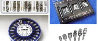
To perform tooth extraction surgery, special tools are used: forceps and elevators (levers). The forceps differ in their intended purpose:
- for removing teeth of the upper or lower jaw;
- for removing teeth with or without a crown;
- for tooth extraction in adults and children;
- for removing teeth with limited mouth opening.
All of them have elements such as cheeks, handles and a lock. The use of elevators is based on the implementation of the lever or gate principle.
Elevators can be used as the main tool for removing teeth with damaged crowns (roots), impacted and dystopic, and also as an auxiliary tool. In these cases, the first stage of dislocation is carried out using an elevator, and the dislocation and extraction of the tooth are completed with forceps.
