A beautiful, attractive smile is very important for communication in the modern world. When meeting, it is customary to smile, at least out of politeness, and in moments of joy we involuntarily laugh with our mouths open.
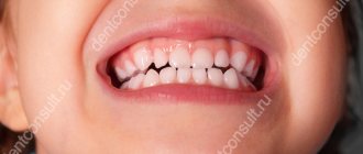
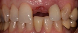
Unfortunately, various pathologies can lead to a noticeable change in the color of one of the central teeth. This significantly reduces the aesthetic perception of the entire smile as a whole. Even one tooth of a different shade involuntarily attracts attention.
Tooth discoloration is a pathological change in color in the area of enamel, dentin, or both. There are two types – external and internal.
External discoloration
The reason is a pigmented plaque that forms on the roughness of the enamel, in microscopic cracks or anatomical cavities. Discoloration of teeth in this case is a consequence of consuming coloring products (tea, coffee, red wine, soda, etc.), smoking, and a reduced level of hygiene. Professional teeth cleaning at the dentist with mandatory enamel polishing, teaching the patient the rules of personal hygiene, and limiting the intake of coloring products will help get rid of it. In some cases, it is possible to use abrasive preparations.
Repairing a chip using composite material restoration
A chipped tooth is a common problem that our patients come to us with. Specialists at the Moscow Region clinic eliminate this defect in several ways, one of which is using a composite material.
The procedure has the following positive aspects:
- The use of modern composite materials that imitate dentin and enamel in the desired color range and transparency.
- The procedure lasts no more than 1.5 hours and takes only one visit to the dentist.
- Affordable price.
- No dental laboratory services required.
- The tooth is processed minimally; only carious tissues and old fillings are removed; healthy tissues are not trimmed.
- If minor defects arise after restoration, they can be easily and quickly eliminated.
However, the method has several disadvantages. Firstly, composite restoration requires systematic monitoring by the dentist. Over time, the shape and color of the corrected surface changes and needs to be adjusted. Composite materials quickly wear out and lose their original shine and color; roughness appears on the surface, which is filled with plaque. Secondly, the composite has low strength; five years after use, cracks appear, which disrupts the adhesion of the material to the tooth. As a result, chips are formed and the risk of re-development of caries. And thirdly, the restored area may differ from the rest of the teeth due to the difficulty of choosing a composite that is visually similar to dental tissue.
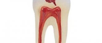
Internal discoloration of teeth
In this case, the color change occurs at a deeper level in the enamel or dentin of the tooth. Whitish erosive spots form on the teeth, which can turn yellow and black over time.
The cause of dental discoloration in this case is the penetration of certain substances into the body. In particular, the increased content of fluoride in drinking water (fluorosis) and the use of tetracycline antibiotics affect it. Violation of mineral and protein metabolism causes enamel hypoplasia - its absence or insufficient development. Age-related changes in the body also lead to thinning of the enamel and, in combination with external factors, yellowing and darkening can develop.
We list the local causes of discoloration:
- “dead tooth” or depulpation (nerve removal). Over time, the tooth becomes darker, grayer, yellower than vital,
- previous root canal treatment using the resorcinol-formalin method. Over time, the crown turns pinkish-brown,
- trauma during the growth of the dental system with the penetration of blood into the mineralization zone,
- Improperly performed orthodontic treatment can damage the ligamentous apparatus (periodontium) of the tooth, resulting in trophic disturbances and loss of vitality.
Types of discoloration and causes of its occurrence
Discoloration of teeth can be external and internal. In the first case, the problem is caused by the formation of pigmented plaque on the tooth enamel. It is formed:
- when smoking,
- improper or insufficient oral hygiene,
- consumption of products with coloring properties - colored carbonated drinks, tea, coffee, carrots, beets, etc.
Internal discoloration of teeth is characterized by changes deep in the enamel and dentin. Changes in the shade of enamel can lead to:
- drinking water with a high fluoride content,
- disturbance of protein or mineral metabolism in the body,
- long-term use of tetracycline antibacterial drugs,
- pulp removal,
- use of resorcinol-formalin treatment method.
Local discoloration in the area of one or more teeth can also be caused by incorrect bite correction and jaw trauma during the period of active growth.
Treatment of dental discoloration
Aesthetic dentistry has various methods of treating discoloration:
intracanal bleaching – will help restore the light shade of a pulpless tooth,- in-office teeth whitening – brightens and evens out the color of enamel by several orders of magnitude,
- home teeth whitening with trays - can be done at home, but under medical supervision,
- installing veneers on teeth in the smile area will help eliminate discoloration due to internal causes of the body,
- installing a crown made of pressed ceramics completely imitates a living tooth in a row and will hide any defect.
Treatment of dental fluorosis
Patients with enamel damage suffer from aesthetic discomfort and the inability to chew food normally. In order to restore teeth and their color, the center’s staff sometimes have to make non-standard decisions. Their implementation is possible thanks to the availability of modern equipment in the clinic and the professionalism of doctors who strive to make the treatment of dental enamel hypoplasia as effective as possible.
Patients of the center receive a full range of services, including diagnosis of the causes of the disease and preventive measures. You can go to the clinic with any degree of illness, confident that the dentists will take all measures to gently and quickly correct defects.
How to treat discoloration at home
You can try to remove pigmented yellow and brown plaque with tooth powder or baking soda. The main thing here is not to overdo it and not damage the enamel layer. Highly abrasive pastes must be used very carefully - paste with charcoal can erase the enamel or leave scratches on it. It is recommended to use such toothpastes no more than 2-3 times a week.
There are ways to combat discoloration online using lemon juice and hydrogen peroxide. Most likely, such methods will not harm the teeth, but the effect will be insignificant.
More effective methods are based on a high concentration of hydrogen peroxide or urea. These are various sticks, strips, mouth guards with gel of dubious production from the mass market. These medications often cause tooth pain and sensitivity. These methods are not suitable for treating internal discolorations; you should visit a dental clinic and consult a dentist.
A method for masking intense discoloration of anterior teeth using direct composite restoration
Most patients come to the dentist to improve the aesthetics and appearance of their smile. As a rule, the dentist can improve these indicators using means such as whitening, direct or indirect dental restoration. But there are situations, for example, intense discoloration of the hard tissues of the tooth due to previous endodontic treatment or trauma, in which it is quite difficult to achieve a good result. Moreover, intracoronal teeth whitening often causes relapses and gradually the dark shade of the tooth returns.
One way to solve such problems is direct composite tooth restoration using camouflage shades of the composite to cover discolored areas of the tooth. This article describes a solution that will help the dentist cope with the camouflage of even significant dental discoloration.
Clinical case
The patient, 30 years old, came to the clinic due to a violation of the aesthetics of the smile, the presence of darker teeth 12 and 11. According to the patient, 11 years ago, after an injury to two central teeth, they were endodontically treated, and then over the course of 5 years, darkening of the teeth gradually occurred and From that moment to this day, the patient is embarrassed to smile.
The examination revealed a significant change in color of teeth 1.1 and 1.2 (shade A6) compared to the remaining teeth (shade A2), as well as a violation of the marginal fit of old restorations (Photos 1, 3). Radiography showed acceptable quality of obturation of the root canals of both teeth (Photo 2).
Photo 1
Photo 2
Photo 3
After an examination and conversation with the patient, a decision was made on direct aesthetic restoration of teeth 11 and 12. Considering that pigmentation affects not only the crown of the tooth, but also its root, it would not be possible to avoid the dark tissues of the tooth being visible through the gums, which the patient was informed about. However, this transillumination will not significantly affect the aesthetics of the smile, because The patient's midline smile and gums are almost invisible during communication.
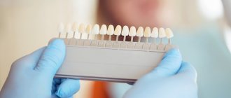
The shade of the A2 restorations was determined based on intact teeth 2.1 and 22. The first stage of work was direct modeling of the shape of the teeth in the oral cavity (mock-up) to recreate the optimal shape of future restorations and taking an impression using a silicone key (Photo 4, 5).
Photo 4
Photo 5
Due to the vestibular position of tooth 22 and the fact that food constantly gets between teeth 21 and 22, the patient asked to change the shape of tooth 12 during restoration and make the interdental contact between 11 and 12 more dense. This is why the shape and position of teeth 12 and 22 will be slightly different upon completion of the restoration.
Upon completion of the first stage, a retraction thread (Ultrapak 00, Ultradent) was inserted into the periodontal sulcus and the hard tissues of the teeth were prepared. Our task was to free teeth from old restorations, carious tissues and create a vestibular space of about 0.7 mm to mask discoloration and restore teeth (Photo 6).
Photo 6
After preparation, the teeth were isolated using a rubber dam (Photo 7). The restoration was performed sequentially, starting with tooth 11. Orthophosphoric acid gel was applied to the tooth tissue for 30 and 15 seconds on the enamel and dentin, respectively, and then washed off with water for 30 seconds (Photo 8). At the same time, the adjacent tooth was covered with Teflon tape to prevent chemical damage to healthy enamel by phosphoric acid. The Single Bond Universal adhesive system was applied to the dried tooth surface for 20 seconds (Photo 9). After drying the adhesive, it was polymerized for 20 seconds vestibular and palatal (Photo 10). The first portions of the composite material were applied from the palatal surface of the tooth under the control of a silicone key, Filtek Z550 composite in opaque shade OA2 (camouflage shade) was used, this helped make the restorations more natural and vibrant. The material was applied in two layers, each of which polymerized for 30 seconds.
Photo 7
Photo 8
Photo 9
Photo 10
Next, using a silicone key, the restoration of the palatal wall of the tooth was carried out using Filtek Ultimate material in enamel shade A2E (Photo 11, 12).
Photo 11
Photo 12
After polymerization of the palatal wall, the proximal walls of the tooth were restored with the same shade of composite. In this case, ultra-thin contoured metal matrices were used (Photo 13). After this, Filtek Z550 OA2 was applied to the discolored area with a layer of 0.5 mm and polymerized for 20 seconds (Photo 14). The dentinal body of the tooth was restored with Filtek Ultimate dentine shade A3D (Photo 15). Vestibularly, the entire surface of the tooth is covered with enamel shade A2E (Photo 16). Upon completion of the restoration of tooth 11, the restoration of tooth 12 was carried out in a similar manner, after which both restorations were pre-ground and polished (Photo 17)
Photo 13
Photo 14
Photo 15
Photo 16
Photo 17
The final processing of the composite was carried out 10 days after complete restoration of the gum tissue. Upon completion of the work, the patient was given recommendations and restoration of tooth 21 was carried out. The photographs (Photos 18, 19) show the appearance of the finished restorations.
Photo 18
Photo 19
conclusions
Even in cases of significant discoloration of the tooth structure due to previous root canal treatment or trauma, the combination of the camouflage properties of Filtek Z550 OA2 and the aesthetics of Filtek Ultimate can easily restore the beauty of a smile (Photo 20). This technique is simple and applicable in clinical practice and provides a predictable restoration result.
Photo 20
Source: Styleitaliano
How is the treatment carried out?
- The patient comes for a consultation. The doctor conducts an examination and, if necessary, prescribes x-rays.
- A plan is drawn up to restore damaged teeth and calculate the cost of the procedures.
- Preparation of materials.
- Treatment.
- Carrying out a preventive examination.
- Recommendations for home care with the selection of hygiene products.
Restoring teeth without installing dentures and jaw surgeries is the reality of modern dentistry. The restoration is carried out in the shortest possible time and painlessly, and the effect satisfies all the patient’s needs and gives him a beautiful snow-white smile.