Safety of X-ray diagnostics
The standard of safe radiation exposure is regulated by the federal law “On Radiation Safety of the Population”. Article 9 of this law “State regulation in the field of radiation safety” states that the average effective dose for the population per year is 0.001 sievert (1 millisievert). In this case, the dose received during one cone-beam computed tomography is 0.09 millisieverts. Accordingly, at least 10 CT scans of teeth can be safely performed per year.
For comparison, during air travel the radiation dose is on average 0.08 millisieverts. The dose of so-called natural radiation is 2.4 mSv. It consists of the effects of cosmic and solar rays, the influence of atmospheric radioactive atoms (radon), radiation from soil and building materials, and food radionuclides. Thus, performing a dental CT scan is absolutely safe for your health.
Cone beam computed tomography (CT/CBCT) using the latest generation SIRONA device
Dental tomograph Sirona (Germany) is an effective equipment for creating three-dimensional X-ray images of the dentition and jaw bones.
Patients are often surprised: why do dentists send them to do a cone beam computed tomography (CBCT) if they already have a panoramic photo in which all their teeth are visible? Of course, the first version in the patient’s brain is the intention on the part of the doctor - to earn more money, and the second is the doctor’s inability to understand and make a diagnosis using a panoramic image (orthpantomogram).
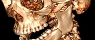
Unlike a panoramic image, CBCT is a three-dimensional image. And since we are three-dimensional objects, we need to look at our teeth and jaws from all sides. With CBCT, the doctor can see the image not only from front to back, but also from above, below, from the side and from any angle. He can twist his jaws in all directions, look into any place in the jaw, evaluate the parameters of the bone, see if there is an inflammatory process hiding behind the root of the tooth, etc.
We often see things on CBCT that are not even close to visible on a flat X-ray.
It's all about the special design of the computed tomograph: during the exposure it takes not one, but a whole series of x-rays of the jaws from different angles. These images are then transferred to a computer program, which processes them and ultimately creates a 3D model of the patient’s dental system. The advanced German tomograph ORTHOPHOS SL 3d implements a number of technological solutions. The anatomical scanning field allows you to cover the entire dental system along with the nasal sinuses. The metal artifact suppression program automatically eliminates interference and ensures high quality and accurate images.
The X-ray machine transmits the scan results to the monitor. The panoramic image in combination with the volume control program allows you to analyze the structures of bone and soft tissue in the treatment area. Based on the analysis, the doctor can make an accurate diagnosis and plan effective treatment.
*Therapy. In therapeutic dentistry, CBCT allows us to identify acute inflammatory processes not only in teeth, but also in surrounding dental tissues; hidden carious process that is not visible during a routine examination of the oral cavity
*Surgery and Implantology. Often, even for simple tooth extraction, CBCT may be necessary, because the surgeon will see in advance how many roots the tooth has, how and where they are directed, how they twist, and what important anatomical structures they border on. Nowadays, it is completely impossible for implant surgeons to work without CBCT. Planning the operation includes precise positioning of the implant in the bone even before the operation, and this is impossible to do in only one plane, because we are three-dimensional objects. On CBCT, the surgeon clearly sees not only the height and width of the bone, but also its thickness and quality, as well as the location of adjacent teeth and the sinuses and nerve canals located in the maxillofacial area that pass through the jaw bones.
*Orthopedics. This image allows you to accurately draw up a treatment plan, assess the condition of the supporting teeth and the quality of the treatment performed, if any, identify teeth with inflammatory foci, preventing complications after orthopedic intervention and the risk of altering the entire denture design in the future.
*Not a single highly professional orthodontist today will undertake to correct a patient’s bite without an accurate analysis of the situation, without special preliminary calculations when planning orthodontic treatment. Calculations are made using special programs and without exact knowledge of how the roots of the teeth are located, whether all the teeth have erupted, whether there are “surprises” in the jaw in the form of undone and supernumerary teeth, etc. it will be impossible to do.
When performing X-ray procedures, a radiation dose of 1000 μSv (1000 microsieverts or 1 millisievert) per year is considered relatively safe.
CBCT of the dental system is performed with a radiation dose of 45-60 μSv. That is, you can take up to 20 such pictures a year without harm to your health. For comparison, during one transatlantic flight a person receives a radiation dose of 80 μSv. Also, the procedure is absolutely safe and is indicated for children from a very early age.
Based on the research data, it follows that computed tomography is an indispensable assistant in the process of diagnosing diseases of the teeth and jaw bones ; it allows you to obtain the most accurate and informative picture, make the correct diagnosis and begin the necessary treatment, without wasting time trying various treatment tactics.
This type of diagnostic test has been widely used by all PRESIDENT dentists in South Butovo since 2010.
The procedure for performing CT scans of teeth in Moscow at Dental Guru clinics
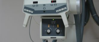
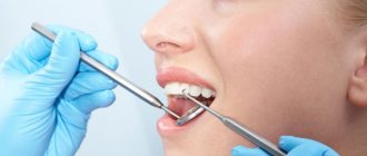
Computed tomography of the jaw, Moscow. Any Dental Guru clinic has a full range of X-ray diagnostics, including a computer tomography machine. No special preparation required. Before the examination, all metal jewelry and items of clothing (earrings, chains, piercings in the dentofacial area, hairpins) are removed. A lead vest or apron is worn to protect against X-ray radiation. To fix the head, the chin rests on the stand and the forehead on the bracket. During the examination, the sensor scans the dentofacial area. At this time, you should refrain from any movements, including swallowing. The patient sits or stands and the examination takes 30 seconds. At Dental Guru clinics, cone beam computed tomography is performed using a multifunctional Rayscan device.
What are the differences between MSCT and CT?
Among traditional diagnostic techniques based on X-ray imaging, multislice computed tomography occupies a special position. The fluorographic method is considered informative in the study of obvious pathologies. The disadvantage of this type of diagnosis is the layering of the tissues being studied in photographic images of the abnormal area. Layer-by-layer scanning of dense tissue structures can only be obtained during MSCT. The difference from CT is only in the technical characteristics of the equipment used.
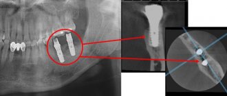
Implant planning using CT
A CBCT scan is necessary before dental implantation. Three-dimensional examination reduces the risk of the procedure, significantly improves the quality of implant installation, and shortens the patient’s rehabilitation period. CT scan shows the condition of the bone tissue, possible inflammatory processes, the location of the maxillary sinuses, nerves and blood vessels, helps to create a model of the implant, calculate the depth of its immersion in the bone and the angle of inclination, and the optimal physiological load on the implant.
Why is 3D examination important before implant placement? An artificial root will stand long and firmly if it is surrounded on all sides by bone tissue. And here an individual approach is needed. The structure of bone tissue varies in shape, width, height, density, and possible defects. For longevity, it is important to choose a site where bone volume and density are sufficient. You need to choose the angle of inclination for acceptable aesthetics. In order to determine the required installation depth, you need to know the location of the nerves and blood vessels. Thus, it is possible to obtain a surgical navigation template for implantation for the most accurate virtual planning.
Performing a CT scan with a radiopaque tray when planning a surgical template
Most patients who come for implants already have fillings, crowns, veneers or other dental restorations. All these materials can produce additional glare during CT scanning, so-called artifacts. To minimize them and ensure the accuracy of the CT examination, this procedure is performed using special radiopaque trays. They can be standard (if there are supporting teeth) or individual (when there are very few or no teeth and guidelines are needed for further work).
In these cases, special radiopaque trays are placed in the oral cavity, a CBCT (cone beam computed tomography of the jaws) is performed, then the trays are removed and sent to a dental laboratory. It is thanks to this type of research that it is possible to achieve accurate results and guarantee that the implant is installed in the area in which its location will be best. If implantation using surgical templates is planned, a CT scan with a radiopaque tray must be done. Before implantation with immediate loading, this study allows the fabrication of a temporary prosthesis to be worn before surgery.
If you do not do a CT scan before implantation, there may be:
- Loss of sensitivity and pain in the teeth and jaw.
- Injury to the gums due to incorrect calculation of the permissible load.
- Bite defects.
- Loosening and loss of the implant.
- Increased costs to correct problems, including installing a new structure.
This cannot happen in Dental Guru clinics. We care about the interests of our patients. All clinic specialists have extensive experience and high qualifications, confirmed by diplomas and certificates. CT scans of the upper and lower jaw are equally important.
Computed tomography of the upper jaw
To diagnose the condition of the maxillary sinuses before implantation, it is important to do a CT scan of the upper jaw. After tooth extraction, thickening of the mucous membrane in the maxillary sinuses often occurs; there may also be manifestations of sinusitis, a tumor process, the presence of a cyst or polyp. A CT scan of the jaw will show whether there is perforation of the maxillary sinus due to the protrusion of the root or crown of the tooth. The term “odontogenic sinusitis” means that the cause of the disease is in the tooth. The bony septum between the oral cavity and the maxillary sinus is quite thin, so infection from dental tissue can cause sinusitis. Therefore, for implantation to be successful, it is necessary to promptly identify and treat possible diseases and eliminate the inflammatory process.
Computed tomography of the lower jaw
A CT scan of the lower jaw, as well as the upper jaw, shows the condition of the teeth and bone tissue: possible diseases, inflammatory processes, granulomas and cysts. A CT scan of the jaw is important to identify hidden processes and timely initiation of treatment. A CT scan of the upper and lower jaw is usually done at the same time, but some clinics can do a CT scan of the upper and lower jaw teeth separately. The lower jaw bears the main load when chewing and speaking; chronic subluxation of the lower jaw is quite common. CBCT of the temporomandibular joint helps to make a diagnosis and distinguish subluxation from dislocation and fracture.
Benefits of the survey
Diagnostics using CT is safe for the Patient, which compares favorably with other methods, for example, classical x-rays. Benefits also include:
- the detail and quality of the resulting image is high, it only takes a couple of seconds to obtain;
- obtaining a three-dimensional computer model;
- detailed study of tissues, jaw apparatus, mucous membrane and bone structure;
- identification of hidden pathologies not visible in other ways;
- assessment of the degree of occlusion, malocclusion;
- detection of foci of inflammation and neoplasms.
Panoramic photo, CT scan of teeth in Moscow, price
For a CT scan of the jaw, the price in Moscow starts from 3,500 rubles. If you search online for “computed tomography of teeth price Moscow” or “computed tomography of jaw price”, then the average price of this examination in the search will be approximately 4,500 rubles. Dental Guru is currently running a unique promotion - a dental CT scan costs only 1,900 rubles. If the question of where to get a CT scan of a tooth is relevant to you, come to Dental Guru. When referred by a doctor, we guarantee non-interference in the patient’s treatment.
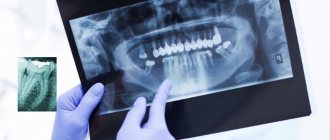
The doctor may also order two-dimensional examinations, such as spot and/or panoramic images. The targeted photograph shows the condition of 3-4 adjacent teeth. An OPTG (orthopantomogram) shows the condition of both jaws, including soft and hard tissues, malocclusions, impacted teeth and tumors, but you need to remember that this is a two-dimensional (single-view) image.
Indications
CT is recommended for use in the following situations:
- identification, precise determination of the position and extent of injuries;
- detection of inflammation, caries;
- study of structure;
- assessment of malocclusions;
- search for formations, cysts, tumors;
- checking for impacted units;
- diagnostics, assessment of the condition before prosthetics, installation of implants;
- preparation before surgical treatment, installation of orthodontic appliances.
Features of the procedure
The diagnostic process using computed tomography includes the following steps:
- Before the procedure, the Patient must remove metal and other jewelry to prevent malfunctions and distortions;
- to protect and reduce the degree of exposure, it is necessary to wear a special vest, all strangers leave the room with the equipment;
- the chin is fixed on a special stand; you cannot move or talk during the examination;
- After switching on, the scanner begins to move around the head, transmitting the resulting three-dimensional image to the computer.
The procedure takes no more than a minute, and the Patient does not feel pain or discomfort. No extensive preparation or anesthesia is required, and images can be taken immediately after the examination is completed.
Contraindications, how often can it be done
The examination has virtually no contraindications, but it is not recommended during pregnancy. Contraindications also include the period during breastfeeding; the procedure is not performed for small children. If a contrast scan is required, you should first check whether the Patient has any allergic reactions to the substances used.
The examination does not pose any danger, but it is recommended to do it no more than 12-14 times a year. Radiation exposure is minimal, for example, with standard fluorography the danger is much higher.
Evaluation of the results obtained
After performing the diagnostics, the Patient receives the results recorded on a flash drive or disk. Additionally, you can print the images or get a transcript if necessary. When assessing, the specialist takes into account the following aspects:
- condition of tissues, internal structure, jaw;
- assessment of the condition of canals and roots;
- occlusion anomalies, possible defects;
- tumors;
- retinalization of individual units.
With the help of CT, you can get the most complete picture, which is necessary for making a full, accurate diagnosis and choosing a treatment regimen.
Prices
The diagnostic procedure using CT costs from 1,500 to 500 rubles. The cost depends on the center where the services are provided and the diagnostic option:
- a photograph of a section of three teeth – 1,500 rubles or more;
- jaw – 2500;
- both rows (one side only) – 3500;
- full panorama – 4000;
- paranasal sinuses – 3000;
- full examination – 5000.
Additionally, you may be charged for recording data on a disk or flash drive, or for printing individual or several images. Therefore, if research is necessary, it is recommended to check with your doctor which images will be taken and for what purposes.
Commentary by the chief physician of the Dental Guru clinics
To understand the clinical picture, it is necessary to make an accurate diagnosis. Dental Guru dental clinics have all the necessary diagnostic equipment for x-ray diagnostics. These are multifunctional devices with the ability to take pictures of the temporomandibular joint, cephalometry, and a digital radiovisiograph for targeted imaging. We take a panoramic photo of the teeth, CT scan of the upper and lower jaw, the price is now unprecedentedly low. We are always glad to see you in our clinics. Call and make an appointment with specialists!
Where is hardware diagnostics carried out?
Not every district clinic provides consultations in this area, which is associated with the high price of equipment and consumables. To quickly find the nearest center, a single city service 36go works. All computer scanning clinics are presented in list form, distributed according to ratings and prices for procedures. In addition to general and contact information, on the website you can find reviews from clients who have undergone diagnostics at the selected medical institution. If you wish, you can leave your own comment about the activities of the center.
To make an appointment for a consultation or session, call the hotline located at the top of the page. Operators provide complete information, answer remaining questions, and provide addresses and prices. Booking a time for the procedure and consultation are provided free of charge.