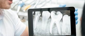
A dental x-ray is a detailed image of the teeth, bones and soft tissues around them, allowing us to identify various dental and oral problems. X-rays can show cavities, hidden dental structures (such as wisdom teeth), bone loss and other abnormalities that cannot be detected during a visual examination and a routine dental consultation. Dental X-rays are usually performed annually. It is also actively used for follow-up monitoring of teeth after treatment. X-rays may be prescribed more often if your doctor monitors the development of a dental problem directly during treatment.
Why do you need dental x-rays?
After the examination, the dentist will refer the patient for an X-ray examination:
- A targeted x-ray of a diseased tooth helps to clarify the degree of caries, distinguish pulpitis from periodontitis, find hidden carious cavities, and clarify the condition of root canals.
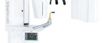
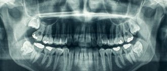
- Panoramic x-ray of teeth (orthopantomogram) - gives a detailed picture of the condition of all teeth, jaws, maxillary sinuses, temporomandibular joints. This study is the “gold standard” in dentistry, as images help diagnose hidden pathologies and inflammatory processes.
Is there an alternative to CT
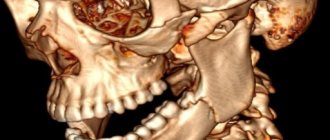
There are many other diagnostic imaging methods (x-ray, orthopantomogram, ultrasound, etc.), but only CT provides the possibility of highly accurate, separate images of all types of tissue at different angles and to different depths. Although a panoramic dental photograph remains an equally important diagnostic tool for a dentist today, it can only provide a general overview. In turn, a 3D tomogram allows you to obtain not a single flat image of the jaw, but a whole series of sequential multiplanar images in different projections and without the distortions inherent in a panoramic image.
Example:
due to the different density of bone structures exposed to X-ray radiation, it is impossible to see less dense bone in a 2-dimensional image; accurate information is provided by a 3- D image of the teeth.
Indications and contraindications for the procedure
An X-ray is necessary in cases such as:
- severe toothache in the jaw when biting or chewing food;
- suspicion of inflammatory processes, cysts, granulomas, abscesses, deep caries;
- checking the quality of root canal treatment.
Diagnostics are also prescribed before orthodontic treatment, dental prosthetics, and x-rays of healthy teeth are taken before eliminating bite problems.
There are few contraindications to the procedure. These are diseases associated with immunodeficiency, pregnancy, pulmonary hemorrhages, and mental disorders.
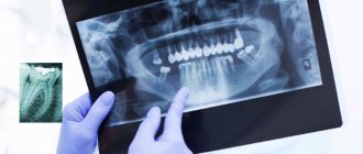
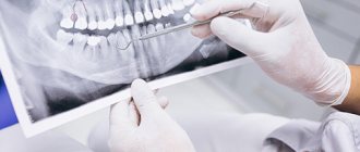
Analysis of targeted dental images –
Analyzing X-rays is not difficult if they are of good quality.
Almost any patient will be able to see signs of periodontitis or cysts in the image, and will also be able to determine how well his root canals are filled. All you need here is skill. But it is worth remembering that absolutely everything cannot be diagnosed using X-rays, for example, inflammation of the nerve of a tooth. You can diagnose from the image -
- caries (but not in full),
- periodontitis (granulomas, cysts),
- quality of root canal filling after treatment of pulpitis and periodontitis,
- perforations and root cracks,
- fragments of instruments in root canals,
- presence of periodontal pockets...
1) A group of images indicating the development of inflammation (periodontitis) at the apexes of the roots of previously untreated teeth. In this case, you will always see a clear or vague darkening at the apex of the tooth root, which can be of different sizes and shapes.
2) A group of photographs taken after root canal filling. The first 2 pictures (Fig. 14-15) show what well-filled root canals look like. The following images show poor quality treatment and complications that arose (read the descriptions in each image).
Price for x-ray (image) of teeth
You can get an X-ray of your teeth in Moscow, find out prices, and make an appointment with a dentist by calling +7. Experienced specialists at Zubastik Dentistry will perform the necessary research and interpret the results in a high-quality manner.
In modern dentistry, increased attention is paid to dental x-rays. Radiography makes it possible to assess the general condition of the dentition, accurately determine the problem of a specific unit, and develop an optimal treatment program. You can take an X-ray of a tooth in Moscow at any Zubastik dental clinic. The procedure does not require special preparation.
Sight image of a tooth –
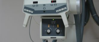
A targeted photograph of a tooth can be recorded either on photographic film or using a special intraoral sensor that detects X-ray radiation and transmits the image to a computer screen (such a device is called a radiovisiograph or simply a visiograph - Fig. 5). In both cases, an X-ray machine is used as a radiation source (Fig. 4), i.e. the only difference is in the way the image is captured - either on X-ray film or using a digital sensor.
Taking an image using a visiograph -
Digital vs Film Photography: Pros and Cons
Targeted dental x-rays using film were once the only examination option in clinics. It must be said that film photographs have a number of disadvantages that have significantly reduced their use. They require expensive consumables (film, reagents), time to develop photographs, there are difficulties with storing photographs, and over time they fade and are lost. There are also differences in patient safety.
Even modern X-ray films require 4-8 times the radiation dose compared to digital X-ray sensors. For example, the radiation dose to a patient for 1 film image is 10-15 μSv (microsieverts), and for a picture on a visiograph it is on average 1-3 μSv (this dose corresponds to the background natural radiation received by each person in 1 day).
The patient's exposure time when using film X-ray is 0.5-1.2 seconds, and using a digital visiograph sensor - 0.05-0.3 seconds. It is precisely by reducing the required exposure time when using a radiovisiograph that the radiation dose is significantly reduced. Thus, in one day of treatment at the dentist you can take no more than 3 film photographs and 5-6 digital photographs. And as you will see below, the use of a visiograph in some cases even allows you to take an X-ray of a tooth during pregnancy, but in compliance with all safety rules and for urgent indications.
How to take a picture of a tooth using a visiograph: video
Important: always try to take digital photos and inform them in advance that you want them saved to a flash drive. Firstly, then you will always have the pictures at hand, and you can always show them to another doctor. Secondly, the photographs taken for control after treatment will be your guarantee that if you received poor-quality treatment, you will always be able to prove it (the clinic will no longer be able to lose your photographs and rewrite the medical record).
Thirdly, if a digital image is printed on a printer, then the quality of the image will depend not only on the quality of the digital image, but on the resolution of the printer (rare clinics have printers that print in high resolution). Therefore, a photo in digital format will have better quality than a photo printed on paper.
Why do they send you for a dental x-ray?
The comfort and effectiveness of treatment of dental pathologies largely depend on an accurate diagnosis. An X-ray of a diseased or damaged tooth is prescribed by a doctor after a visual examination or during therapy. X-ray studies can be local or panoramic:
- A targeted spot X-ray of a problem tooth helps to notice a cyst or tumor at an early stage of development, to detect hidden caries under a filling, an abscess, or a broken root.
- A panoramic X-ray image of all teeth, unfolded in a plane, covers the maxillary sinuses, reflects a complete picture of the state of the dental system, and allows you to quickly identify the source of the inflammatory process.
Indications and contraindications
When examining a patient at an appointment, the dentist sees only obvious pathologies in the oral cavity. However, most problems are hidden in places inaccessible to the eye - spaces between teeth, canals, roots. In many cases, it is possible to correctly respond to a patient’s complaints only by having an X-ray of the teeth in hand. Indications for radiography are:
- sharp pain in the jaw when chewing;
- change in tooth angle;
- deep caries, including under fillings and crowns;
- inflammation and bleeding of the gums;
- mobility, crowding, fan-shaped discrepancy of teeth;
- checking the quality of tooth resection and canal treatment.
Prices for x-rays of problem teeth in Moscow are low. They allow you to take all the necessary images for the provision of medical services such as prosthetics, implant installation, and bite correction.
! X-rays are not recommended during pregnancy, low immunity, bleeding, or mental disorders.
Types of dental x-rays
In the network of dental clinics "Zubastic" you can undergo the following types of x-ray examinations:
- traditional orthopantomography with recording of a detailed image of the jaws on a CD;
- intraoral bitewing radiography;
- dental computed tomography (CT) with obtaining a volumetric image of the maxillofacial area;
- teleradiography (TRG) of the skull in frontal and lateral projection.
How much dental x-rays, CT and TRG will cost can be found in the price list for services presented on the dentistry website.
Features of radiography
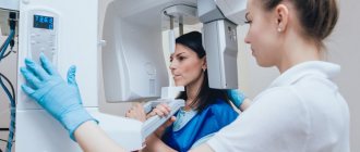
- All types of X-ray examinations in the clinic are carried out in a specially equipped room.
- To protect vital organs, the patient is wearing a special apron with lead inserts.
- The radiologist gives concise, clear instructions on how to behave during the procedure. Offers to remove metal jewelry.
- To obtain clear images, it is not recommended to move. At the request of the specialist, you need to hold your breath for a few seconds.
- In terms of time, the shooting process takes 1-2 minutes, transillumination – no more than 2-3 seconds. The sequence of the procedure depends on the type of x-ray.
When creating a panoramic image, the subject's head is fixed with a special device. The patient clamps the plate placed in a disposable cover with his teeth and remains motionless while the functional part of the X-ray machine rotates around his head.
X-ray of teeth in dentistry Zubastic: advantages
Expert X-ray equipment and highly qualified clinic specialists who are proficient in advanced techniques allow examinations to be carried out without risk to the patient’s health. We offer the best quality/price ratio for dental x-rays in Moscow, ensuring the safety of the procedure and quick results. Interpretation of CT images using artificial intelligence guarantees the complete elimination of diagnostic errors.
A good dental x-ray is the main source of reliable information about the condition of the dental system. It makes it possible to start therapy at an early stage of the disease, speed up treatment, and avoid complications and unforeseen consequences.
Is it possible to take a dental x-ray during pregnancy?
Is it possible to take a photograph of a tooth during pregnancy? This issue is regulated by the recommendations of SanPiN 2.6.1.1192-03. These recommendations do not prohibit taking dental x-rays during pregnancy, but it is strongly recommended to use x-rays only in truly necessary cases, for example, in case of acute pain and the provision of appropriate emergency care.
It should be noted that over the past 20 years, the radiation doses received by patients with 1 x-ray of the teeth have become tens of times less, thanks to the advent of radiovisiographs and ultra-sensitive photographic films, which require significantly lower x-ray radiation (24stoma.ru). Therefore, the risks of fetal pathologies have decreased significantly in recent years.
Of course, if possible, X-ray examination of teeth should be avoided, but there is nothing terrible about this today, because The radiation dose of 1 image on a radiovisiograph is approximately equal to the radiation dose of any person to natural background radiation in 1 day. In addition, the irradiation time on the visiograph will be only 0.05-0.3 seconds, which, if protective measures are observed (lead apron), can significantly increase the safety of the procedure.
Additionally, it is worth considering the following recommendations –
It is better not to do a dental x-ray during pregnancy in the early stages, because at this most important time for the laying of organs and tissues of the fetus. And if x-rays are taken, then it is in the second half of pregnancy, because the risks to the fetus during this period are significantly reduced. Please note that you can only take pictures using a modern digital radiovisiograph of the latest generation, because Their radiation dosages are significantly lower than those of outdated digital radiovisiographs and, even more so, film devices.