Full text of the article:
Working with diseases of the internal organs is especially difficult because they cannot be seen. Previously, doctors had to treat patients literally “blindly,” since it was possible to examine a person’s internal organs only during surgery. Nowadays, doctors don’t have to pick up a scalpel; various types of scans help make a diagnosis. However, patients are wary of this type of examination. This is due to the high cost of some procedures and the fear of radiation. Let's try to figure out what the features of certain scans are and when to resort to them.
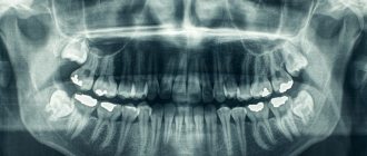
X-ray
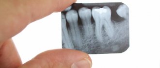
The oldest and most common method of visualizing the human body. X-rays are used everywhere, from surgery to dentistry. The method is simple and clear: a person is irradiated with special rays that easily pass through soft tissues and linger in hard ones. Thanks to this principle, an image is transmitted to a photographic film or sensor located on the side opposite the ray source, and radiography or fluoroscopy is available to the doctor.
Main advantages
such an examination: speed and cost. Almost all hospitals are equipped with X-ray machines; the procedure is quick and inexpensive.
Main disadvantages:
exposure and image quality. When performing radiography, the patient is irradiated, and the picture is two-dimensional. The doctor can hardly see the internal organs individually, since their shadows overlap each other. It is also impossible to see the cartilage tissue and brain in detail. The cartilage practically does not block the rays, the brain is securely closed by the skull. X-rays are not suitable for their examination.
It will be most effective to carry out radiography for damage to bones, joints and teeth.
Expert opinions
Ilya Gipp, Ph.D., Head of MRI-guided therapy:
— Many of these devices can be used for treatment. For example, a special installation is attached to an MRI machine. It focuses ultrasound waves inside the body, increasing the temperature in a targeted manner, and burns out tumors - for example, uterine fibroids.
Kirill Shalyaev, director of the largest Dutch manufacturer of medical equipment:
- What seemed impossible yesterday is reality today. Previously, during CT scans, a drug was administered to slow down the heart. The newest CT scanners make 4 rotations per second, so there is no need to slow down the heart.
| What radiation doses do we receive* | ||
| Action | Dose in mSv** | Over what period of time will we receive this radiation in nature? |
| X-ray of a hand | 0,001 | Less than 1 day |
| X-ray of a hand using the very first machine in 1896. | 1,5 | 5 months |
| Fluorography | 0,06 | 30 days |
| Mammography | 0,6 | 2 months |
| Mammography with MicroDose characteristic | 0,03 | 3 days |
| Whole body CT scan | 10 | 3 years |
| Live in a brick or concrete house for a year | 0,08 | 40 days |
| Annual norm from all natural radiation sources | 2,4 | 1 year |
| Dose received by liquidators of the Chernobyl accident | 200 | 60 years |
| Acute radiation sickness | 1000 | 300 years |
| Epicenter of a nuclear explosion, death on the spot | 50 000 | 15 thousand years |
| *According to Philips **Microsievert (mSv) is a unit of measurement of ionizing radiation. One sievert is the amount of energy absorbed by a kilogram of biological tissue. | ||
Fluorography
Another type of examination that all residents of our country regularly undergo. Fluorography was “invented” almost a hundred years ago. This is a kind of accelerated radiography. Scientists proposed photographing a screen with an image obtained from radiography. This made the procedure faster and more widespread. Screening tests began to be administered to everyone in order to detect latent pulmonary tuberculosis.
Main plus
The procedure is fast,
the main disadvantage
is the image quality. The patient also receives a dose of radiation, and the doctor has a rather blurry picture, so fluorography is recommended to be supplemented with questionnaires and laboratory tests for the presence of tuberculosis.
Mammography
A separate type of radiography designed to diagnose breast diseases, which is why women undergo mammography. There is no consensus on the recommended age for the procedure. Mammography helps ensure the absence of a malignant tumor with an accuracy of 89%. It is believed that women should be screened regularly starting at age 39, although some cancer societies recommend screening at a younger age.
Mammography is prescribed to diagnose breast cancer, the procedure is quick
, this is a plus, but the patient
is irradiated
, and the risk of an incorrect diagnosis remains, this is a minus. Mammography can be digital or film; digital mammography provides a clearer image.
Computed tomography (CT)
Computed tomography is also carried out on the principle of radiography, but as a result the doctor receives not a flat two-dimensional picture, but a three-dimensional image. This is achieved by simultaneously taking a large number of images, which are assembled into a single image. CT scanner sensors are highly sensitive and distinguish a huge number of shades, so the doctor can examine in detail all the bones and organs of the patient. The image quality can be further improved by injecting the patient with a special substance, the so-called “contrast”. The contrast helps to distinguish healthy tissues from altered ones and detect abnormal structures in the body, and also makes it possible to study the condition of blood vessels in detail. CT with contrast is not prescribed in every case; often a simple computed tomography is sufficient.
CT scan is done quickly
, with its help to screen for lung cancer. You can also use computed tomography directly during surgery.
Disadvantages of CT
can be considered a high radiation dose to the patient. Therefore, CT is not prescribed to pregnant women, children and overweight patients (more than 200 kilograms).
Ultrasound examination (ultrasound)
X-ray is not the only way to “look inside” the human body; another technology is ultrasound. Some animals, such as bats, use sound waves to orient themselves in space. People have also learned to use waves to solve some problems, including in medicine. A picture of the internal organs can be obtained by sending a sound wave into the human body and monitoring its return. The computer helps process the results and present them in the form of a three-dimensional picture.
The main advantage of this research method is safety.
. Ultrasound can be performed even on pregnant women; in addition, ultrasound devices are mobile and can easily be placed in the patient’s room to monitor the condition of organs and blood flow in real time.
However, ultrasound cannot provide a high-definition image, so the use of this method of research is limited, for example, gastrointestinal diseases cannot be diagnosed using ultrasound.
What is the difference between X-ray and digital X-ray?
The principle of digital diagnostics in medicine is completely similar to the method that is used for research in industry, construction and even, for example, at customs, in veterinary medicine. Modern X-rays in any advanced clinic or hospital are a special method of radiation diagnostics, during which all images are processed digitally.
Radiography of this type has many advantages, thanks to which it is successfully displacing analogue techniques from absolutely all areas of medicine. Even hospitals are striving to acquire such equipment, not to mention advanced paid medical centers, which have long forgotten about the old analog equipment.
The essence of the digital radiography method in medicine
The main and, perhaps, the most fundamental change affected the very method of capturing the image. If previously a special material with a special photosensitive coating was needed, now such a need has disappeared completely. The image is immediately generated in electronic form and is ready for viewing and processing.
During the shooting process, a special system of converters and detectors is used, which allows you to display the image immediately on a computer monitor. The sensitivity of flat panel detectors is several times higher than that of film, so a specialist can reduce exposure time and the radiation dose itself, and still get an image of exceptional quality.
Magnetic resonance imaging (MRI)
The principle of MRI is based on the property of atomic nuclei to respond to a strong magnetic field. The calculation is based on the reaction of hydrogen nuclei, of which there are many in the composition of water molecules, and the human body, as is known, consists of 60% water. When entering a magnetic field, the nuclei of atoms are oriented along it; they can be excited and the energy that they will give off when the influence weakens can be recorded, i.e. “relaxation”. Computer analysis allows you to convert the information received and determine the location, density and structure of tissues in the body.
MRI allows you to “see” cartilage, soft tissue and the human brain without causing harmful effects
, so the procedure can be performed by anyone and as many times as desired.
However, the examination takes a long time
, and closed-type tomographs can cause attacks of claustrophobia. True, there are open-type devices. MRI procedures should not be performed on people who have electrical devices (such as pacemakers) or metal implants implanted in their bodies.
MRI will be effective in studying tumors, brain and vascular abnormalities.
Digital X-ray: capabilities and practical applications in medicine
Digital technologies in radiography open up quite broad horizons for physicians. The technique is absolutely not inferior to the analog method of research and is excellent for:
- Assessing the condition of soft tissue and bone tissue after injury or any negative impact.
- Checks for various types of neoplasms. Can be used to work with both benign and malignant tumors.
- Search for foci of active inflammatory processes.
- Assessment of pathological deviations from the norm, abnormal scenarios of development of the congenital type.
- Diagnosis of the condition of the spinal column.
- Finding the location of foreign bodies if they are swallowed. This is especially true when working with young children, for whom such situations are not uncommon.
- In preparation for surgical treatment.
For example, by checking the head area, a specialist can detect metastases, lesions, strokes, or even a hematoma. When checking the lungs using digital radiography, bronchitis, changes due to oncology or tuberculosis, and fibrosis can be detected. When assessing the condition of the spine, the doctor will see a hernia, disc displacement, cancerous lesions and many other abnormalities.
Digital X-rays can be used not only to quickly make a diagnosis, but also to track progress in the treatment process. Timely assessment of dynamics allows you to quickly adjust prescriptions, avoid side effects and achieve the best possible result.
The use of digital x-rays today not only simplifies the work of specialists and reduces diagnostic costs. With its help, it is possible to detect those pathologies for the search for which previously it was necessary to use more serious invasive methods.
Scintigraphy, SPECT, PET
Perhaps these are one of the rarest procedures on our list. These examination methods are based on radiation diagnostics, but it is used in reverse. The patient is not irradiated from the outside, but a special radioactive drug is injected into him to make him “glow from the inside.” First, scientists invented and tested scintigraphy. With its help, it was possible to obtain two-dimensional images. Then research went further and single photon emission computed tomography ( SPECT)
), followed by positron emission tomography (
PET
). The difference between these methods is rather technical; they use different radiopharmaceuticals and different types of detectors that record radiation from the patient’s body.
The question arises: “Why such difficulties?” The fact is that thanks to these procedures, you can see formations in the pictures that are not visible in pictures obtained by external irradiation. Metastases and tumors can appear inside bones or organs and remain silent for a long time. The radiopharmaceutical is injected into the body and accumulates in the tissues, which allows it to “illuminate” certain areas.
Main disadvantage
of this examination method is the cost. The radiopharmaceutical is developed individually for each patient; in addition, the patient receives radiation exposure, and the procedure itself is more complex than those we described earlier. However, in some cases it cannot be avoided, for example, in cases of oncological and neurological diseases, diagnosis of heart and thyroid diseases.
X-ray and general information about it
Many health problems cannot be identified without an accurate diagnosis or x-ray. For example, when we suspect pneumonia, we often hear from the doctor: “we need to do a fluorography.” What is an X-ray examination? And why do doctors often recommend it?
Fluorography is the most popular type of x-ray.
What is an x-ray? In professional medical terms, an x-ray is a detailed study of the internal structure of the body by shining through it with x-rays and recording the image on a special film or digital detector, i.e. x-rays penetrate through the tissues of the body without damaging them and form a picture of the condition of human organs.
What does an x-ray show? In the pictures you can see (depending on the purpose of the device) various pathologies: inflammation, fractures, neoplasms (tumors), degenerative-dystrophic changes, destructive changes, developmental anomalies, etc. X-ray methods are used to examine the lungs, bones, soft tissues, and internal organs (stomach, kidneys, etc.).
After an X-ray examination, the doctor can make an accurate diagnosis in a number of complex diseases.
The color of the image of organs depends on their density. Different tissue captures X-rays differently. Bones, muscles, lungs - will appear differently; the denser the tissue, the lighter it will be on the x-ray image.
How often can an x-ray be taken?
X-rays can be preventive or diagnostic. For preventive purposes, fluorography or radiography of the chest organs is done (at least once a year), mammography (at least once every two years). Diagnostic x-rays (including fluorography) are done if the presence of any diseases is suspected; it is prescribed by the attending physician. Radiation dose limits for patients (and, accordingly, the number of x-ray procedures) for diagnostic purposes are not established (SanPiN 2.6.1.1192-03).
What norm is acceptable?
The standard for preventive radiation during preventive medical X-ray examinations is 1 mSv per year. Radiation dose limits for patients for diagnostic purposes are not established (if the doctor considers it necessary, then it is necessary). When the accumulated dose of medical diagnostic radiation to a patient reaches 500 mSv (the average dose for one x-ray examination is approximately 0.001-0.5 mSv), measures should be taken to further limit his radiation exposure if radiation procedures are not dictated by vital indications.
Is it necessary to remove radiation from the body after an X-ray examination?
After X-ray examinations, there is no need to remove radiation, since the radiation dose is negligible. Even after scintigraphy, in which a radioactive drug is injected into a vein, it is only recommended to drink more fluids.
An important role is played by high-quality modern equipment and competent work with a specialist’s apparatus.
At Sanas MC, X-rays are taken using the best new generation Japanese equipment, Shimadzu SONIALVISION G4. This is the best in its class and the only multi-complex in the Far East, which, in addition to standard radiographic functions, has unique functions - tomosynthesis (layer-by-layer examination) and SLOT radiography (panoramic image of the spine or lower extremities). Provides the highest quality images and detailed transmission of information with a minimum radiation dose.
7 indisputable advantages of Shimadzu SONIALVISION G4 over other devices:
- SONIALVISION G4 is a premium universal remote-controlled X-ray diagnostic complex . The all-in-one multi-purpose system sets new standards for flexible imaging systems, increasing radiology room productivity compared to conventional systems.
- SONIALVISION G4 recognized as the best in its class universal X-ray unit. Independent analysis company KLAS awarded Shimaszu Medical Systems the “2015 Best in KLAS award” in the X-ray equipment segment.
- The world's first remote-controlled device with the function tomosynthesis is a radiographic research method that produces a layer-by-layer image of the area under study with a slice thickness of 0.5 mm, which allows you to see the smallest pathological changes up to 1 mm. The diagnostic capabilities of this method are much wider than with conventional digital radiography.
Tomosynthesis significantly expands the detection limits of smaller pathological changes than traditional radiography. 74% of focal-like shadows (focal-like shadows can occur with tumors, metastases, tuberculosis and other pathological processes) are detected with tomosynthesis compared to 25% with standard radiography, which indicates a threefold increase in detection sensitivity with tomosynthesis. Digital radiography failed to detect metastatic changes in the lungs in 21.3%, which were determined by tomosynthesis. The information value of tomosynthesis in detecting peripheral lung cancer has been proven by scientists at the Research Center for Cancer Prevention and Screening (Tokyo, Chiba).
- The low radiation dose allows tomosynthesis to be used as a screening method, in contrast to computed tomography. In the low-dose mode (20 sections), the dose does not exceed 0.001 mSv, which corresponds to radiation safety standards.
- Another advantage of tomosynthesis over computed tomography is the ability to examine patients with metal implants without artifacts.
- SLOT radiography – (also known as panoramic radiography, slit radiography, axial radiography, teleradiography). This method allows you to produce a panoramic image of the entire spine including the pelvis or lower extremities including the pelvis in one image with one pass of the X-ray tube. The image is obtained with true anatomical dimensions, in contrast to the image stitching method. Slot radiography is effectively used to diagnose: scoliosis, shortening and deformation of the lower extremities, distortion and rotation of the pelvic bones. This method is necessary for the work of orthopedic doctors and chiropractors.
- The well-thought-out design of the device ensures that all studies are carried out without moving the patient; the head-to-foot coverage is 202 cm.
The Sanas MC employs experienced doctors - radiologists and x-ray technicians who will take and describe high-quality X-ray images of the desired organ and give truthful information about the state of your health.
Hybrid Imaging Techniques
Probably, science would not be science if it did not constantly move forward and try to create something new from the old. So, doctors began to combine different scanning methods to obtain even more detailed and high-quality images. PET and SPECT are combined with CT, MRI complements PET. Such experiments are not cheap, but can sometimes help make decisions about further treatment for the patient.