Hello! Today we will talk about a procedure used in dental practice, x-rays. And the most common question we hear from our clients is how safe is this procedure?
Our clinic performs two types of X-ray examinations using a radiovisiograph and a computed tomograph. Using them, we can take a picture of one or several teeth, as well as a panoramic picture of the entire oral cavity.
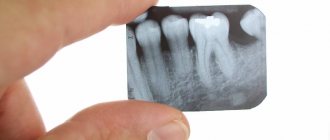
What is the difference between a radiovisiograph and a computed tomograph? On a radiovisiograph we see all teeth in a two-dimensional projection. It's essentially a photograph of a tooth. On a computed tomograph we can see all organs in a three-dimensional projection.
What can a doctor see on an x-ray?
He can see:
- total number of teeth;
- anatomical features of the structure of the maxillofacial region;
- level, bone height;
- possibility of installing implants.
X-rays are also done for the purpose of establishing a diagnosis, when the doctor cannot make one based solely on the patient’s complaints.
X-rays are also done to monitor the doctor’s work. It shows how:
- canals are sealed;
- tooth removed;
- an implant was installed.
What you wanted to know about dental x-rays
Modern dentistry boasts a wide variety of diagnostic methods used to identify dental pathology. Thus, computed tomography of teeth in Krasnoyarsk has recently become very popular.
An OPG image of teeth in Krasnoyarsk is no less informative, and therefore today experts will share with you the most important information about these research methods.
CT dental scan in Krasnoyarsk
Dental X-ray using computed tomography allows the doctor to get a complete picture of all the structural features of the patient’s dental apparatus. The method is used not only to identify pathological changes, but also to assess the individual anatomical and physiological characteristics of the jaw structure.
A dental CT scan in Krasnoyarsk, in Russia, is carried out in just a few minutes and at the same time has impressive information content. Due to the fact that the study is completely safe for the patient’s health, such a diagnostic procedure can be carried out repeatedly.
The images obtained during the study are detailed and created in good resolution. If the doctor is sufficiently professional, deciphering the results does not present any difficulties, which means that in the shortest possible time you will be given a diagnosis and prescribed the necessary treatment regimen.
Experienced radiologists and dentists work here. The joint and well-coordinated work of several specialists at once allows us to improve the quality of service and make dental treatment as effective as possible while minimizing the time spent on treatment.
The images obtained during computed tomography are recorded on disk and can be used in the future in the treatment of other dental diseases.
In what cases is there a need for a dental CT scan?
Dental X-rays using computed tomography have a wide range of indications and are performed in dentistry in the following cases:
- within the framework of existing treatment protocols, which provide for a CT scan immediately after consultation with a specialist for any detected dental problems;
- in the presence of jaw injuries;
- in the presence of congenital pathologies of the development of the dentofacial apparatus;
- in preparation for subsequent orthodontic treatment;
- as part of preoperative preparation;
- before dental implantation;
- in the presence of any kind of neoplasm of the facial area;
- in order to identify hidden, deep-lying foci of caries;
- to visualize the root system of teeth and determine the exact anatomy of the dental canals;
- for the purpose of objective assessment and control over previously performed treatment.
Like any other dental procedure, computed tomography also has contraindications. Despite the fact that the radiation exposure during dental CT is significantly lower compared to X-rays, a small dose of radiation is still present. Of course, it will depend on the area being examined and the number of images required.
Computed tomography is contraindicated for women in the first trimester of pregnancy and during lactation. Other contraindications are relative, and the question of the advisability of conducting the study is decided by the attending physician individually based on all data previously received from the patient.
To perform a dental CT scan, no special preparation is required. The patient is in a standing or sitting position. The chest and neck are covered with a lead-based apron to prevent radiation exposure to these areas.
Following the instructions of the radiologist, the patient remains in a motionless position for about 15–30 seconds, during which the computed tomography machine takes pictures in various projections. Within a few minutes, the doctor has access to the constructed model of the patient’s dentoalveolar structure and, accordingly, has all the information necessary to make an accurate diagnosis.
To summarize, let's say that computed tomography has found wide application not only in therapeutic dentistry: it is an indispensable diagnostic method in implantology, periodontology, orthodontics and even endodontic therapy.
For periodontists, computed tomography is perhaps the only way to determine the condition of periodontal tissues.
Using CT scans, orthodontists can draw up a plan for the correction of the dentition, as well as determine the possibility of extracting teeth that have not fully erupted from the gums.
In implantology, using tomograms obtained, doctors specify the installation location of the future implant, and also assess the condition of the jaw bone tissue at the intended location of the implant.
And finally, in endodontic therapy, computed tomography is used to visualize the physiological bends of the dental canals and determine their size. CT images of teeth make it possible to detect diseases at the earliest stage of their development, without losing sight of even the slightest deviations.
Modern computed tomography devices are installed, the use of which maximizes the quality of diagnosis and treatment of dental diseases.
Orthopantomogram
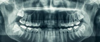
As mentioned earlier, OPTG is no less informative. The study involves the creation of panoramic x-rays of the lower part of the facial skeleton, where the upper and lower jaw, maxillary sinuses, joints and bone structures are located.
Few people know that dentists recommend performing an orthopantomogram at least once a year. It has been proven that when performing studies with such frequency, the likelihood of early detection of any dental pathologies increases significantly.
Among the advantages of the method, it is necessary to note the utmost reliability of the results obtained, high quality of images, speed of examination, as well as comfort and convenience for the patient during it.
Hidden cavities, deep-lying caries, anatomical and physiological features of the structure of teeth, features of filled and unfilled root canals, the condition of bone tissue, the quality and tightness of installed crowns - all this and much more is assessed based on the results of OPTG.
You will receive a comprehensive and high-quality diagnosis of dental diseases. Thanks to the professionalism of the staff and the presence of modern European-level equipment within the clinic’s walls, the diagnosis of any disease becomes as simple and fast as possible.
Where is computed tomography of teeth performed in Krasnoyarsk
You can get a CT scan or OPG of teeth in Krasnoyarsk at reasonable prices at. We care about our clients and try to make quality dentistry accessible to everyone by regulating prices for dental CT scans and other services in Krasnoyarsk.
Differences between a panoramic dental image and a computed tomography scan in Krasnoyarsk
An orthopantomogram is a panoramic photograph of teeth in Krasnoyarsk, giving a single-plane visualization of the structure of the patient’s dentofacial apparatus. Computed tomography, in contrast, allows the doctor to see the image in 3D format.
Both diagnostic methods are widely used in the daily practice of doctors. Specialists make the choice in favor of OPTG or CT based on the goals pursued and the available data.
Our Center has Gendex and OP300 Maxio 3D tomographs with a radiation dose 40 times less than a standard X-ray machine.
Orthopantomograph Orthophos Plus DS automatically controls exposure and adapts the focal length depending on the structural features of the patient’s jaw. Such characteristics allow the device to create clear images of the OPG of teeth in Krasnoyarsk, thereby facilitating the diagnosis of diseases.
You are guaranteed the correct diagnosis. The result is a quick start to treatment of identified pathologies and a healthy, beautiful smile in a short time.
If you have any questions, please contact the administrator by contacting us at the specified phone numbers, in instant messengers, or by leaving a request for a call back.
What is an x-ray?
This is one of the types of radiation. If you think that X-ray radiation is present only in the X-ray room, then we can say right away that you are very mistaken. Let's remember physics lessons. We receive small doses of radiation every day:
- outdoors, from a natural source of radiation - the sun;
- at home from household sources located in the apartment: from a TV, various gadgets, a refrigerator, etc.
Radiation is measured in sieverts. What is the rate of radiation that a person can consume? Acceptable is 1000 sieverts per year.
Now let's remember mathematics and calculate how many X-rays can be taken for a person. A targeted image of one tooth is equal to 2-3 microsieverts. You can take 300-500 x-rays per year. A panoramic photograph (all teeth) is equal to 16-18 microsieverts. You can take 60-70 x-rays per year.
Now let's look at computed tomography. 1 photo taken on this device is equal to 60-80 microsieverts. You can take 16-20 x-rays per year.
We are now considering the maximum dose limits for X-ray equipment. Our clinic has a radiovisiograph and a computer tomograph of the latest generation made in Germany. The load on the devices is reduced to a minimum.
Previously, when taking an x-ray, the exposure time (you hear it as a beep) was approximately 2-3 seconds. Now this time has been reduced to 0.05 seconds.
Now let's talk about household radiation. If you watch TV for 3 hours at a distance of less than 2.5 meters from the screen, you get approximately 0.5 millisievert, i.e. Watched TV for 5 days, get 1 x-ray. If you sit in front of a computer monitor for more than 3 hours, you get 1 microsievert, 1 x-ray. We flew by plane to Turkey, where the flight is 3-3.5 hours, you get 10 millisieverts or 5 targeted shots. And add radiation from TVs, refrigerators, microwave ovens and you will understand that you will not receive terrible radiation in the X-ray room.
Why do you need a panoramic photo?
A panoramic photograph of the dentition is taken to diagnose the general condition of the oral cavity. The procedure is not painful. The only thing the patient faces in this case is the need to open his mouth wide while standing.
The procedure has its own indications, which are determined by the attending physician. Usually they are provided for in the future of any serious procedures and are as follows:
- dental implantation;
- its alignment;
- treatment of periodontal disease;
- inflammation of the soft tissues of the oral cavity;
- assessment of tooth growth rate;
- surgical intervention.
It is worth considering that before an orthopantogram you should remove all objects that interfere with the passage of x-rays (jewelry, etc.).
Patient protection is mandatory during the procedure. To carry it out, he wears an apron made with lead and a special collar on the neck area. This helps avoid radiation exposure and subsequent problems from the harmful effects of X-rays.
It is worth understanding that the safety of x-rays is achieved only when all necessary measures are followed. In addition, the better the device, the faster and better quality the picture will be. The X-ray of a digital orthopantomograph is better than that of a film one.
How is X-ray performed?
As already mentioned, the patient must stand. His chest should be pressed against the platform, which determines the correct position in space. The conditions of the procedure stipulate that he will need to bite a special plastic stick, after first pressing his tongue against the soft palate. Lips must be closed.
The doctor may ask you to change the position of the head so that the x-ray can cover the required area and the diagnosis is correct.
Currently, the procedure is performed using a safer PaX-i3d “Green” tomograph. Its main differences from devices of previous generations are that it is safer and scans an order of magnitude faster, protecting not only the patient, but also the clinic staff from exposure to X-rays.
Can X-ray examinations be performed on pregnant women?
In the first half of the term, the study is done only according to strict indications. In the second half of the term, you can do as much research as you like.
Another interesting fact. People often ask X-ray technicians to wear more protective lead aprons. Let's say right away that this is useless. You receive the same dose of radiation from covered and exposed parts of your body.
And remember that a doctor will never order an x-ray just out of curiosity. X-ray is one of the types of diagnostics of the area of proposed treatment and the anatomical features of the oral cavity.
Come to our clinic and we will make every effort to make your stay comfortable and enjoyable!