The concept of “eye” teeth does not relate to medical terminology - it is, rather, a popular term that refers to the upper, and with them, the lower canines. There is an assumption that this name arose due to the proximity of the facial nerve, the impact of which is accompanied by severe painful sensations radiating in the fundus of the eye. Fang extraction is a complex procedure that requires the use of strong painkillers.
Grabbing and biting
In the front part of the dentition there are incisors, four each on the upper and lower jaws.
They have a relatively sharp edge that, when pressure is applied, can cut even fairly dense food. Also in the oral cavity there are fangs - pointed teeth, inherited by humans from predatory ancestors. They are involved in holding food and tearing small pieces from it.
The loss of incisors and fangs leads to the fact that without its correction a person can only eat liquid or pre-cut foods into small pieces.
The structure of the fangs
The canines of the upper jaw are shaped like an irregular cone. The cutting edge is shaped like a triangle, which is limited by three teeth - a pronounced middle one and two outer ones. The area of the tooth facing the lip (vestibular) has a convex shape and gradually approaches the lingual.
On the lingual part of the tooth there is a longitudinal ridge that divides the dental crown into two facets: the lateral part has the largest area. The upper canine has a pronounced crown curvature and root deviation.
The root of the tooth is quite thick and has a shape slightly compressed from the sides. Wider in anteroposterior dimension. The lateral and medial surfaces are very wide and have barely noticeable grooves.
The tooth has one, wide enough and accessible if treatment is necessary, root canal.
The canines located on the lower jaw are smaller in size than the upper ones, and the shape of the crown is close to the upper lateral (side) incisors. They are slightly narrower and longer than the upper canines. The medial surface is in a straight line with the root line. The cutting edge is less pronounced. The distal part of the cutting edge is longer than the medial one.
The labial surface of the lower canines is less convex and pronounced than that of the upper ones, and the lingual surface can be either straight or slightly concave.
The lower fangs have a slightly shorter tooth root than the upper ones, but they have absolutely no differences in shape. In rare cases, anatomical pathology occurs in which the root is divided into two parts.
The canines of both jaws have quite pronounced external differences in the dental crown, but at the same time the dental cavities are very similar.
Grinding food
The human stomach is capable of fully digesting only finely ground food, which is abundantly moistened with saliva. For this purpose, there are premolars and molars in the mouth, which act like millstones - they grind large pieces of food, while simultaneously mixing it with the secretion of the salivary glands.
The absence or loss of one of these teeth leads to the fact that unprocessed large particles of food enter the gastrointestinal tract, which are difficult to respond to enzymes and can cause various chronic diseases of the stomach and intestines.
Aesthetic objectives: canines in the lateral incisor position
Very often, dentists do not think about the long-term consequences of their treatment. An incomplete treatment plan or poor treatment choices (often dictated by the patient) usually lead to more serious problems years later. If only we could know what will happen in the future, it would make the task much easier.
However, dentistry can be a predictable subject if approached with proper function and aesthetics in mind. Each dental student receives a phantom - a model that shows the ideal condition and position of the teeth. All dentists are taught the principles of balanced occlusion and the importance of reducing destructive forces.
This article will demonstrate what happens when the factors of time and aging are not taken into account when choosing a treatment method. Very often, what patients themselves do not notice or consider unimportant, about which they do not complain, can turn into a real problem only several years later. If we, dentists, approach the patient from the standpoint of individuality and at the same time as a component of one population, if we carry out careful planning and elaboration of parameters, striving for that same phantom model, then we come to a predictable and long-term quality result. The clinical case discussed here concerns a woman with congenital absence of lateral incisors. When she was a teenager, the maxillary canines were orthodontically moved into position as lateral incisors. In her younger years, her appearance was quite harmonious and attractive, but over time the picture began to change.
Absence of lateral incisors
It is reported that approximately 2% of the population are congenitally missing one or two lateral incisors. Paired absences are more common, and if only one is present, it is usually a microdent. Performing OPTG at an early age makes it possible to find out which of the permanent teeth have not formed.
It is very important to learn about the congenital absence of a tooth at an early age, so that the entire sequence of therapy can be correctly coordinated to restore aesthetics and function. Treatment of congenital absence of lateral incisors is an orthodontically and therapeutically complex task, which is based on the tooth-dental arch size relationship. Numerous studies have been published comparing the method of mesial repositioning of canines and distalization of canines followed by prosthetic restoration of missing incisors.
From a modern point of view, the most logical way is, of course, to open up space and replace missing teeth with prosthetics. The phantom from student life, at least, definitely had his own lateral incisors.
Moving the canine to the lateral incisor position may have several aesthetic and functional disadvantages:
- The canine, a fairly wide tooth, begins to replace the naturally narrow incisor. The color of the canines is usually somewhat darker than that of the lateral incisors, so they stand out from the general plan if they are not in the corners of the smile.
- The level of the gingiva of the canines is approximately similar to that of the central incisors, so moving the canine to the place of the lateral incisor causes visual disharmony.
- Insufficiency appears in the processes of occlusion and articulation, since after movement the canine guidance of the lower and upper canines is not carried out together.
- In the absence of canine protection, the risk of abrasion of the remaining teeth increases, usually consisting of small cracks and microfractures. Over time, periodontal problems and increased sensitivity may appear.
- Long-term retention with an orthodontic retainer will be required to retain the canine in the lateral incisor position.
- The patient may begin to experience TMJ discomfort, muscle tension, grinding, and headaches. This may occur due to effects on muscles that are not anatomically intended.
- The tissues of the vestibule of the oral cavity and the bony prominence in the area of the canine root do not look natural in the area of the lateral incisor. Without the presence of a tubercle in the corners of the mouth, the tissues do not have sufficient support, which leads to recession of the cheeks and narrowing of the buccal corridors. Over time, fewer and fewer teeth become noticeable when smiling.
Orthodontic vision
Traditionally, orthodontists have not encountered people in need of restorations, as they have primarily worked with younger patients. Young people rarely need major restorations. However, in the 21st century, orthodontists often began to take on patients with the need for restoration or due to periodontal disease in the latter. The absence of a lateral incisor is an aesthetic indication, so the orthodontist should treat such a case in this aspect.
The goals of orthodontic treatment often vary depending on the final goal and the need for restoration:
- Positioning of teeth can occur in order to improve the performance of any restoration, for example, to replace a lateral incisor or another tooth.
- There are some advantages to performing permanent or temporary restorations before, during or after orthodontic intervention. This restoration allows you to create the desired shape and at the same time gives an idea of the required space and dimensions. Tooth wear, fractures, underdevelopment in the form of barrel-shaped incisors, etc. are the main indications for restoration before orthodontic treatment.
- Orthodontists sometimes need to reposition a tooth to improve oral hygiene
- Orthodontic treatment may be performed due to periodontal problems, such as insufficient gingival margins, absent gingival papilla, or bone loss.
Today, the goals of orthodontic treatment have diversified significantly, as the aesthetic and functional vision of problems has expanded. Properly planned orthodontic treatment can achieve stable and functional occlusion, improve the health of periodontal tissues and improve dental and facial aesthetics. Orthodontists simply need to study facial aesthetics. Modern specialized literature, research and training always have a positive effect on a specialist and, accordingly, his work results. However, in the past, very little attention was paid to facial aesthetics and periodontal tissue health. A successful orthodontic treatment can be considered an intervention that results not only in the ideal articulation of the models (as well as the achievement of ideal cephalometric relationships and sizes), but also in the restoration of facial aesthetics and harmony in a given position of the teeth.
A smile analysis should include the following: the vertical position of the teeth at rest and when smiling; the transversal (horizontal) dimensions of the smile; the characteristics of the smile arc; and the vertical relationship of the gingival margins to each other. Taking into account all the data, it becomes desirable to move the canines to their natural position and replace the lateral incisors through prosthetics.
Interdisciplinary treatment planning
Innovations in aesthetic dentistry have led to developments in all dental specialties. In today's standard of dental rehabilitation, specialists proceed, first of all, from the individual characteristics of the patient's face and his needs. Each treatment plan begins with an aesthetic assessment. During the analysis, the patient's lips, skin, and cheeks are examined. We must always refer to the position of the teeth in the upper jaw and the level of the gums in relation to the face, and then determine what corrections need to be made for a given appearance. We cannot achieve correct occlusion until our final vision of aesthetics is determined. Aesthetics dictates where the teeth should be located, what the vertical position, guidance and relationship should be.
The main person in the team is the restorative dentist, and the success of teamwork is achieved through detailed discussions of each problem. The main specialist must achieve real therapeutic goals (take into account the economic component, expectations), create an aesthetic vision of the final result, determine the sequence of treatment and restore poorly formed teeth to ideal proportions. The restorative dentist acts here as a link among all specialists, uniting and controlling the manipulations carried out and the pursuit of the final goal.
Clinical case
Diagnosis and treatment plan
A 40-year-old woman came to the clinic (in 2003) in need of cosmetic correction of treatment performed in adolescence.
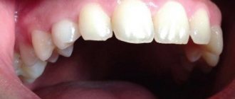
The patient had a congenital absence of lateral incisors, so she underwent orthodontic treatment at the age of 14 years to solve this problem. Relying on the orthodontist and unaware of other treatment options, the parents chose (or allowed) to move the girl's canines to the lateral incisor position. After the treatment, the smile did not look attractive enough, but the patient was able to get used to it. But as she reached middle age, the woman began to notice that her interlocutors were increasingly staring at her teeth. This made her insecure. The canine was darker, had a rounded contour and a gum level that was in disharmony with the rest of the teeth. The buccal corridors were narrowed to close the empty space, creating the appearance that the cheeks were somewhat sunken (Photos 1 and 2). The patient remained deeply dissatisfied with her appearance.
Photo 1: In 2003, a 40-year-old patient presented with an aesthetic problem.
Photo 2: The canines were moved to the position of the lateral incisors, which stood out from the overall appearance.
With the advent of the 21st century, there have been significant changes in dental technology and materials. Aesthetic manipulation has become common and routine. In this situation, should we have moved the canines to their correct position and replaced the lateral incisors? The patient was open to any options.
After weighing all our options, we decided not to move the canines back and place titanium implants. Although this would be the most common solution to the problem, this path did not seem entirely justified to us. Such treatment would take approximately 18 months, and there was also the possibility of resorption and the need for bone grafting. Since the patient clearly wanted to change the shape and color of her front teeth, an easier and more predictable alternative was in development.
After analyzing radiographs, photographs and working models, we decided that after flattening and narrowing the canine, we could use light orthodontic extrusion of the teeth, which would realign the ridge bone and create a new harmonious appearance at the soft tissue level. By leaving space distal to the canine, we can create porcelain veneers to correct the shape of the canines to resemble the lateral incisors and the first premolars to resemble the canines. Placing veneers on the side teeth will also be possible if the patient so desires. According to the plan, the treatment was supposed to take approximately 6 months. It is important to evaluate all the benefits and risks of any of the treatment options, and the chosen option already needs final approval by the patient himself.
Since the patient expressed a desire to install veneers on the teeth of the anterior zone, all we had to do was correct the position of the lateral incisors and canines.
Clinical protocol
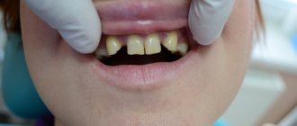
The labial surface of the canines was flattened and the mesial and distal contours were tapered using a flame-shaped diamond bur (Dental Savings Club) followed by finishing with strips (Integra Miltex). Ormco Saphire (Ormco) braces are fixed on the upper jaw up to the first molar (Photo 3). Using Ni-Ti wires of increasing diameter from 0.13 to 0.16 with an extrusion force of 15 g, it was decided to carry out labilization of the central incisors and extrusion of the canines to correct the gingival level (Photo 4). The use of orthodontic extrusion to modify soft and hard tissues was first described in the literature by Heithersay and Ingber. Low-intensity forces (up to 30 g) stimulate marginal positioning of the bone crest, behind which movement of the gums occurs. Restructuring of the alveoli occurs along with the movement of the root.
Photo 3: Preparation for orthodontic treatment: the labial surface of the canines was slightly flattened, and the contours on the proximal sides were narrowed.
Photo 4: Ni-Ti arch with a force of 15 g is used to labilize the central incisors and extrusion of the gingival level of the canines.
Extrusion of the canines by 1 mm was carried out over 3 months, followed by stabilization for 3 months with a Ni-Ti arch 18x25. The total time spent on treatment coincided with the plan and amounted to 6 months. The gingival level of the canines is corrected and lowered, the canines are ready for restoration according to the proportions of the lateral incisors. For the formation of “fangs” from premolars, space is left distal to the true canines (Photo 5a-6). The braces are removed, leaving the teeth in an ideal position for further restoration with porcelain veneers (Photo 7).
Photos 5a and 5b. Reduced gingival level at the lateral incisor.
Photo 6: Space is left at the distal surface of the canine for restoration of the premolar in the shape of the canine.
Photo 7: After removing the braces, the teeth are left in an ideal position for further restoration.
A vinyl polysiloxane (VPS) impression was made, followed by a diagnostic wax-up of the anterior 6 teeth (Figure 8). At this stage, it was decided to evaluate the esthetics of the anterior sextant before widening the buccal corridor.
Photo 8: Diagnostic wax-up creating the ideal appearance of the 6 front teeth.
Conservative preparation (0.3 mm) was carried out (Photo 9) and the final VPS impression was taken (Honigum DMG America) using conventional and lightweight techniques. Bite registration was carried out by Luxabite (DMG America). Using the classic VITA scale, shade A1 was determined. Temporary structures made of bis-acrylic material (DMG America), made on the basis of a diagnostic wax-up matrix, are fixed to the prepared teeth. Fixation was carried out using Temp Bond Clear (Kerr). Next, the appearance of the structures was corrected with soft diamond and Flexi discs (Cosmedent) (Photo 10).
Photo 9: The teeth were prepared by 0.3 mm to install ceramic veneers.
Photo 10: Luxatemp temporary structures (DMG America), creating a natural smile appearance.
Models are cast from plaster (Photos 11a and 11b) and then porcelain veneers are made (Photos 12a and 12b).
Photos 11a and 11b. The Geller technique allows you to emphasize the contour and level of the gums.
Photos 12a and 12b. Veneers made from Creation Porcelain (Jensen Dental).
The veneers were fixed with colorless cement (Variolink Veneer Ivoclar Vivadent) in 2004. The patient was satisfied. Ultimately, she adjusted her smile to the desired result. The size and shape of the front teeth are made according to golden proportions, so it is almost impossible to determine the congenital absence of incisors. Premolars are shaped like canines. At this stage, the slightly reduced buccal corridor did not bother the patient at all.
After some time
Ten years later, in 2013, the patient returned to the clinic. Now the lateral teeth caused aesthetic dissatisfaction (Photo 15). The upper second premolars and molars on both sides were prepared (Photos 16a and 16b), then a suitable shade was selected (Photo 17). The restorations were made from Creation Porcelain (Photo 18). The structures were fixed using transparent cement for veneers (Variolink Veneer). The resulting result looked uniform, as if created at the same time (Photos 19 and 20). The patient was satisfied.
Photo 13: Completed treatment in 2004.
Figure 14: Despite the narrowing in the lateral areas, the patient was satisfied with the aesthetics of the anterior segment.
Photo 15: By 2013, the patient wanted fuller buccal corridors.
Photos 16a and 16b: Preparation of second premolars and first molars.
Photo 17: Shade selection according to the VITA scale.
Photo 18: Making ceramic restorations (Creation Porcelain).
Photos 19 and 20: Completed treatment in 2013.
Final comment
People often change their minds, so it is difficult to predict what a patient who was treated as a teenager will want. A careful treatment plan helps overcome many of the negative factors associated with subsequent gradual aging. A careful discussion of all the details allows for high-quality rehabilitation, as well as building a trusting relationship with the patient.
Author: Elliot Mechanic , DDS, BSc
Indicative
Teeth are an indicator of the general condition of the body. The presence of any metabolic disorders, chronic diseases of the endocrine, digestive and other systems, vitamin deficiencies, unbalanced nutrition - all this negatively affects the condition of the dentition - the teeth change their shade and begin to crumble.
Therefore, caring for your smile includes not only regular hygiene procedures and visits to the dentist’s office, but also treatment of concomitant pathologies.
As you can see from the article, teeth are not only about comfortable eating. The loss of at least one element of the dentition is fraught with possible problems with digestion, speech, as well as the development of complexes against the background of a cosmetic defect.
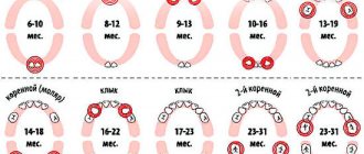
Eruption of canines in infancy
The order of formation of the primary occlusion provides for the appearance of fangs after the incisors and the first chewing units have erupted. The upper elements emerge at the age of 1.2-1.5 years, which is slightly earlier than the period of formation of the lower molars. Late eruption is determined by the deep position of the units in the structure of the jaw, which also determines the characteristic painful sensations that may occur during growth.
To relieve the discomfort experienced by the child, the following techniques are recommended:
- Massage of gum tissue - before the procedure, you need to thoroughly wash your hands, then use your index finger to massage the area above the canine for a couple of minutes. It is recommended to repeat the massage several times a day;
- The use of teethers that provide a cooling effect - a product filled with distilled water does not pose a danger to the baby’s health even if the baby manages to chew the shell;
- Applying anesthetic gels that relieve inflammation within a few minutes;
- For nasal congestion, use children's nasal medications that promote vasoconstriction;
- At elevated temperatures (more than 38 degrees) - take fever medications, available in the form of syrup or suppository.
It is worth emphasizing that the use of medications must be agreed with a doctor, who will determine the appropriate list, as well as the duration of the course.
Description, structure, functions, name and location of human teeth
It is not without reason that human teeth have different shapes, structures and sizes. Each tooth performs certain functions, and its purpose is, one way or another, reflected in the name:
- The incisors are the front teeth that fall into the smile zone. They are quite sharp as their function is to tear or cut food, hence the name. Normally, every person has lateral and central (larger) incisors.
- Canines are cone-shaped teeth that can be used to tear and hold food. They are located immediately behind the incisors.
- Premolars are the small back molars located behind the canine teeth.
- Molars are the back teeth necessary for mechanical processing of food. Wisdom teeth refer specifically to molars.
The listed types of teeth are also found in animals. But in the process of their evolution, some dental units were slightly modified:
- elephants' upper incisors became tusks;
- Poisonous snakes have poison in their fangs.
Many animals have front teeth similar to human ones, but since they are called differently, they cannot always be identified with the usual incisors and canines.
The structure of human teeth
Human teeth consist of a crown and a root part. The first is located above the level of the gum, the second is inside it. The top of the crown is covered with enamel, the strongest tissue in the body. It protects the inner layer of the tooth – dentin – from external influences. Under the dentin there is a hollow chamber with nerves and blood vessels - the pulp.
Despite its strength, enamel is vulnerable to bacteria. If left untreated, caries can affect the dentin and pulp. When the pulp chamber, which contains nerve fibers, is damaged, pulpitis develops, accompanied by acute throbbing pain. If the infection spreads to the root of the tooth, it is highly likely that it will have to be removed.
The basis of the lower part of the tooth is the root canals, which also contain arteries, veins and nerve fibers. Through the apical foramen, all of these structures are connected to the main neurovascular bundle.
The lower part of the tooth is covered with dentin and cement, which is attached to the periodontium using collagen fibers. The roots of teeth are hidden in the alveoli - depressions in the jaw bone.
What is a dental formula
A dental formula or diagram is a brief description of the dental structure of the human dentition using numbers and Latin letters.
The letters are abbreviations from the Latin names of teeth accepted in medicine:
- I – these are dentes incisivi or incisors.
- C is for dentes canini or fangs.
- P stands for dentes premolars or premolars.
- M stands for dentes molars or molars.
The alphanumeric dental formula is common in many countries, including America, which is why it is sometimes called American. After the letters in the formula there is always a digital fraction, where the numerator indicates the number of teeth on the upper jaw, and the denominator on the lower jaw.
The dental formula of a healthy adult with a normal bite and 32 teeth looks like this:
This formula indicates that on each human jaw there is:
- 2 pairs of incisors;
- 1 pair of fangs;
- 2 pairs of premolars;
- 3 pairs of molars.
Dental formula in the absence of some teeth
It is not difficult to correctly number dental units with a healthy dentofacial apparatus; it is much more difficult to indicate the location of the teeth if a person does not have any molars, incisors, canines or premolars. In this case, the dentist indicates the number 0 opposite a certain group of teeth, rather than shifting the number series.
If the patient has any anomalies in the development of the dentofacial apparatus, for example, teething in the wrong place (hyperdontia, polydontia), the dentist prescribes an individual formula. In addition to numerical designations, the doctor generates a detailed report on the structure of a person’s dentition.
Correct names of baby teeth
In Latin, baby teeth in a child are called the same as permanent teeth in an adult. But children do not have all the dental units characteristic of older people. Milk teeth are divided into:
- central incisors;
- lateral incisors;
- fangs;
- first and second molars.
There are different names for teeth, but it is better to focus on Latin terms and serial numbers. In this way, you can identify teeth even without serious knowledge of dentistry.
Alternative names for human teeth
In addition to the official names, there are also alternative names for human teeth. They are not written in dental records, but have long been used in informal communication, since these teeth in people are located or grow in certain places or at certain times.
Eye teeth
The upper canines are called eye teeth because of their close location to the branches of the facial nerve. When they become inflamed, the pain radiates to the eye area and upper face. How close the incisive canals and the dental nerve are located to each other is shown in the figure:
Wisdom tooth
The wisdom tooth is the posterior third molar. He was called “wise” because he grows up in adulthood - when a person has already gained wisdom (by about 20 years).