Almost every person is afraid of dentists - fear of pain and unpleasant sensations makes them put off going to the doctor for many years. Finding a patient who does not experience discomfort from drilling in the jaw is almost impossible. And no matter how hard experts fight this fear, it is difficult to force a person to visit the dentist regularly. However, experts from the American Medical Center identified as many as 10 facts that few people know about teeth and the oral cavity in general. Some of them may influence the decision to postpone visiting a specialist and take more careful care of the condition of your teeth. And here are just a few little-known secrets that our teeth keep:
The most important thing is to brush your teeth properly
Whether heredity is good or bad, if you do not take proper care of your oral cavity, problems will inevitably arise sooner or later. Some people have caries, others have periodontitis, but there will be problems! Satisfactory oral hygiene is the key to healthy teeth, even those with not very good heredity.
We do not take into account serious diseases when the formation of teeth fails even before they erupt. These diseases include:
- Enamel hypoplasia.
- Enamel hyperplasia.
- Drug-induced disorders of tissue development.
- And also hereditary disorders.
We are talking about the average person with normal health. Each person has one or another predisposition, but if prevention is carried out correctly, the disease itself can be prevented
. Nowadays, to be healthy, you need to make a lot of effort. Proper nutrition, exercise, personal hygiene, self-care, etc., all this requires not only time, but also willpower, which, unfortunately, not many can boast of.
Unilateral canine agenesis: clinical case and literature review
Dental agenesis or hypodontia is one of the most common anomalies of the human dental system, which is characterized by the initial absence of one or more teeth. This condition can occur in association with a certain genetic syndrome or separately (non-syndromic). The absence of one or more permanent teeth without systemic pathology is the most common phenotype of hypodontia.
Both external conditions and genetic factors can cause disruption of the development of the tooth germ, although hypodontia mainly occurs on the basis of the genetic aspect.
Recent reports have shown that in the Caucasian population, the incidence of hypodontia in permanent teeth (excluding third molars) is in the region of 4.5-7.4%.
In syndromic oligodontia, permanent canines are often missing. Congenital absence of permanent canines in non-syndromic patients is described sporadically in severe hypodontia and oligodontia, while isolated cases of absence of permanent upper canines are quite rare. Previous studies have revealed the approximate frequency of such pathology – 0.07 and 0.13%.
This clinical case demonstrates a unilateral absence of a permanent upper canine tooth. A review of the literature on the frequency, etiology and diagnosis of pathology was also carried out.
Clinical case
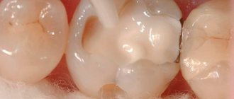
In October 2013, an 18-year-old woman came to the Hacettepe University Clinic for a regular dental examination. The general health condition was satisfactory, no systemic pathologies were identified. Intraoral examination revealed a class I relationship between the molars, a correctly positioned central line and a slightly increased vertical overlap. Between 22 and 23 there was a 2 mm trema. Clinical examination of the oral cavity revealed a small canine with signs of wear on the right side, the tooth was immobile (Photo 1). There is a small carious lesion on the distal surface of the tooth. To detect the permanent canine, palpation of the transitional fold and palatal surface of the alveolar process was performed. Palpation is ineffective.
Figure 1: Intraoral view of the upper dental arch showing a small canine with signs of wear.
An OPTG was performed to identify the location of the permanent canine and other possible anomalies. The x-ray showed that the permanent canine bud was absent, and that in the dental arch there was a primary canine with a characteristic short root, a small crown and a thin enamel structure. The primary canine had grade 1 root resorption, and all third molar buds were also absent (Photo 2). According to the patient, no teeth were removed and there was no history of trauma. The family history is also without any peculiarities or similar pathologies. No other dental anomalies were identified. However, in the area of the left third molar of the mandible, the radiograph revealed an irregularly shaped area of radiopacity (Photo 2). The lesion was diagnosed as osteosclerosis on a targeted photograph (Figure 3).
Figure 2: OPTG showing the absence of the right permanent canine and all third molars. A primary canine with signs of root resorption is located in the dental arch. Pay attention to the radiopaque lesion in the area of the left third molar of the mandible
Figure 3: Spot X-ray of the left postmolar area showing the area of osteosclerosis
The patient was sent to the Department of Therapeutic Dentistry for treatment of a carious tooth.
Discussion
Many studies have examined developmental disorders and explained these conditions using anatomical and evolutionary models. According to Bolk's theory of terminal reduction, due to the phylogenetic evolution of the human species, reduction of distal elements of the dental arch is much more common than mesially positioned teeth: thus, the most commonly missing second premolars, upper lateral incisors and third molars. In adapting Burler's concept to human occlusion, Dahlberg defined the canine as a key tooth that sits on its own in a specific location and has great stability and is therefore rarely congenitally absent.
The causes of congenital absence of teeth are very different. Severe hypodontia is usually associated with genetic disorders such as Witkop syndrome, ectodermal dysplasia, and Rieger syndrome. Mild and moderate hypodontia can occur due to improper formation of the bud, various injuries, Down syndrome and syndromes associated with cleft lip and palate.
The etiology of dental agenesis is subject to active debate. Graber states that congenital absence of teeth is an inherited phenomenon and is possibly transmitted to other generations as an autosomal dominant trait with incomplete penetration and variable expression. Brook proposed a multifactorial theory of hypodontia with a combination of polygenic and external influences. The role of genetics has been confirmed, but there is controversy on the subject of environmental influences due to a study on monozygotic twins: Markovic studied cases of hypodontia in twins and identified one pair in which unilateral canine absence had the appearance of a mirror image, which spoke primarily of a genetic basis for the phenomenon hypodontia. However, Kindlan demonstrated that genetic coding is not the only controlling factor in dental agenesis in twins, as it revealed differences in facial features and the extent of hypodontia. It has recently been suggested that isolated hypodontia of permanent canines is associated with mutations in the WNT10A gene.
The absence of a permanent canine rudiment is more common in women and mainly affects the upper jaw on the left. Statistics on agenesis of the upper canines vary from study to study. Harris and Clark found only two cases of congenital absence of the upper canines among 600 black American citizens (0.4%), while they could not find a single case among 1,100 whites. Cho described the absence of permanent canine rudiments in 32 cases out of 69,852 Chinese children. Fakuta Found only 42 cases with a frequency of 0.13% among 35,927 studied. Davis also reported five similar cases among 1093 Chinese children (0.46%). In European studies, Fekonja reported one case of agenesis of the upper permanent canine among 212 patients who received orthodontic treatment (2.1%), while Rozsa found an incidence of 0.27% in his study. Sisman showed a rate of 0.37% in an analysis of Turkish orthodontic patients.
It has already been said that the absence of one canine is more common than multiple canine agenesis, and basically this anomaly is combined with congenital agenesis of other teeth, microdontia, delayed tooth development and eruption, supernumerary teeth, odontomoma, taurodontia and additional cusps of teeth. In our clinical case, no additional anomalies of the dentofacial system were identified.
If clinically permanent canine teeth are not found in the oral cavity, several other conditions should be considered. For example, if palpation on the buccal side is ineffective in an 11-year-old child, it is possible to assume ectopic eruption or impaction of the tooth. Bone diseases, cysts, tumors can cause ectopic eruption and delay this process. In a rare situation, transposition with first premolars or lateral incisors is possible. Migration of the upper canine past the midline is known as transmigration, an uncommon anomaly that may occur when the canine is absent from the mouth on examination. Early diagnosis of permanent canine agenesis helps to carry out timely rehabilitation, reduce treatment time, complexity and avoid complications. Thus, if a canine tooth is absent in the oral cavity during a clinical examination, it is necessary to conduct an X-ray examination to clarify the condition and identify associated anomalies.
Congenital absence of upper permanent canines requires a specific treatment plan. Many factors must be taken into account: the condition of the primary teeth, the patient's occlusion (crowding or tremors, midline shift), type of facial growth and patient preference. Treatment may include removal of primary teeth to facilitate spontaneous or orthodontic closure of the site, or retention of primary teeth until growth is complete to preserve alveolar bone and create a favorable situation for implant placement without bone grafting.
In this case report, the main goal was to preserve the temporary canine. It was decided to observe the patient, since tooth root resorption had already begun. In the near future, the girl will need orthopedic rehabilitation.
Authors: Nagihan Koc, L. Berna CaLJrankaya, Nursel Akkaya