Treatment of
jaw
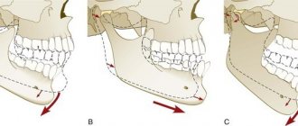
fractures is a characteristic of conservative and surgical treatment methods. Treatment of any injury must begin with first aid. For fractures of the maxilla, it is necessary to restore the prominence and height of the face and recreate the pre-traumatic occlusion. The jawbone is often stable, so treatment is carried out by fixing the upper and lower teeth together. Various options for osteosynthesis of facial bones with titanium mini-plates. When the upper jaw is fractured, its fragments are displaced downward, disrupting the usual relationship of the teeth of the upper and lower jaw and somewhat lengthening the face.
PROVIDING ASSISTANCE FOR FRACTURES OF THE UPPER AND LOWER JAWS
Qualified assistance is provided before admission to a specialized hospital.
When providing qualified surgical care, the surgeon must:
1) anesthetize the fracture site;
2) inject antibiotics into the wound and administer antibiotics internally;
3) carry out simple transport immobilization, for example, apply a standard transport bandage;
4) make sure there is no bleeding from the wound, asphyxia or its threat during transportation;
5) carry out anti-tetanus measures according to the instructions;
6) ensure proper transportation to a specialized medical institution, accompanied by medical personnel (determine the type of transport, the position of the patient);
7) clearly indicate in the accompanying documents everything that was done to the patient.
Patients with complex and complicated facial injuries should be referred to a specialized department if there is a need for primary plastic surgery of soft tissues and the use of the latest methods for treating facial bone fractures, including primary bone grafting.
Temporary (transport) immobilization of fractures of the lower and upper jaw. It is carried out outside a specialized medical institution or at the scene of an incident by paramedics, doctors of other specialties, sometimes in the form of mutual assistance.
For temporary immobilization use:
1) Circular bandage parietal-chin bandage
2) Standard transport bandage
3) Soft chin sling Pomerantseva-Urbanskaya
4) Intermaxillary ligature fastening
Circular bandage parietal-chin bandage.
Circular tours of the bandage, passing through the chin of the lower jaw and parietal bones, do not allow fragments to move during transportation of the victim. For this purpose, you can use a mesh elastic bandage.
Standard transport bandage.
A standard transport bandage consists of a rigid chin sling and a support cap (dimensionless). The latter has 3 pairs of loops for fixing rubber rings, which tightly press the sling to the chin area. Under the loops there are fabric pockets for cotton pads, which allow you to remove the rubber rings from the swollen soft tissues of the face and prevent their injury. The cap is placed in such a way that it tightly covers the occipital protuberance, and its straps are tied on the forehead. A rigid chin sling is made with a cotton-gauze liner so that it covers the edges of the sling along its entire perimeter. This prevents direct contact of the rigid structure with swollen soft tissues, and can also serve as a protective bandage in case of damage to the skin of the chin area. Depending on the number of pairs of rubber rings used in the cast, the sling can hold the fragments without pressure or apply pressure to them. For fractures of the lower jaw behind the dentition or for a fracture of the upper jaw, a standard bandage can be applied using 3 pairs of rubber rings (as a pressure bandage)
In case of fractures of the lower jaw within the dentition, it should be applied only to support the fragments. Excessive pressure on displaced fragments will lead to even greater displacement with the risk of developing asphyxia. However, such a differential approach is only possible in a specialized department where there is a dental surgeon. Non-specialists should be advised to apply a standard transport dressing as support.
Soft chin sling by Pomerantseva-Urbanskaya.
Its chin part is made of several layers of canvas or calico. The intermediate section is represented by two wide elastic bands (haberdashery), which go into the peripheral section of the bandage, made of the same material as the chin part. The latter has lacing, which allows you to adjust the degree of tension of the rubber strips of the sling. This bandage is comfortable for patients, easy to use and provides good fixation of fragments
Types of conservative methods of immobilization of jaw fragments
There are temporary methods of immobilization (including transport) and permanent (therapeutic). Temporary methods for securing jaw fragments are divided into: - extraoral (bandage, chin sling, improvised bandages using improvised means); — intraoral (methods of intermaxillary ligature fastening, splints-spoons with “mustaches” of different designs). Permanent (therapeutic) methods of immobilization are divided into : - non-laboratory splints (individual dental splints made of metal or other material, standard dental splints); — laboratory-made splints (Weber’s supragingival splint, simple or with an inclined plane, Vankevich and Vankevich-Stepanova splints, various dental aligners, Port’s supragingival splint).
Temporary (transport) immobilization
Indications for imposing temporary (transport) immobilization: - lack of conditions for permanent (therapeutic) immobilization and the need to transport the victim to a specialized medical institution; — lack of specialized personnel who can carry out permanent immobilization; - lack of time required for permanent (therapeutic) immobilization. This usually happens during combat operations or other emergency situations (earthquake, accidents with a large number of casualties, etc.), when a large flow of victims and wounded with trauma is simultaneously observed; - severe general somatic condition (traumatic shock, coma, intracranial hematoma, etc.), which is a temporary relative contraindication for therapeutic immobilization. Temporary immobilization is imposed for a period of no more than 3-4 days (the maximum time required to transport victims to a specialized institution or call a specialist to the patient), since with its help it is impossible to achieve the required long-term immobility of fragments. In exceptional cases, this period is extended due to the severe general condition of the patient, in which therapeutic immobilization is temporarily contraindicated. Temporary immobilization can be performed both outside a medical institution and in a specialized clinic. If it is imposed while the victim is being transported to a medical facility, it is called “transport”. Typically, temporary immobilization is applied by junior or nursing staff, as well as in the form of self- or mutual assistance. Some methods are performed only by specialists (intermaxillary ligature fastening).
Extraoral methods of temporary (transport) immobilization.
— A simple bandage parietal-chin bandage . It is applied for fractures of the upper and lower jaw. A wide gauze bandage is used, circular tours of which are carried out through the chin and parietal bones. You can use improvised material: a headscarf, scarf, etc., which is less convenient. A simple bandage does not stay firmly on the head and must be adjusted frequently. — The parietal-chin bandage according to Hippocrates is securely fixed on the head and does not require correction. Used for fractures of the upper and lower jaw.
Parietomental bandage according to Hippocrates
— Standard soft chin sling by Pomerantseva-Urbanskaya. Used for fractures of the upper jaw and lower jaw. It consists of a chin sling, to which wide elastic bands are sewn on both sides, turning into fabric ribbons with holes for a lace. The sling is convenient and versatile, but is not used for fractures of toothless jaws and the absence of dentures.
Standard soft chin sling Pomerantseva-Urbanskaya
— Standard bandage for transport immobilization (rigid chin sling) for fractures of the lower and upper jaw. This bandage consists of a standard dimensionless cap and a rigid chin sling with slots and protrusions used to fix rubber rings and the victim’s tongue, as well as to drain wound contents. Intraoral methods of temporary (transport) immobilization.
Standard bandage for transport immobilization
— Standard transport splint for immobilization of the upper jaw . Consists of a standard cap and a standard metal spoon splint with extraoral rods (“whiskers”) firmly fixed to the spoon splint. — Intermaxillary ligature fastening . It is used most often in clinical practice. For immobilization, wire ligatures are used, which should be easy to bend, not oxidize and be inexpensive. Bronze-aluminum wire with a diameter of 0.5-0.6 mm meets this requirement. To apply an intermaxillary ligature fastening, take pieces of bronze-aluminum wire 7-10 cm long and tools (crampon forceps, Billroth-type hemostatic clamps, scissors for cutting metal wire, anatomical tweezers). Indications for the application of intermaxillary ligature fastening are to prevent displacement of fragments and eliminate intra-wound trauma during the transportation of the victim and during his examination, until the moment of therapeutic immobilization. General rules observed when applying intermaxillary ligature fastening: immobilization is carried out under local anesthesia, tartar is first removed, mobile teeth and teeth located in the fracture gap are not used for intermaxillary ligature fastening, stable antagonist teeth are used, wire ligatures are twisted clockwise. There are a large number of different methods of intermaxillary ligature fastening of jaw fragments.
INTERDENTAL AND INTERMAXILLARY LIGATURE BINDING:
Requirements for using the method:
1) on each fragment there are at least two adjacent stable teeth and two antagonist teeth;
2) the bandage should not include teeth located in the fracture line, with signs of periodontitis and pulpitis, and having pathological mobility.
Contraindications to the application of intermaxillary ligature bonding:
1) concussion;
2) the possibility of bleeding in the oral cavity;
3) danger of vomiting;
4) transportation of the patient by water or air.
Among the many types of intermaxillary ligature fastening, the following are most often used: simple, figure eight, according to Ivey.
a) simple ligature binding.
With a simple intermaxillary ligature fastening, the end of a ligature wire 5-6 cm long is passed into the interdental space, covers one of the teeth included in the bandage from the lingual side and returns it through the other interdental space to the vestibule of the mouth. On the vestibular side, both ends of the wire are twisted together. The twisted wire tightly wraps around the neck of the tooth. The second ligature is fixed in the same way on the adjacent tooth. These two wires are then twisted together, combining the two teeth into one bandage. A similar bandage is applied to the teeth of the second fragment, then to the antagonist teeth. Having reduced the fragments, they are brought into contact with the teeth of the upper jaw and fixed in this position by twisting the wire extending from the teeth of the lower and upper jaws with each other on each side in turn. The ends of the wire are cut with scissors for cutting metal and bent so that they do not injure the mucous membrane of the cheek and gums.
b) figure eight ligature binding.
When fastening in the form of a figure eight, both ends of a ligature wire 6-8 cm long are passed into the interdental spaces from the vestibular side to the oral side so that the wire covers two teeth included in the bandage at once. Then both ends of the wire are returned to the vestibular side, passing them through the gap between the teeth included in the bandage. In this case, one end is passed over the wire covering the teeth from the vestibular side, and the other - under it. On the vestibular surface, the ends of the wire are twisted together. Then the same bandage is applied to the teeth of the second fragment and the antagonist teeth. As in the previous case, the wire fixed to the teeth of the upper and lower jaws is twisted together. The excess is cut off with scissors.
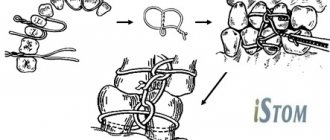
c) ligature binding according to Ivey
When fastening according to Ivy, a wire 10 cm long is first bent into the form of a hairpin, leaving one end 1-1.5 cm longer than the other. At the end of the “hairpin” a loop with a diameter of about 0.2 mm is formed. To do this, you can use a small piece of aluminum wire, crampon tongs, or tweezers. Both ends of the wire are passed from the vestibular side to the oral side between the teeth included in the bandage. The long end of the wire is returned to the vestibular surface through the interdental space located posterior to the loop and passed through it. The short end is brought out to the vestibular side through the interdental space located anterior to the loop and twisted with the long end. The excess wire is cut off by bending the remaining end about 0.5 cm long so that it does not injure the mucous membrane of the cheek. The same bandage is applied to the teeth of the second fragment, the antagonist teeth. The fragments are reduced and fixed to the teeth of the upper jaw with wire passed through the loops of the ligature bandage on each side.
This method has some advantages over the simple one: it is less traumatic, it allows you to examine the oral cavity without removing the entire structure, but only by cutting off the ligatures connecting the teeth.