Symptoms of this disease will be:
- hyperemia, swelling, burning and hemorrhages from the gums;
- hypersensitivity to cold, hot, sour foods;
- aesthetic defect of the gums.
Often, hypertrophied gums act as a mechanical obstacle when chewing food, which contributes to injury, penetration of pathogenic microorganisms into damaged areas and aggravation of the inflammatory process. Diagnosis of hypertrophic gingivitis is carried out through examination, palpation examination of the patient’s gums, determination of dental indices, as well as based on the results of additional studies (radiography and CT of the dental system). Therapeutic tactics include the use of local anti-inflammatory drugs, physiotherapeutic procedures, sclerotherapy, diathermacoagulation of the gum papillae and surgical excision of hypertrophied tissue (gingivectomy).
According to statistical data, the children's category of the population suffers from this disease most often, especially children and adolescents in the pre-pubertal and puberty period (from the beginning to full puberty), since the phenomenon of hypertrophic gingivitis is often associated with hormonal development.
Hypertrophic gingivitis is one of the forms of gingivitis (inflammation of gum tissue) and is less common in comparison with other forms (catarrhal, atrophic, ulcerative necrotic).
Classification of gingivitis according to ICD-10
- K05.0 Acute gingivitis
- K05.1 Chronic gingivitis
- K05.11 Hyperplastic
- K05.18 Other specified chronic gingivitis
- K05.19 Chronic gingivitis, unspecified
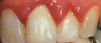
Normally, the gum reaches the crown of the tooth without covering it. It is pale pink, does not hurt or bleed. There is no noticeable distance between the tooth and the gum, however, with hypertrophic gingivitis, due to the enlargement of the gum, a “false pocket” appears between the tooth and the gum, i.e. The gum is attached to the tooth, and its enlarged part forms a pocket.
Ulcerative-necrotizing gingivitis
Otherwise, the disease is called Vincent ulcerative-necrotizing gingivitis. It occurs acutely or occurs chronically.
The source of the development of the disease is said to be a sharply increasing amount of bacterial plaque at the border of the gums and tooth enamel, with a predominance of spirochetes and fusobacteria in the biomass. In this case, there is an initial ulceration of the mucous membrane, and then the tissues die. The provocateurs of ulcerative gingivitis are often a decline in immunity and serious illnesses with a chronic clinical picture.
The acute form is accompanied by high fever and bleeding from under the gums. Loss of appetite, the smell of rotting flesh emanates from the mouth. The gums look affected, with ulcers, the interdental papillae acquire a gray tint and become necrotic. In a chronic course, the same thing happens, but the picture is blurred and not expressed so intensely. Changes are developing gradually.
It is impossible to treat ulcerative gingivitis at home; a visit to the dentist is required to establish a diagnosis and prescribe antibiotic drugs. Dental plaque is removed using professional means. It will not do without applications and rinses. To regenerate the mucous membrane, active gels are prescribed, for example Solcoseryl.
Causes of hypertrophic gingivitis
Hyperplastic gingivitis can be caused by both internal disorders and local factors.
Local factors
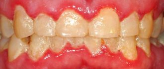
- copious amounts of tartar and plaque
- trauma to the gums due to the edge of a crown, filling, or denture that puts pressure on the gum
- improper use of dental brushes and toothpicks also leads to injury
- malocclusion
- orthodontic devices (braces, plates): they irritate the gums and complicate oral hygiene
- old and low-quality fillings, crowns or veneers put pressure on the gums and cause them to enlarge
General factors
- taking medications to treat epilepsy, diabetes, reproductive system diseases and heart disease
- taking contraceptives
- physiological hormonal changes in adolescents, pregnant women, and women after menopause
- vitamin deficiency, especially vitamin C
- blood diseases
- endocrine diseases such as diabetes
Diagnostic features
Gingivitis can be recognized visually—sometimes one examination by a doctor is enough. But you should make sure that there are no more serious pathologies, so diagnosis may include not only a visual assessment of the condition of the oral cavity, but also other measures:
- collecting anamnesis, assessing the condition of structures in the oral cavity;
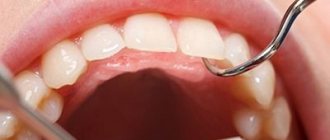
- probing of periodontal pockets if present;
- determination of tooth mobility;
- electroodontodiagnosis to determine the condition of the dental pulp;
- panoramic x-ray or targeted x-ray - to exclude periodontitis, periostitis and other pathologies of deep structures, jaw bone tissue, etc.
It is important to take into account the presence of chronic diseases and medications. Only with complete information can a doctor make an accurate diagnosis and develop an effective treatment regimen.
Classification of hypertrophic gingivitis by severity2
| DEGREE OF SEVERITY | A COMMENT | ||
| severity: | Lightweight | a comment: | The gum covers less than half the height of the tooth crown |
| severity: | Average | a comment: | The edge of the gum covers the crown up to half |
| severity: | Heavy | a comment: | The gum covers more than half of the crown |
Hypertrophic gingivitis – Prices
| Services | Price |
| Periodontist consultation | 1100 rub. |
| Consultation with a periodontist with drawing up a treatment plan | 1000 rub. |
| Medicinal treatment of periodontal pockets in the area of 2 teeth | 200 rub. |
| Surgical treatment of periodontal pocket 1 tooth | 600 rub. |
| Tooth extraction according to DS: Periodontitis, with bone grafting (preparation for implantation using biomaterials) | 20,000 rub. |
| Opening a periodontal abscess | 1750 rub. |
| Flap surgery on the periodontium of one jaw without the use of biomaterials | from 3000 rub. |
Classification of hypertrophic gingivitis by location2
| DEGREE OF SEVERITY | A COMMENT | ||
| severity: | Localized form | a comment: | The gums are enlarged only in a certain group of teeth; it occurs due to trauma, crowded arrangement of a separate group of teeth |
| severity: | Generalized form | a comment: | The entire gum is affected and occurs when taking medications or diseases. |
Classification
There is no generally accepted classification. Morphologically, catarrhal, ulcerative-necrotic, hypertrophic and atrophic forms of Gingivitis are distinguished; according to the nature of the course - acute and chronic gingivitis. According to the prevalence of the process, a localized form is distinguished (the so-called papillitis - inflammation of individual gingival papillae) and a generalized form (the so-called marginal Gingivitis - inflammation of the entire gingival margin).
According to the clinical course, they are distinguished: mild form - the inflammatory process is localized in the cervical part of the gums (there is no pathological dental-gingival pocket); moderate-severe - inflammation of the gingival papilla and the mucous membrane of the alveolar part of the gums and the presence of a pathological dental-gingival pocket up to 4 mm deep, as well as a severe form, when the entire mucous membrane of the gums is inflamed up to the transitional fold and the depth of the dental-gingival pocket is more than 5 mm. Hypertrophic G., in addition, is divided into the first, second and third degrees of hypertrophy of the gingival papillae.
Based on the etiological basis, they distinguish between G. traumatic, infectious, allergic, G. due to drug intoxication, systemic diseases (hypo- and avitaminosis, endocrine, gastrointestinal tract, blood system, etc.)” and also G. due to periodontal disease.
To express the prevalence of the inflammatory process of the gums in numbers or percentages, the so-called. papillary-marginal-alveolar index (PMA), which is calculated by adding up the assessment of the gingival condition of each tooth; at the same time, the number of teeth at the age of 6 to 11 years is conventionally considered equal to 24, from 12 to 14 years - 28 and from 15 years - 30; inflammation of the papilla is assessed as 1, cervical part of the gum - 2, alveolar part of the gum - 3: RMA (in%) = (sum of indicators × 100)/(3 × number of teeth) Etiology. The most common local causes of G. are unsatisfactory hygienic condition of the oral cavity and hron, mechanical trauma: tartar, decayed tooth, overhanging filling, poorly fitting crowns. G. can be caused by physical. damage to the mucous membrane (acids, alkalis, thermal burns, ionizing radiation, etc.), as well as microorganisms contained in dental plaque. Ulcerative-necrotic G. occurs with the participation of spindle-shaped rods (Bac. fusiformis) and spirochetes (Borrelia vincentii). Common causes include bacterial and viral infections, diseases of the gastrointestinal tract. tract, endocrine changes during puberty, pregnancy, allergic reactions, intoxication with heavy metal salts, vitamin deficiencies, blood diseases. A predisposing factor is a decrease in the body's resistance.
Symptoms depend on the form of the disease:
Edema form
The gums between the teeth are red, swollen, glossy, and a false pocket forms between the enlarged gum and the tooth. There is a change in the appearance of the gums, it looks swollen, bleeds, hurts when brushing teeth, and there is a feeling of burning and swelling. The edematous form develops as a complication of catarrhal gingivitis under the influence of an irritating factor (crown edge, fillings, braces, etc.). During pregnancy, hypertrophic gingivitis develops if the woman already had catarrhal gingivitis before pregnancy. The disease may resolve spontaneously after childbirth.
Fibrous form
The gum, as normal, is pale pink, there is no pain or bleeding, but its growth is observed, there are false pockets between the tooth and the gum. Abundant dental deposits are found above and below the gum. The fibrous form often develops when taking medications or in the presence of general diseases, i.e. this is the reaction of the gums to internal changes.
Principles of treatment
Treatment of gingivitis begins with hygiene: professional teeth cleaning. It is important to remove soft and hard dental plaque. For this, hand tools, an ultrasonic scaler, and the powder blasting method can be used. Subgingival dental plaque can be removed using the Vector device.
It is important to eliminate foci of infection - to fill teeth affected by caries, to undergo endodontic treatment in the presence of pulpitis, to remove the roots of teeth that cannot be restored and are not involved in the prosthetic process.
It is necessary not only to pay attention to the causes of the disease, but also to reduce the influence of harmful factors:
- stop smoking;
- consume food and drinks only at a comfortable temperature;
- remove spicy and smoked foods, marinades, especially those with vinegar, from the diet;
- During the treatment period, try to eat less solid food so as not to injure the loose gum tissue.
If the source of injury to the mucous membrane is the sharp edges of teeth or dentures, the doctor will immediately take measures to prevent further damage or recommend contacting a dentist of another profile - an orthopedist, an orthodontist.
Colored plaque
The brightest color is painted on the denser plaque; it is in these areas that teeth are most often not cleaned, which leads to gum inflammation.
After the final diagnosis, the dentist selects treatment tactics depending on the identified form of gingivitis.
Main symptoms
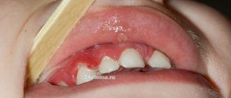
- Severe growth of the gum edge and gingival papillae, to the point that they cover almost the entire tooth crown, preventing normal chewing of food;
- pain, swelling and bleeding at the slightest touch or while brushing your teeth;
- bright red color of inflamed gums (characteristic of the edematous form of hypertrophic gingivitis);
- pain when eating, a sharp pain reaction to cold, hot or sour foods.
Since the gums react with pain to the slightest touch, brushing your teeth becomes an extremely painful procedure. This leads to the accumulation of plaque on the surface of the teeth and gums, which only aggravates the complexity of the disease. Therefore, it is important to start treatment in a timely manner to clean the surface of the teeth and gums from plaque, minimizing the bacterial factor.
Pathohistology
Catarrhal Gingivitis is characterized by thickening of the epithelium of the gingival papilla with the phenomena of parakeratosis (see) and acanthosis (see). The cytoplasm of the cells of the spinous layer is granular, the clarity of the boundaries between cells is disrupted. In the nuclei of cells, the phenomena of vacuolar degeneration are often detected (see). In the connective tissue of the stroma there are limited or diffuse lymphoid-macrophage infiltrates with an admixture of plasma cells, in the acute period - with an admixture of segmented granulocytes. In acute cases - swelling of the connective tissue of the gums, expansion of capillaries, venules, swelling of the endothelium, parietal hyaline thrombi. Signs of connective tissue disorganization with mucoid swelling are determined.
In ulcerative-necrotic G., a picture of an acute inflammatory process is observed with the presence of surface defects of the epithelium, pronounced acanthosis, parakeratosis, and infiltration of segmented nuclear granulocytes. Lymphoid-macrophage infiltrates and necrobiosis phenomena are detected in all layers of the gums (see Necrosis). In the stroma there are foci of moderately pronounced plasmorrhagia, the number of capillaries is increased, their lumen is unevenly expanded.
Hypertrophic G. is characterized by hyperplastic growths of the epithelium with pronounced vacuolization of the cytoplasm of the cells of the spinous layer. In the connective tissue - edema, plasmorrhagia of the walls of blood vessels, perivascular infiltrates consisting mainly of lymphoid and plasma cells, an increased number of mast cells with degranulation phenomena, fibrosis in the deep parts of the stroma.
Atrophic G.: pronounced thinning of the epithelial layer, the phenomenon of vacuolar degeneration of the cytoplasm of the cells of the spinous layer; in the connective tissue - thickening of collagen fibers with moderate plasmorrhagia, less often with fibrinoid deposits, moderate lymphoid-macrophage infiltration with the presence of plasma cells, single leukocytes and mast cells.
Gingivitis – gum disease
Gingivitis is the most common gum disease, which is characterized by redness, bleeding of the gums, and swelling. According to statistics, only 3% of the population have completely healthy gums. To get into this percentage, you need to take care of your gums and regularly see a doctor so as not to miss the initial signs of the disease.
Gingivitis is a stage of gum disease when it is still possible to prevent the development of the inflammatory process and completely stop it. The next stage is periodontitis, in which inflammation spreads to the following periodontal tissues. After the onset of periodontitis, a complete cure is no longer possible; here we are talking about relieving the symptoms, bringing the disease into remission and delaying the situation when it is necessary to remove teeth. In this regard, it is necessary to be attentive to the health of the gums, to prevent their disease, and if problems arise, immediately consult a doctor to prevent serious damage to the periodontium.