Implantation, the most successful and effective method of restoring a natural smile, is constantly being improved, as is dental treatment in general. Patients are offered innovative techniques that provide better results or reduce the duration and cost of treatment. One of the major achievements in recent years has been the barrier membrane, which is installed in conditions of bone tissue deficiency.
Alveolar ridge atrophy is almost inevitable in people who require implantation. Some people delay therapy for a long time, which is why the bone has time to dissolve, while others have congenital structural features of the jaw apparatus. In any case, performing dental implantation in the event of atrophic phenomena in the bone tissue becomes impossible. Such an error threatens the following adverse consequences:
- Loosening, retraction or even loss of the implant (sometimes its loss is quite traumatic, involving the patient’s bone tissue);
- Impossibility of performing bone grafting in the future due to excessive thinning of the bone (any intervention risks fractures).
Moreover, jaw bone resorption is so seriously detrimental to the patient that even without implantation, bone grafting remains a necessary surgical procedure.
Gradually progressive atrophy leads to the fact that the face becomes asymmetrical and ugly: wrinkles worsen, the functioning of facial muscles is disrupted, and so on. The chewing function of the jaws is significantly affected, which is why patients eat poorly and have problems with the gastrointestinal tract. In advanced situations, speech and swallowing are noticeably impaired, and breathing movements are less likely to suffer. Therefore, bone grafting with additional installation of a barrier membrane must be performed immediately after the problem is detected.
In what cases are barrier and protective membranes applied?
Membrane linings perform several functions:
- Protective - protect the operated area from external factors.
- The role of the reinforcing material is to fix the osteoplastic material in a suitable position.
Applicable:
- when strengthening mobile units;
- to protect bone from atrophy after tooth extraction;
- when it is necessary to fix granules of osteoplastic material during osteoplasty before or during implantation;
- when building up the jaw bone with blocks.
The decision to use barrier membranes is made by the dentist after a preliminary examination.
To increase bone
An osteoplastic material is fixed under the gum, on top of which a protective pad is placed. It protects the bed and its contents from growing soft tissues. Eliminates washing out of the bone graft from the wound through the suture line. More reliable treatment results are provided by the use of a bone substitute along with two types of membranes. One of them covers the defect and osteoplastic material, and a collagen plate is placed on top of it.
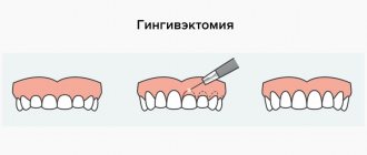
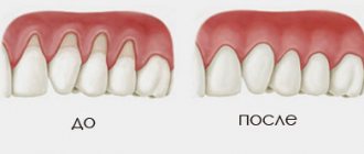
Gum extension (augmentation) during gum recession
Deficiency of soft tissues as a result of atrophy is characteristic of patients with long-term absence of teeth. Significant bone loss in such cases is accompanied by a proportional decrease in gum volume, which causes significant discomfort. Augmentation is more expensive and more difficult to install an implant. Gum augmentation can be performed with non-resorbable membranes, usually based on a non-porous material and resorbable, with a high rate of tissue compatibility. The method promotes rapid healing, reduces the risk of relapse and soft tissue swelling.
Types of barrier membranes
The barrier membrane is an ultra-thin elastic film and is attached to the jaw with small titanium pins. Located between the gum and the bone, the membrane performs not only a separating, but also a retaining function. It fixes the graft in the desired position and provides its reliable protection from mechanical damage. In modern dentistry, two types of such films are used:
1) Removable barrier membrane.
It does not require surgery to remove it, as it resolves after a certain period of time.
2) Non-removable barrier membrane.
This is a dense, non-absorbable film that at a certain stage requires surgical removal.
| Collapsible barrier membrane | Non-removable barrier membrane |
The material for the barrier membrane is natural collagen; it is absolutely neutral for the immune system and does not cause inflammation or rejection by the body. In addition, the film prevents the penetration of pathogenic bacteria, thereby minimizing the risk of complications.
Types of biomaterials used
The method of directed tissue regeneration in surgical dentistry is based on the use of two main groups of membranes: resorbable (collagen, vicryl, polylactic acid, etc.) and non-resorbable (Gore-tex or titanium mesh, with titanium reinforcement, Teflon and others).
Resorbable
Installation of absorbable membranes prevents their removal after the osteoplastic material has engrafted. Such linings do not always remain stable over a long period. To enhance the prolonged action, their composition is supplemented with slowly resorbing substances. One of the main advantages of membrane linings is the possibility of enriching their structure with drugs that enhance osteogenesis, anti-inflammatory, antimicrobial agents, etc.
Types of resorbable membranes:
- Synthetic polymers (polyactides, polyglycolides) and their chemical modifications (for example, Guidor, EpiGuide, Vicryl) - decomposition into microscopic fragments that undergo phagocytosis occurs under the influence of hydrolysis. Among the disadvantages of the products, weak tissue integration and a high likelihood of developing swelling during resorption due to changes in pH in the tissues are noted. Advantages of polymer plates:
- no risk of transmitting an infectious disease;
- hypoallergenic;
- long-term effect (5-6 months).
Non-resorbable
They contribute to the effective restoration of bone in a given volume and trajectory, there are:
- Frameless - for restoration of bone tissue less than 4 mm.
- With a titanium frame - for bone restoration in large volumes, in all directions.
Main advantages:
- high mechanical strength;
- pronounced barrier properties.
These types of pads should be removed after 6-9 months . Consisting of titanium, they are effective in situations where there is a large load on the bone and it is necessary to protect implants or osteoplastic material. The most common indication for use is vertical ridge augmentation. They are removed after restoration of soft and bone tissues damaged during the operation.
Main disadvantages:
- risk of exposure - during suturing of the flap, the insert must be completely closed;
- absence of the process of periosteum formation under the overlay;
- the patient needs to visit the dentist frequently (every 2 weeks).
Statistics show that the most effective membranes are polytetrafluoroethylene (PTFE) GoreTex and aliphatic polyetherurethane (Bone-up). Less commonly used are mesh forms made of calcium sulfate (Capset) or titanium (Frios, BoneShield, Tiomesh).
general characteristics
Bone demineralized collagen, in the form of an elastic membrane plate, has a pronounced mechanical barrier property that prevents the migration of soft tissue into the bone defect to ensure the directed development of new bone tissue. Prevents apical migration of gingival epithelium.
Contains sGAG - connective tissue growth modulators that promote cell adhesion and specific binding of growth factors and molecules necessary for tissue reconstruction.
Fixing elements of different types
The membrane is adjusted to the size of the defect, or the required dimensions are transferred to it using sterile templates in the package. The edges should overlap the boundaries of the bone defect by at least 2-3 mm . Fixation can occur in one of several ways:
- Before installation, the resorbable pad is pierced and placed on the implant using the rubber dam principle. The edges are secured with screws or pins into the bone, in which holes are previously created with burs. Additionally secured with sutures to the periosteum. When used on the upper jaw, titanium pins with a wide head are used for fastening.
- The self-curing mixture or film with an adhesive composition is fixed directly to the alveolar process.
- Non-resorbable plates are fixed with titanium pins or microscrews. The edges of the barrier must be tightly adjacent to the bone to prevent soft tissue ingrowth. When suturing the mucosa, small diameter suture material is used. The thread should pass along the edge of the membrane in contact with the neck of the implant. The stitching is done in the middle of the overlay between the edge stitches.
Opinion of a dentist-implantologist : “The success of implantation depends on how accurately patients follow medical recommendations. The operated area should be protected from hard, hot foods. Hygiene must be regular. Medications are taken only in consultation with a doctor.”
- Complete restoration of the dentition in just 4 days!
more detailsRoott Pterygoid Implants Sinus lift is no longer needed!
more details
Once and for life! Express implantation in 4 days with a permanent ReSmile prosthesis
more details
All-on-4, All-on-6, ReSmile, Zygomatic implantation We use all modern methods of dentition restoration
more details
Use of titanium membranes for guided bone regeneration
K. N. Khabiev
PhD, certified implantologist of the European Association of Osseointegration, expert of the international research center MINEC, president of the group
The problem of restoring bone tissue around an implant immediately after its installation is one of the most pressing. The use of titanium membranes for these purposes gives good lasting results. To obtain a long-term aesthetic result, the thickness of the bone tissue around the implant, especially on the vestibular side, must be at least 1.8 mm. But, in addition to the thickness of the bone around the implant, it is also necessary to take into account the height of the bone above the implant. It should also be at least 0.5-1 mm.
Both horizontal and vertical tissue volume can be restored using titanium membranes with a special fixation system. To fix the membrane over the implant, a special flat abutment is used, which rises 1, 2, 3 mm above the implant platform.
A pre-curved titanium membrane is placed over this flat abutment and secured to the abutment with a plug or former. A feature of the I-Gen membrane is that it is pre-curved and therefore allows one to fairly accurately predict the volume of hard tissue restoration both vertically and horizontally (Fig. 1-3).
Rice. 1. The AnyRidge implant is installed in a narrow ridge. Rice. 2. The I-Gen titanium membrane is fixed with a flat abutment. Rice. 3. After 3-4 months, the horizontal and vertical volume of the bone is restored.
Clinical case
Patient G. came to the Dental Guru research clinic with complaints of a missing front tooth. The tooth was removed 2 years ago. A targeted X-ray image showed a vertical bone defect; on a tomogram, the thickness of the alveolar edge was 2.9 mm (Fig. 4). Since the vertical bone defect was cup-shaped, which means there was good potential for new bone formation, it was decided to install an implant simultaneously with bone grafting.
An AnyRidge implant with a diameter of 3.5 mm and a length of 11.5 mm was installed (Fig. 5). A mixture of allogeneic and xenogeneic osteoplastic materials was used as an osteoplastic material. The I-Gen system was used to hold the graft on the vestibular side (Fig. 6).
To do this, a flat platform 2 mm high is fixed to the implant, an I-Gen membrane is put on it, which is then fixed with a gum former. Restoring aesthetics during the healing period was performed using temporary crowns, which were fixed to adjacent teeth (Fig. 7). 1.5 months after the operation, partial exposure of the titanium membrane occurred, however, the membrane was removed only 3 months after the start of treatment (Fig. 8).
Due to the fact that after removal of the membrane a defect formed in the area where it was exposed, a month later plastic surgery was performed with a free tissue graft from the palate (Fig. 9-10). The gum profile was formed with a temporary crown on the implant (Fig. 11-14). The successful formation of soft tissue in this case is due to sufficient support from the bone, so an optimal aesthetic result was obtained.
Since, thanks to the use of I-Gen technology, not only the thickness, but also the height of the bone tissue was restored, long-term aesthetic and functional results can be predicted.
Conclusion: to obtain a good functional and aesthetic result with a long-term prognosis, it is recommended to use the technology of fixing a titanium membrane 2-3 mm above the implant platform, which allows you to obtain not only horizontal, but also vertical tissue volume.
Rice. 4. Sighting x-ray before surgery. An AnyRidge implant with a flat abutment is installed. Rice. 5. The I-Gen membrane is fixed on a flat abutment using a healing abutment. Sighting x-ray immediately after surgery. Rice. 6. Partial exposure of the membrane 1.5 months after surgery. Rice. 7. Removal of the I-Gen membrane can be performed without detaching the mucoperiosteal flap. Rice. 8. A temporary crown is fixed on the implant. Rice. 9. A free tissue graft from the palate was transplanted. Rice. 10. Immediately after transplantation and fixation of the tissue graft. Rice. 11. Condition of soft tissues 2 months after the 2nd operation. Rice. 12. Condition of soft tissues before taking impressions to make a permanent structure. Rice. 14. Sighting x-ray with a temporary crown. Rice. 14. A combined zirconium abutment and a crown on a zirconium oxide frame are fixed to the implant.
The list of references is in the editorial office.
How membranes take root during dental implantation
Collagen membrane overlays reduce the risk of complications due to loss of soft tissue. Non-stitched - due to the healing of soft tissues by secondary intention, the wounds are protected from the general inflammatory process. Both types integrate well into the jaw tissue. The bone under the surface of the barrier plate is regenerated in a manner similar to fetal bone growth.
When using absorbable overlays, the postoperative wound heals without the use of additional medications. Often the healing process of non-resorbable membranes is accompanied by complications, in particular, exposure of the structure. This can lead to infection and inflammation, which means it will have to be removed prematurely.
Often, under the barriers a favorable background is created for the development of microbes, so it is not always possible to achieve the desired result.
PRF membrane: what is it and why is it needed?
The PRF membrane provides natural support for the process of tissue restoration - bone and soft, while the period of regeneration of epithelial cells after surgery is significantly reduced.
The PRF membrane is used in dentistry. The decoding of three Latin letters (PRF - Platelet Rich Fibrin) literally sounds like “platelet-rich fibrin”. This is exactly what it represents - the patient’s own fibrin, enriched with its own growth factors. The membrane is manufactured directly in the clinic. During the appointment, blood is drawn from a vein from the patient, which is then placed in special tubes in a centrifuge, where the blood is separated into red blood cells, plasma and fibrin clot (PRF). Subsequently, it is cut and placed in a special box to be converted into a membrane of the required thickness.
The PRF membrane itself is ultimately a fibrin matrix containing growth factor-rich platelets. They will be released on the surface of the wound and speed up the healing process.
Membrane exposure after bone grafting - consequences and treatment
The non-resorbable membrane pad is exposed in 1% of cases , causing the following consequences:
- wound infection with the development of purulent inflammation;
- rejection of osteoplastic material during vascularization of the mucous membrane (in 40-50% of cases).
In such situations, it is recommended to remove the implanted materials, carry out treatment, and then repeat bone grafting. Typically, a second layer of soft tissue is formed under the incision using a mucoperiosteal flap. To do this, it is peeled off, split, modeled, and then sutured in layers.
The exposed resorbable pad should be treated with 3% hydrogen peroxide and 0.12% chlorhexidine. Sometimes this allows you to postpone the removal procedure by 8 weeks.
Opinion of a dental surgeon : “Most of my patients are smokers. Many people do not immediately realize the complexity of the situation caused by their bad habit. Smoking causes a sharp narrowing of the blood vessels in the mouth. This weakens the flow of blood filled with oxygen and nutrients. Due to their lack, the healing process is delayed. Therefore, you need to completely stop smoking at least a couple of weeks before implantation and for several months after it.”
Why are collagen membranes reliable?
Collagen membranes are very popular for a number of reasons. But the first and main thing is the ideal biocompatibility of collagen with the tissues of the human body. This fact has long been scientifically proven. Collagen is present in large quantities in the protein compounds of human gingival tissue, which is why it is so popular in dental practice.
Collagen membranes for dentistry are made from porcine, bovine, calf dermis, bovine tendon and material of human origin. The latter type of membrane has not been studied enough, so animal collagen is usually used. It is highly antigenic and takes root well in the human body.
Collagen has proven itself ideally in hemostatic processes, in cases of membrane exposure - healing occurs exactly with secondary intention, completely eliminating the risk of inflammation and supporting the healing process of the walls of blood vessels and capillaries.
In relation to such positive dynamics, the Genoss collagen membrane - Collagen Membrane - showed excellent results. Today, the Genoss collagen membrane (South Korea) is the absolute leader among all analogues of collagen membranes in the world.