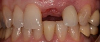
The roots of our teeth are located in the bone tissue of the jaw. Due to the fact that during chewing food the bone receives load from the teeth, it functions fully and is saturated with nutrients. But as soon as the tooth is removed, an empty space forms in its place - the bone cells grow quite slowly and do not have time to fill this space. As a result, the bone decreases in size, besides, metabolic processes are disrupted and cells stop receiving oxygen and nutrients. The bone atrophies.
Bone atrophy is a big problem when many teeth are missing. Indeed, in this case, not only aesthetic and psychological problems arise (facial skin sags, wrinkles appear near the lips, cheeks sag, lips become thinner and sink into the oral cavity), but also functional ones - after all, it is unlikely that you will be able to eat fully and efficiently without teeth .
Directed bone tissue regeneration is a surgical operation, the purpose of which is to restore the volume of the jaw bone, that is, to increase it to the previous size established by nature. This operation is mainly carried out as a preparatory stage before dental implantation - after all, in order to implant and securely fasten implants (artificial metal roots), a sufficient volume of strong bone is required. Sometimes it can be carried out together with the installation of implants.
Special offers!
Bone grafting, open sinus lift - RUB 35,000. Closed sinus lift (area of 1 tooth, excluding the cost of bone substitute) - RUB 10,000. Guided regeneration of bone tissue (area 1 tooth) - RUB 10,000. Guided bone regeneration (GBR) - RUB 35,000.
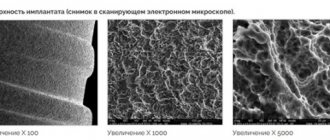
Materials for bone tissue augmentation
The word “regeneration” itself translated from Latin means “rebirth” or “renewal”. The operation is performed surgically and its essence boils down to replanting bone material in an area where the volume of the jaw bone is insufficient.
Various materials can be used for bone tissue regeneration:
- the patient's own bone: the most preferable option, since in this case 100% result of engraftment of the material is achieved. A piece of bone is usually borrowed from the chin area, the angle of the lower jaw (where the outermost wisdom teeth are located) or the hip area. The obvious disadvantage is that it is often necessary to perform two surgical interventions simultaneously,
- artificial synthetic materials: developed in laboratory conditions. A fairly common option, today it is used everywhere, in most cases as an additional material when grafting the patient’s own bone. Has good survival rate,
- related bone: This is typically cleaned cadaveric bone from another person that is kept in a sterile jar. It also takes root well and does not require additional surgical intervention.
- animal bone: the material can be presented in the form of bone blocks or in the form of shavings. It takes root well and is almost not rejected.
Bone regeneration: how is it done?
The operation lasts 1-2 hours, depending on the volume and complexity of the work. After 3-4 months, when the implanted piece has fused with the natural bone, it will be possible to proceed to the installation of implants. In some cases, it may take longer to engraft, depending on the individual characteristics of the organism. The technology for bone tissue regeneration is as follows:
- anesthesia is administered, in some cases, if serious surgery is required, it is possible to perform the operation under anesthesia,
- access to the bone is created: a piece of gum is cut and peeled off, with the help of protective membranes, which dissolve on their own over time, a cavity is formed to be filled with bone chips,
- bone material is introduced: the cavity is filled with artificial or natural bone material. After 2-4 months, the natural bone cells will penetrate deep into the implanted material, and it will turn into its own bone tissue - then implantation can begin. On top of the new bone is covered with a protective membrane, which will protect against pressure from the gums,
- The gum is sutured - sutures are fixed on the mucous membrane, which are removed on average after a week.
Expert opinion
Emir Romanovich Omerelli
Maxillofacial surgeon, implantologist
Experience: more than 13 years
If, based on the results of the examination, the doctor diagnoses a lack of bone tissue, it is necessary to increase its volume before implantation. Placing implants in low or narrow bone carries serious risks. The bone may fracture or the implant itself may fall out after a short period of use. To prevent this from happening, before classical implantation of the upper teeth, it is necessary to carry out a sinus lift, and for the lower teeth, extension through osteoplasty. The only exception is the basal protocol, according to which implants can be installed without bone grafting and without risks.
If the bone tissue is not wide enough, that is, it is very thin, the operation can be performed by splitting the alveolar ridge: first the gums are exfoliated, then the bone is cut along the length and artificial or natural bone material is placed inside.
Also, the problem of narrow bone tissue is solved by transplanting a bone block borrowed from another bone of the patient himself. To do this, a piece of bone is placed under the gum, fixed with special screws, sprinkled with bone chips around it, covered with a barrier membrane and gum - the mucous membrane is sutured. This option is more preferable, since in most cases the own bone survives.
Bone tissue augmentation surgery, depending on the indications, can be performed simultaneously with the installation of classical implants without their subsequent loading. That is, the artificial roots are fixed in the bone, but are covered by the gum - the dentures will be fixed in 3-4 months. In order for classical implants to take root in the bone, it is necessary to ensure their rest and relieve any pressure.
Complications after bone tissue regeneration
The operation of bone tissue augmentation is considered one of the safest in dentistry, complex, but if carried out correctly, without any unpleasant consequences. In the first days after the operation, it becomes clear whether the block will take root or the operation will have to be performed again. Among the unpleasant sensations are slight tissue swelling and pain in the area where the bone block was taken and the site where it was transplanted. But after 2-4 days, all unpleasant sensations go away on their own.
After bone augmentation
After the bone regeneration procedure, the patient must take precautions: be sure to take medications prescribed by the doctor to quickly restore the body, and do not ignore painkillers. You should refrain from eating food for 2-3 hours after surgery; afterwards it should be warm, but not too cold or hot. It is recommended to chew foods on the side opposite the surgical procedure. In addition, it is necessary to give up physical activity for a while and provide the body with rest for a couple of days after the operation. Do not forget to visit your doctor for a preventive examination and monitor the results of treatment.
Other jobs
Currently, injuries are among the top five leading causes of death in the world, threatening economic and social development. Fractures of long bones occupy a leading place in the structure of injuries in recent decades and, according to various authors, account for up to 80% of all injuries to skeletal bones.
Therefore, the problem of treating bone fractures has remained relevant in recent years, although progress in the field of traumatology is undoubted. A number of issues related to the so-called osteogenic failure remain unresolved.
Despite the use of modern medical technologies, the percentage of complications resulting from the treatment of long bone fractures remains high.
These include delayed consolidation, formation of false joints, nonunion of bone fragments and other problems.
Thus, impaired consolidation of bone fragments in fractures of long bones ranges from 15 to 50%, and the frequency of false joints varies from 4 to 33%.
According to the American Orthopedic Association, of the two million long bone fractures in the United States each year, about 100,000 result in nonunion.
According to domestic researchers, violations of the consolidation of bone fragments during fractures of the limbs account for about 25% of the disability structure of victims of mechanical trauma.
The percentage of failures in the treatment of such disorders using classical methods reaches 33%, which is almost twice as high as the number of unsatisfactory anatomical and functional results of treatment of fractures.
In recent years, there has also been a hidden increase in disability due to injuries and an increase in the consolidation period in every third case.
The problem of treating disorders of reparative bone tissue regeneration that arise after diaphyseal bone fractures remains relevant for modern orthopedics and traumatology. Cases of the development of such pathology range from 2.5 to 18%.
In the structure of the consequences of injuries to long bones, pseudarthrosis of the femur accounts for 10-30%, of the tibia - 15-50%, and of the humerus - 1-10%.
It is noteworthy that among the population with impaired reparative bone regeneration, people of working age predominate.
This pathology is marked by permanent disability in 5 people per 10,000 population, and the resulting anatomical and functional disorders of the limb are the cause of permanent disability in 12-45% of patients.
Long-term treatment of patients with high material costs, low efficiency, and a high level of disability allow us to consider complications from fractures of long bones as the most important social problem, which many victims find difficult to cope with without social assistance.
It follows from this that the treatment of long bone fractures complicated by reparative disorders is an urgent economic and medical-social problem.
The main task of modern traumatology is to improve existing and develop new, economical and effective treatment methods.
What is bone tissue regeneration?
Bone is a complex organ that performs mechanical and biological functions in the body and has a complex hierarchical structure.
Bones participate in metabolic processes due to the content of a significant percentage of the body's minerals, and also create a specific microenvironment for red bone marrow blood precursors.
Bone tissue is a dynamic system in which, during the life of an organism, two interconnected, opposing processes occur that make up the natural remodeling cycle - resorption (destruction) and osteogenesis (synthesis).
Therefore, the conditions of reparative osteogenesis are constantly in the field of view of practicing doctors - traumatologists and orthopedists.
It has long been known that fractures in the area of compact (cortical) and spongy (trabecular) bone tissue differ in the timing of consolidation, which is associated with the peculiarities of its structural organization.
The prospects for healing of cancellous bone fractures are more favorable, since its structure contains the elements necessary for the formation of a regenerate. In turn, compact bone tissue is characterized by a low vascular density compared to spongy bone tissue, and also has peculiarities of blood supply.
Reparative regeneration is defined as the process of tissue repair after injury. The mechanisms of reparative and physiological regeneration are the same and are based on common biological principles.
Reparative regeneration is considered to be enhanced physiological to one degree or another. Reparative regeneration of bone tissue - also known as reparative osteogenesis - is an important theoretical and practical problem in traumatology.
Indeed, ideally, consolidation of the fracture should lead to the formation of renewed bone tissue identical to the old one that existed before the injury. However, fracture healing in practice is a rather complex, long-term, multi-stage process, which is influenced by many factors.
Bone has a very high reparative potential.
The problem is that the processes of reparative osteogenesis can be accelerated by activating metabolism only to a very small extent (on the order of weeks). On the other hand, it is very easy to slow down the process by disrupting physiological conditions, which often occurs due to insufficient understanding of bone physiology.
According to the definition of Rutsky and Tkachenko, reparative regeneration is a complex process caused by the destruction of bone structures, which quantitatively exceeds the permissible limits of physiological regeneration and is aimed at completely restoring the anatomical integrity and function of the bone.
According to researcher A. Korzh, bone regeneration processes are a complex interweaving of general influences at the systemic level and local changes in tissue metabolic processes, including changes at the molecular level.
D. Sarkisov and co-authors present reparative osteogenesis as physiological regeneration, which occurs under conditions of extreme influences on the body and is characterized by a greater intensity of manifestations.
Other researchers argue that, unlike physiological regeneration, which is actually an adaptation, reparative osteogenesis is a compensatory process that restores the structure after the death of part or the entire organ.
Reparative regeneration of each tissue type has unique characteristics, but always includes the following processes:
- Destruction of damaged cells and structures
- Proliferation of viable cells in the defect area
- Differentiation of viable regenerate cells
- Formation of intercellular connections and restructuring of the regenerate.
Reparative regeneration of bone tissue can be incomplete or complete.
Complete regeneration is characterized by replacement of the defect with tissue that matches the old tissue. It is customary to speak of incomplete in cases where a bone defect is replaced by connective tissue or scar.
Reparative osteogenesis is a multicomponent process, the main stages of which are cell differentiation, proliferation, resorption of dead tissue and bone formation with its remodeling, the formation of an organic extracellular matrix and its mineralization.
The described processes occur in parallel, but one of them can become dominant at certain stages of reparative osteogenesis.
Practice confirms that bone tissue is truly unique, since it is capable of completely restoring even large defects.
Causes of disorders of reparative bone regeneration
It is known that reparative regeneration of bone tissue is a complex, genetically programmed process. The stage-time characteristics of this process depend on the action of a number of endogenous and exogenous factors.
The course of the osteoreparative process is associated with the following factors:
- Features and intensity of injury
- Nature of bone and soft tissue damage
- Degree of post-traumatic disorders of peripheral blood supply
- Quality of primary and qualified medical care
- Features of rehabilitation treatment
- The presence of concomitant pathology.
One of the first places among the reasons contributing to the development of disorders of reparative osteogenesis is the untimeliness and inadequacy of the provision of qualified medical care.
It has been experimentally proven that in cases of delayed immobilization of fragments from fractures of the forearm bones, signs of disruption of the bone repair process appear already on the 3rd day with an increase in the consolidation period by 1.5 times.
Delayed comparison of shin bone fragments after 14 days leads to the formation of a false joint, and in the absence of stable fixation of the fragments, the time required for the formation of periosteal bone fusion increases by at least 4 times.
Further experimental studies showed that violation of the conditions of reparative regeneration in the form of comparison of bone fragments delayed up to 14 days leads to the formation of a pseudarthrosis on the 50th day.
When a fracture heals slowly, bone restoration does not occur within the usual time frame, and remodeling and callus maturation are inhibited.
As a rule, healing in such situations occurs after a year or more.
A fracture is regarded as non-union if after 6 months there are no radiological signs of healing, or when there is no positive dynamics within three months of observation.
When a fracture fails to heal, reparative processes are inhibited and healing stops. The concept of “delayed consolidation” is considered to be relative, since the timing of fracture healing in each patient is individual and depends on many factors.
Conventionally, these factors can be divided into general and local.
General factors are associated with the general condition of the body, concomitant somatic pathology, drug therapy, the presence of bad habits, and the like.
The factors of the second group include the lack of reliable immobilization of the segment, insufficient reposition, fixation and impaired blood supply to bone fragments, traumatic surgery, violations of treatment tactics, and the use of massive metal implants.
An important link in reparative regeneration is the condition of the bone tissue at the time of injury, as well as the endemic condition of the region where the injured person lives.
Recently, the number of studies regarding the influence of unfavorable environmental factors on reparative osteogenesis of bone tissue has been increasing. Domestic and foreign authors have studied the influence of the human ecological environment on the structure and metabolism of bone tissue, on which the course of reparative regeneration depends.
Clinical studies demonstrate that increased fluoride intake is accompanied by bone fragility, while the process of reparative osteogenesis slows down and false joints are more likely to form.
After conducting several in vitro experiments, scientists came to the conclusion that radiation has a destructive effect on bone tissue, which also slows down regeneration processes and increases the number of complications.
Therefore, when treating bone fractures, it is necessary to take into account the impact of unfavorable environmental factors on bone tissue regeneration.
Currently, the topic of scientific discussions is the issue of the dependence of the frequency of violations of osteoreparative processes on the mechanism of injury. According to clinical studies, the etiological factor of injury affects the wound healing process.
The course of the reparative process is influenced by both the type of traumatic agent and the nature of the damage to bone tissue. According to a number of authors, in 55% of patients with impaired reparative osteogenesis, the injury was caused by the action of a high-energy traumatic agent.
Experimental studies have revealed that when exposed to high-intensity traumatic force, a significant number of cellular sources of osteoreparation die, and the surviving cells undergo such significant morphological changes that they are unable to maintain normal tissue functioning.
E. Pobel and co-authors cite a number of reasons that lead to disruption of post-traumatic osteoregeneration processes:
- Patient age
- Nature of traumatic injury
- Pathological condition of bone tissue (osteoporosis)
- Complicated medical history (chronic liver and kidney diseases, obesity)
- Decreased osteoreparative potential (deficiency of growth factors, osteocalcitonin, active form of the hormone vitamin D3)
- Traumatic nature of the metal osteosynthesis procedure.
These changes lead to disruption of the processes of consolidation of bone fragments and require measures to optimize reparative osteogenesis.
Bone is a highly specialized tissue that exists in close interaction with the circulatory system. The interconnection of hemocirculation pathways in bone tissue is manifested both in local nutritional processes, maintaining the general mineral balance of the internal environment of the body, and directly in the physiological and reparative regeneration of bone tissue.
Therefore, a significant factor causing disorders of reparative osteogenesis is impaired blood circulation in the area of damage.
Insufficient blood supply to the fracture zone due to massive damage to soft tissues, bone, detachment and trauma of the periosteum, and impaired medullary circulation lead to the activation of chondrogenesis and the inferiority of the process of fusion of bone fragments.
The studies carried out revealed the features of the structural and functional state of blood vessels in cases of disorders of reparative osteogenesis. These disorders may be caused by structural and functional changes in the vessels of the injured limb, which determine the characteristics of its course.
Of great interest is the conceptual model of the mechanism for compensating for disturbances in the regional blood supply during bone fractures, which was developed by researcher G.V. Gaiko and his colleagues.
Conducting experimental and clinical studies of peripheral blood supply during fractures, the authors came to the conclusion that the restoration of regional blood supply after a bone fracture is based on the redistribution of blood circulation and tissue revascularization.
Compensation of regional and local blood supply, as a rule, occurs within a few hours after injury in fractures without displacement of bone fragments, without extensive damage to soft tissues and great vessels.
Incomplete compensation is accompanied by hypoxia, promotes the development of fibrous connective tissue and stimulates fibrous fusion of bone fragments.
At the site of decompensation, necrosis of bone and soft tissue occurs, which is the reason for prolongation of treatment, the formation of false joints and the occurrence of purulent complications.
Stages of reparative bone tissue regeneration
The stages of reparative osteogenesis in their interrelation have been developed, in which each stage is characterized by a certain morphological cellular and tissue composition.
The study of morphological changes during the treatment of a bone fracture using various methodological approaches made it possible to distinguish two stages in regeneration:
- Construction of connective tissue callus and its replacement with immature callus.
- Restructuring into a callus precursor formed by mature bone.
According to the results of pathomorphological analyzes of the healing of bone fractures, the following stages of restoration of the bone defect are distinguished:
- I – destabilization of cellular elements
- II – intense cell proliferation
- III – differentiation of various tissues (cartilaginous, fibroblastic, osteoblastic, undifferentiated tissue similar to mesenchyme, fibroblastic connective tissue)
- IV – epigenesis of osteogenic tissue, in which processes of direct metaplasia, atypical enchondral ossification and osteoid modification are observed
- V - sponging of osteoid tissue and formation of osteons
- VI – formation of lamellar bone.
In another variant, 4 stages are distinguished: proliferation of osteoblastic cells, formation of collagen fibers, formation of an amorphous carbohydrate-protein substance and impregnation of the intercellular substance with mineral salts.
Using new data from molecular biology, biochemistry, morphology, immunomorphology and genetics, N. Korzh and co-authors identified 5 stages of the reparative process: inflammation, cell differentiation and the formation of tissue-specific structures in the area of injury, reorganization of tissue structures and their mineralization, remodeling and completion of the recovery process.
Many famous foreign authors share the same opinion.
In general, bone repair is a complex biological process that requires changes in the expression of several thousand genes.
More often, consolidation of fractures occurs through indirect bone repair and consists of several successive stages - inflammation, soft callus formation, hard callus formation and remodeling.
The inflammation phase begins immediately after the injury and lasts up to 5 days.
This stage includes the formation of an inflammatory hematoma due to rupture of blood capillaries in the fracture zone, migration of mesenchymal cells, neutrophils and macrophages into the inflammatory zone to remove fragments of damaged tissue, which differentiate into fibroblasts, osteoblasts and chondroblasts, followed by the formation of a cartilage matrix.
During the second stage, which lasts up to 40 days, the hematoma is replaced with fibrocartilaginous tissue through the differentiation of mesenchymal cells into chondrocytes, as well as the synthesis of a matrix from type 2 collagen.
Next, the matrix gradually calcifies and is replaced by bone tissue synthesized by osteoblasts.
During the third stage of the process, ossification occurs and bone bridges are formed between fragments of the broken bone.
The process of reparative regeneration ends with the remodeling stage, when its original shape, structure and mechanical strength are restored.
Disturbance in the course of any of these stages can lead to a slowdown in the process of osteoreparation as a whole or even non-union of bone fragments.
Histologically, there are two types of fusion of bone fragments: primary (direct) and secondary. Primary healing occurs due to the active proliferation of osteogenic cells against the background of tissue hypervascularization at the fracture site. Secondary healing occurs in several stages.
To date, several classifications have been developed that describe in detail the stages of fracture healing.
There is also known data on the stages of fracture healing, which are based on specific morphological transformations in the area of the bone defect:
- Thrombosis of hematoma
- Blood clot organization
- Formation of fibrous precystic regenerate
- Formation of a full-fledged bone tissue regenerate
- Formation of secondary bone regenerate
- Functional reconstruction of the regenerate.
A number of authors, based on the totality of structural, morphological and biochemical changes occurring in bone regenerate, identify the following phases of bone regeneration:
- I – catabolic, with disintegration and degradation of surrounding structures
- II – progressive cell proliferation and differentiation of cellular elements with secretion of the organic basis of bone regenerate
- III – complex biochemical, biophysical and physiological processes leading to the appearance of the primary bone structure
- IV - formation of a lamellar bone structure, ensuring restoration of the shape and function of the bone.
When compared with the above studies, the variant of classification of the stages of bone fracture healing (inflammation, phagocytosis, fibrous callus, primary and secondary callus) looks original, in which the authors did not sufficiently substantiately separate phagocytosis from inflammation.
Based on systemic concepts and evaluation of the results of numerous clinical and experimental observations of the dynamics of fusion of bone fragments, A.T. Brusco et al. proposed their staging system for reparative osteogenesis. Here it is considered as a unidirectional process occurring with a natural sequence of morphological changes in the regenerate.
The authors identified the following stages of healing of bone fractures:
- I – reparative reaction
- II – formation of fusion of bone fragments
- III – fusion of fragments, in which the following options are possible: a) primary bone fusion, b) fibrocartilaginous fusion, c) secondary bone fusion
- IV – functional restructuring of callus and consolidated fragments with the formation of the organ structure of the bone.
As we see, the problem of bone tissue regeneration occupies a special place in biological and medical knowledge. At the moment, the key features of bone tissue regeneration have been identified and certain successes have been achieved in elucidating the biological mechanisms underlying reparative osteogenesis, and the main trends in the development of science have been formulated.
Examples of work “Before” and “After”
Restoration of all teeth on the upper and lower jaw - basal implantation
Case: partial absence of teeth on the upper and lower jaw, complicated by severe periodontitis (tooth mobility).
Complex one-stage implantation of the lower jaw
Case: there was a loose bridge of 4 front teeth on the lower jaw; after diagnosis, removal of the remaining teeth and coplex basal implantation were prescribed.
Restoration of all teeth using basal implantation method (March 2012)
Case: partial adentia, exposed roots of natural teeth, periodontitis, increased tooth mobility, severe atrophy of bone tissue in some places beyond the possible norms for classical dental implantation.
Restoration of anterior teeth using basal implantation method (April 2012)
Case: partial absence of front teeth and destruction of supporting teeth under the prosthesis, the relief of the gums and interdental papillae is disturbed.
Sources of regeneration
Restoration of bone integrity occurs through the proliferation of cells of the osteogenic layer of the periosteum, endosteum, insufficiently differentiated pluripotent cells of the bone marrow, as well as due to metaplasia of giaraosseous tissues.
Modern ideas about the processes of bone tissue regeneration combine the concepts of neoplastic and metaplastic theories. Preosteogenic cells are considered osteoblasts, fibroblasts, osteocytes, pericytes, histiocytes, lymphoid, fat and endothelial cells, cells of the myeloid and erythrocyte series.
During the fusion of broken bones, a staged pattern of reparative osteogenesis has been established, which is conditional. The division into stages is not of fundamental importance, since they overlap in dynamics.
Even with ideal reposition and fixation of fragments, differentiation of various cells occurs simultaneously, and therefore the stages of the reparative process are difficult to distinguish. But to choose the optimal treatment tactics for patients, you need to have an idea of the patterns of reparative osteogenesis.
Types of membranes
The membrane technique used by the surgeon is needed to isolate the space from fibrous tissue - creating conditions for normal tissue regeneration. There are several requirements for membranes:
- biological compatibility;
- strength;
- prevention of migration of epithelial cells;
- corresponding resorption period.
In practice, two types of membranes are used:
- Resorbable.
They dissolve on their own 6-24 weeks after extension. Membranes are used for small defects (when you need to build up no more than 2 mm) - they do not hold their shape as well as the next type. - Non-resorbable.
They do not dissolve - they are eliminated several months after surgery.
To quickly fuse the artificial material with the jaw bone tissue, in some cases membranes from the patient’s blood plasma are used.
Indications for bone grafting
When a doctor informs about the need for bone grafting, the patient wonders: why is this necessary? Reasons for jaw bone augmentation may be as follows:
- long-term absence of one or more teeth: with the loss of dental roots, the bone tissue ceases to function correctly in the process of chewing food, remains without the proper load, therefore becomes thinner and decreases in volume, i.e. atrophies,
- Jaw injuries: These may include damage to teeth as a result of domestic accidents, accidents or participation in dangerous sports. As well as complications after tooth extraction,
- inflammation in the oral cavity: cysts, granulomas, periodontitis, which lead to bone tissue resorption,
- removal of the tooth along with the root - the extension procedure is necessary in order to preserve the volume of the bone,
- Carrying out dental implantation: to install implants, it is necessary that there is enough bone tissue - otherwise the structures simply cannot be fixed. However, there are implants that are attached to other parts of the bone - read about such treatment approaches below.
If you do not resort to bone grafting on time and do not restore your teeth, the patient may experience the following problems:
- the external aesthetics of the smile will deteriorate: neighboring teeth will begin to change their position and bend, gaps will appear between them, the bite will be disrupted,
- The oval of the face will change: wrinkles will appear, the lips will recede into the mouth, the lower jaw will become visually smaller,
- there will be discomfort in communication: diction and articulation will deteriorate,
- Digestion will be disrupted: high-quality chewing of food will become impossible.
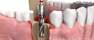
How is bone grafting performed and what materials are used?
Dental bone grafting or bone augmentation is a surgical operation whose purpose is to replenish the existing volume of the jaw bone. First of all, bone grafting is necessary for dental implantation. Before carrying out such an operation, careful preparation is required: examination by a doctor, taking tests, identifying and excluding acute stages of chronic diseases, i.e. contraindications to surgery.
During the operation, anesthesia will be performed, an incision of the periosteal tissues will be performed to provide access to the bone, fixation of the bone material, and suturing of the tissues. Afterwards, the patient needs to tune in to the recovery period - on average it takes about 2-4 months.
After bone augmentation, it takes time for the grafted bone to completely fuse with the existing one, that is, to become one with it. Only after this can you proceed directly to the installation of implants. However, in a number of situations, if the initial volume is sufficient for the primary attachment of the implant, then bone grafting can be carried out simultaneously.
Various materials are used to replace lost bone tissue. These can be donor tissues of animal origin obtained by collection from cattle; human tissues taken from the chin or jaw (the main option) or grown in a laboratory - alloplasts1 or artificial bone analogues. There are several bone grafting techniques that are used depending on the indications and individual characteristics of the patient.
Bone block grafting method
It is used in cases where it is necessary to increase the volume of the bone not only in height, but also in width. The blocks are attached with special screws and can take up to six months, after which the screws are removed (or they dissolve on their own) and implantation is carried out. In this situation, the patient's own donor material is used. The advantage is that it will not be rejected by the body, this virtually eliminates the risk of complications with the survival of the planted blocks. However, the method is quite traumatic, because it involves the healing of two wounds at the same time, one at the site of extraction of the donor material, the second in the area of extension.
Guided bone regeneration
During surgery, a synthetic bone analogue is implanted into the bone. On top it is covered with special protective membranes. Additionally, platelet-rich blood plasma obtained from your own donor material can be used. It stimulates bone growth. The process of fusion between artificial and natural bone can take from 3 to 6 months. In this case, the membrane either resolves or is removed surgically.
Sinus lift
The procedure helps increase bone volume by changing the position of the floor of the maxillary sinus. It is performed exclusively on the upper jaw. If a large volume is required, an open operation is performed. If a few millimeters of bone are missing, it is closed, i.e. simultaneously with implant installation. In any case, the technology is as follows: through an incision in the gum and bone, the bottom of the sinus is lifted with a special tool, and artificial bone material is placed inside. The advantage of this method is the effective restoration of bone tissue. The disadvantage is the length of rehabilitation. It is also necessary to take into account that the treatment requires high professionalism of the doctor. A poor-quality operation can lead to the development of inflammation and damage to the mucous membrane of the nasal sinus, as well as pushing the implant inside it.
Methodology
The work was performed on 30 male Wistar rats (weight 330 – 360 g, age 3.5 months). The rats were kept under standard conditions, 5 animals per cage, with controlled temperature (24 oC) and lighting (for 12 hours) and with free access to water and food. The operation to simulate the injury was performed under general anesthesia. First, the animals were anesthetized with light ether anesthesia. For deeper anesthesia, chloral hydrate was used intraperitoneally at a dose of 300 mg/kg. Then the animal was fixed on the operating table, the hair in the left thigh area was trimmed, and the skin and muscle tissue was cut with a scalpel. The femur in the area of the hip joint was exposed, the femur was drilled to the medullary canal 8-10 mm distal to the hip joint using a miniature dental drill (bur diameter 0.8 mm). After bone damage, layer-by-layer suturing of soft tissues was performed. The animals were divided into two groups. Control (15 rats) and experimental (15 rats). In experimental animals, using the Osteon-1 device, stimulation of the injury area was performed for 5 minutes daily for 7 days. (op7), 14 days. (op14) and 21 days (op21). To do this, the animals were fixed on operating tables and a mixed signal of two pulse voltages of different duty cycles, one of which was modulated at a higher frequency, was supplied through surgical needles inserted under the skin. The signals were unsynchronized relative to each other, unipolar with varying frequencies and amplitudes. The cathode was located in the area of injury, the anode was placed distally on the same paw. Electrical stimulation was carried out under general anesthesia (chloral hydrate at a dose of 190-200 mg/kg, intraperitoneal).
Three groups of animals with trauma served as controls (k7, k14, k21); they were also placed on dissection tables; surgical needles were inserted into them, but stimulation was not carried out. In each experimental variant, 5 animals were used. The next day after the end of the study, the animals were removed from the experiment by cervical dislocation. Then the femurs of all 6 groups were removed, which were used for histological analysis and assessment of the severity of the pathological process and the process of osteogenesis.
Animal bones were fixed for 24 hours at room temperature in 10% formalin prepared in phosphate-buffered saline (PBS, 0.02 M, pH 7.6). They were decalcified in 5% trichloroacetic acid for 48 hours, then washed in PBS and frozen in isopentane at -400C. Next, sections with a thickness of 5 μm were prepared. The sections were dried at room temperature for 1 hour and stained with hematoxylin-eosin. For histological analysis, 100 to 200 serial sections of the femur were prepared from each animal in the transverse and longitudinal directions. Using an Olympus microscope (approx. 10x, ob. 4x). Microphotographs of bone tissue sections were taken. Morphometric analysis of bone tissue inside the wound channel (intermediary callus) was carried out using the ImageJ program [19]. The area of bone tissue measured on 3-5 sections from each animal was expressed as a % of the canal area. To determine the significance of differences between the experiment and the control, the nonparametric two-sided Mann-Whitney test (U-test) was used.
What is Guided Bone Regeneration
Guided bone regeneration is an operation to compensate for the lack of bone tissue with a substitute, which is secured with a barrier membrane. After the procedure, a framework is formed around the transplant (it is formed by vessels and osteocyte cells that produce new tissue). Natural bone is gradually replacing artificial material.
Extensions using the NDT method are carried out according to classical parameters:
- thickness of the vestibular bone wall (at the cheek) - up to 2-2.5 mm;
- bone thickness between the implant and the root of the adjacent unit - up to 2.5-3 mm;
- The thickness of the bone wall between two titanium roots is up to 3 mm.
Can it be done simultaneously with implantation?
Two operations are combined into one if the following conditions are met:
- the shape of the defect allows you to correctly install the artificial root in any of the planes;
- when installed, the implant is located within the bone contour (does not go beyond the tangent drawn to the bone line);
- eliminate a horizontal (not vertical) defect.
The implanted implant can become a barrier to the growth of blood vessels and inhibit the formation of new bone. If the implant protrudes beyond the contour of the bone tissue, the risk of developing fibrous tissue with inclusions of the implanted material cannot be ruled out.
What bone materials are used
Osteoplasty involves the use of bone replacement materials. Previously, transplants were used that were not widely used due to frequent rejection of foreign material:
- Allografts
are donated bone from other people. The material was obtained from corpses, processed, sterilized, and stored in bone tissue banks. - Xenografts
are animal bones (cattle, pigs). The material was freed from proteins by heating (to eliminate the possibility of an allergic reaction after transplantation).
We do not use outdated techniques. Nowadays, many synthetic components have been developed that promote the growth of our own bone, which we use to build bone tissue.
Synthetic materials have a high degree of affinity and compatibility with natural bone and are its analogues. These are granular formulations based on:
- calcium phosphates;
- chondroitin sulfate;
- bioglass.
The effectiveness and safety of artificial materials has been confirmed by numerous studies. They are easy to use, take root well, promote the regeneration of your own bone, and are hypoallergenic.
With osteoplasty, a combination of an artificial substitute and a person’s own bone in the form of chips (autograft) is possible. The technique helps preserve the volume of built-up bone tissue and speed up the rate of recovery. The same method helps if you have to grow a large amount of tissue.
If there is a slight shortage, you can only get by with synthetic materials.
Advantages and disadvantages of bone grafting
In ancient times, the profession of “dentist” did not exist: if a tooth needed to be removed, people were wary of going to amateurs in the field of medicine who had tools suitable for such manipulations - to hairdressers and blacksmiths. Nowadays, it is possible to solve any dental problems painlessly, effectively and professionally, but for many, a “primal” fear awakens. Bone augmentation surgery is no exception.
Fear of the procedure can be a serious obstacle to restoring your full smile. If a patient needs bone grafting, it is important to understand the benefits that surgery provides, namely the restoration of full bone volume. This makes it possible to carry out implantation, which will subsequently solve deeper problems, namely, restore the aesthetics and functionality of the teeth. As for the disadvantages, they are associated with material costs (the operation is quite expensive), duration (the procedure delays implantation for at least 2-3 months), as well as difficult rehabilitation. In the first days after bone grafting, you may experience pain, swelling and bleeding. Symptoms can be easily relieved with local anesthetics and antiseptics. In addition, there are restrictions on flights, nutrition, and physical activity. According to statistics, the probability of serious complications after surgery is only 1-3%, and only if the patient is negligent about the recommendations of his attending physician, or the operation was performed unprofessionally.
With a competent and professional approach, the patient will not know what complications are after bone grafting. But the question is different - is it necessary to carry out this procedure if today there are implantation techniques that, with equally high results, allow you to do without bone augmentation? We successfully use methods of dental implantation with immediate loading - in 90% of all cases bone grafting is not required