5957
Teeth grinding is a procedure aimed at achieving two goals at once: on the one hand, there are fewer bacteria on smooth enamel, the rate of plaque formation is reduced, on the other hand, teeth become lighter.
That is, this manipulation is another step towards a healthy and attractive smile.
Process technique
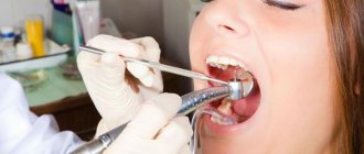
In dentistry, grinding refers to the leveling of enamel through mechanical action . You should not try to perform a similar procedure at home, because... Without professional skills, it is easy to damage the enamel.
This manipulation is part of a single complex for treating the surface of the teeth: it follows immediately after professional cleaning , thanks to which the oral cavity gets rid of contaminants.
At the same time, the procedure precedes the polishing stage , with which it is closely related.
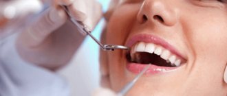
Professional cleaning and grinding of the teeth place local stress on the enamel. Polishing, on the contrary, thanks to the application of special compounds, acts as a protective manipulation for the surface.
Polishing effect
After professional hygiene, the oral cavity is considered completely cleansed and will have high resistance to various pathogenic influences for some time. The final polishing stage has the following effects:
- noticeable teeth whitening;
- uniform enamel color due to the removal of all stubborn dirt;
- smooth, pleasant surface of the teeth;
- visible shine of enamel;
- further prevention of inflammation and plaque deposits;
- improving the quality of restoration;
- correction of tooth shape after filling;
- getting rid of unpleasant odor;
- restoration of the natural properties of enamel.
As a result of polishing, teeth gain the ability to cope with negative influences and food loads, as well as avoid oral infections.
In what cases is it necessary
Grinding of teeth followed by polishing is carried out to achieve the following goals :
- The need for post-processing after cleaning from unwanted plaque and tartar: the procedure is an effective method in a preventive complex designed to prevent the occurrence of such problems in the future.
- Achieving the most comfortable state for the patient .
The surface of the dentition, which has undergone professional cleaning, becomes noticeably rough. This is the result of exposure to abrasive particles used in dentistry. To avoid discomfort, roughness must be removed by grinding. - Grinding at the separation level is a necessary part of treatment in orthodontics: it is performed before and after installing braces.
In addition, grinding accompanies the filling procedure to give the desired shape to a tooth that has undergone any physical impact from the dentist.
When installing fillings, cements and pastes that have a plastic consistency are used. However, when hardened, these compositions do not take on a shape that would correspond to the natural anatomical one.
To remove protruding particles, selective grinding is used, which is especially important on the front teeth.
Failure to do this procedure may lead to negative consequences.:
- patient complaints that he feels “something extra” in his mouth;
- Curvature even at the level of one unit can lead to malocclusion.
- irritation of the mucous membrane, because with the increased sensitivity of these tissues, any foreign object, even special dental compounds, can cause damage and contribute to the development of inflammatory processes.
Read how you can strengthen your gums at home in a new publication.
In this material we will tell you what to do if a piece of front tooth enamel breaks off.
Here https://www.vash-dentist.ru/krasota-i-uxod/otbelivanie/opalescence.html we’ll talk about whether the Opalescence system is suitable for home teeth whitening.
This procedure has a fairly short number of contraindications; it is impossible to carry out if you have:
- viral infections (like any other dental procedure, with the exception of extremely urgent ones),
- inflammation of the oral cavity,
- untreated pathologies (caries, bleeding),
- too thin enamel.
In any case, you should first contact your dentist for advice.
Contraindications
The condition of the enamel and teeth directly affects the indications for polishing. However, the ability to grind all teeth may be limited for a number of reasons:
- acute viral diseases;
- serious cardiovascular diseases;
- early and late stages of pregnancy;
- inflammation of tissues in the mouth;
- extensive caries;
- severe bleeding gums;
- periodontitis and progression of tooth mobility;
- thinning and fragility of enamel;
- problems with kidneys and salt metabolism;
- individual or allergic reaction to the composition of pastes and other drugs.
In any case of contraindications, exceptions are possible. The safety of the procedure is determined by the dentist or other supervising physician. It is usually recommended to first eliminate the cause or wait for a favorable course of the disease.
Sequence
Grinding of the tooth surface is most often done using classical technology .
The traditional approach involves the use of a drill and special pastes with a granular structure. The dentist has a number of special instruments, as well as ultrasonic attachments and drill tips.
Pastes are used in a certain sequence - from higher to lower grain levels:
- first, a coarse paste is applied, with which the dentist cleans off the largest fragments;
- further – medium-grained;
- at the final stage, a fine-grained composition is used, which removes the smallest particles of abrasive material.
This contributes to the most adequate smoothing of irregularities. This method is relevant for correcting the shape of a tooth.
For the purpose of grinding enamel, other sets of auxiliary materials are also used. For example, fine-grained burs are used, as well as corundum heads.
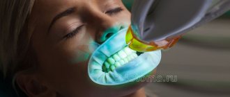
The dentist also resorts to using a set of special heads, strips and discs during the subsequent stage - polishing. Specialized paste-like compositions are used. At the end of the procedure, fluoride preparations are used.
The process is not inherently complicated: a special composition is applied and the surface is processed using a tool (brush, cap, tip, etc.).
Traditional grinding is gradually being replaced by the technology of using air masses for the same purpose. The equipment is built on the principle of functioning of a mini-sandblasting tool for grinding.
The result of the impact is the removal and removal of tissues that have become a “victim” of the reaction with abrasive particles.
Professionals note that air grinding has a number of advantages over classical grinding:
- It is devoid of all the physical disadvantages of the drill, which cause discomfort for the patient and partly for the dentist himself: temperature, pressure, noise and vibration do not accompany this procedure.
- There is no need to inject large amounts of anesthesia. Local impacts are possible.
- Less risk of damage to tissue and tooth enamel.
- The procedure is faster and requires less effort on the part of the doctor.
Thus, air grinding is a progressive method in dentistry. However, the option of partially exaggerating the positive features of the method for advertising purposes cannot be ruled out.
Already today, opponents of this approach note a number of disadvantages, for example, a significant increase in the sensitivity of the enamel after the procedure, the impossibility of use in deep cavities reaching the level of the pulp.
Also, only composite material is susceptible to the influence of air, while compositions using silver cannot be adjusted in this way.
For another type of tooth enamel grinding – stripping, watch the video:
What devices are used
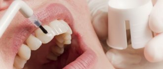
Polishing is carried out using a drill with replaceable attachments. The head is selected individually for each patient. Polishing is a comprehensive cleaning that includes exposure to a device and a special paste with abrasive particles.
There are several types of drill attachments:
- Rubber cup. Ideal for cleaning flat surfaces.
- Cone-shaped brush. Designed for teeth with lumpy edges.
- Strips (strips) with aluminum or diamond coating. This attachment cleans the most difficult to reach places, namely the area between the teeth.
Restoring tooth enamel
Tooth enamel is the hardest structure in the human body, as it is exposed to mechanical damage every day - during chewing and gnawing hard food, brushing and processing.
During the procedure, the doctor first applies the coarsest abrasive paste, and finishes with a medium or fine one. This polishing effect is achieved due to silica, crushed zircon and silicate in the composition of the products. They also have a disarming effect thanks to fluoride and xylitol in their composition. Pastes with grinding particles have the following degrees of abrasiveness (indicated by the RDA index on the packaging):
- 250 – blue color. High abrasiveness for tough dirt.
- 170 - green. Medium polishing effect.
- 120 - red. Paste with fine particles for the middle or final stages of polishing.
- 40 - yellow. A soft paste designed to complete the procedure.
Is it painful to do?
Unfortunately, teeth grinding is a rather unpleasant procedure. Painful sensations cannot be avoided even with the use of modern technologies.
It is necessary to understand that this manipulation involves the use of drugs that will temporarily desensitize the area of the oral cavity that will undergo the intervention. Local anesthesia is usually used .
About ways to care for teeth with braces, our next review.
In the next article, we will discuss whether lemon essential oil can be used to whiten teeth.
At the link https://www.vash-dentist.ru/krasota-i-uxod/narashhivanie/klyikov.html you will find a video of how the canine extension procedure takes place.
This applies to cases where the body of the unit itself is sharpened. At the same time, removing and smoothing out excess particles that appeared when using filling material is not a painful procedure, but rather unpleasant.
It is worth considering: for some time after the procedure, tooth enamel will be more sensitive than usual. The duration of the period depends on the individual characteristics of the organism.
Eating very hot or cold, solid foods may be problematic during this time.
Ideally, you should try to protect the enamel from such influences until the end of this stage.
Is there any harm to enamel?
If the technology is followed properly, there is no need to worry about the condition of the oral cavity in general, and enamel in particular, as a result of this procedure. She is absolutely harmless.
Moreover, periodic visits to the dentist for professional cleaning followed by grinding and polishing are necessary to avoid the formation of tartar and the subsequent development of pathologies.
Thus, teeth grinding provides professional care and is not harmful to the enamel . A pleasant “bonus” for undergoing the procedure will be the acquisition of a more attractive smile appearance.
Aftercare recommendations
The recommendations are mainly due to the fact that teeth become sensitive after the procedure. That is, you should avoid eating foods that seem very hot or cold.
It is also worth giving up too hard foods for a while. All this can be adjusted by trusting your own feelings regarding pain and comfort.
There are also specific tips. Thus, doctors recommend minimizing the consumption of foods that have a “coloring” effect. We are talking not only about certain berries, but also, above all, about tea and coffee.
This measure must be observed for two weeks in order to maintain the pleasant appearance of the cleaned enamel. You can at least switch to green tea at this time. You should also try to minimize smoking.
How to reduce the risk of pain?
Simple measures will help reduce discomfort after cleaning and unsealing:
- give up too hot, cold, spicy, sour and sweet foods - they will irritate inflamed or damaged tissues,
- clean the affected area of the jaw more carefully, try not to chew hard food with it - any mechanical impact can slow down recovery and increase pain,
- Try to eat right and get enough rest - this will speed up recovery.
If the treatment was carried out correctly, the discomfort should disappear after a few days.
Price
The teeth grinding procedure is far from the most expensive in dentistry. For one jaw, its price varies around 1000 rubles, which for one unit is usually 100-200 rubles.
However, you need to understand that the impact of this type on one tooth is usually included in the complex of treatment of this unit, most often its filling.
Enamel polishing , on the contrary, is usually highlighted in the price list and is provided as a separate service. However, it would be reasonable to decide on a one-time payment for comprehensive oral hygiene. In this case, you will need to pay a little more than two thousand rubles for one jaw.
Reviews
Patients who have successfully completed the procedure note that the result debunks the myth about subsequent tooth decay. Moreover, they often write that teeth begin to look whiter and more beautiful.
Sometimes they complain of increased sensitivity, as a result of which they cannot live a normal life, for example, drink hot coffee in the morning. But a small percentage of negative reviews, most of which were the result of the dentist’s unprofessionalism, suggests that in general this procedure is useful and safe.
You can share your opinion in the comments to this article.
If you find an error, please select a piece of text and press Ctrl+Enter.
Tags: tooth enamel professional teeth cleaning ultrasonic teeth cleaning teeth cleaning
Did you like the article? stay tuned
Previous article
Biohorizon implants: features of installation and engraftment
Next article
What determines the service life of dental implants?
Write a comment
Ksenia
August 8, 2016 at 6:38 am
I went to the dentist for a grinding procedure. The doctor with whom I had dental treatment advised me to do it; he said that grinding is also used to clean the surface of teeth from complex plaque and stones. And since I drink a lot of coffee, I need this procedure. Yes, I can’t say that the procedure is pleasant, after all, these are medical, albeit hygienic, manipulations, but I really liked the result. The teeth became smoother, shine appeared, plaque and stones were cleared. It is worth considering that the effect is quite individual, many complain that tooth sensitivity appears, I took this point into account and specifically a week before grinding, and a week after, I brushed my teeth with a special paste that reduces tooth sensitivity.
Alexander
August 9, 2016 at 4:27 am
I've seen enough Hollywood films and I also wanted a snow-white smile. Friends said that it was harmful, that they would erase the enamel and then you would suffer for the rest of your life. First I consulted with a specialist, and then I spoke with the person who did the procedure. It turned out to be not harmful, but on the contrary, useful. Now I walk around with a mouth full of dazzling teeth and a smile from ear to ear. Now there's nothing to hide.
Vladimir
October 11, 2016 at 02:08 pm
Since childhood, I have a terrible stomach for dentists, but from smoking and coffee, which I drink 5-10 times a day, my teeth have become not just yellow, but ugly. As a modern person, I immediately went to the Internet for advice and information, and immediately came across the answer to the question I posed. I plucked up courage and went to the dentist. This procedure turned out to be completely safe, and in some places it was even funny to watch the doctor’s actions. In general, I was pleased with the result, and after the instructions of my doctor, I began to take better and more careful care of my oral cavity!
Anastasia
October 24, 2016 at 8:55 am
I did this procedure once, when I discovered a cinnamon coating on my teeth, I thought it was caries. I ran to the doctor, and they told me that it was just tartar and that this problem is common. They offered to do ultrasonic cleaning - I really liked the result, no plaque, firstly, the procedure was painless and also inexpensive.
Denis
November 22, 2016 at 6:24 am
My teeth grinding went well. There was no particular pain, but mostly it was periodically unpleasant to feel the machine buzzing in your mouth and scraping something. The doctor recommended grinding at least once a year. Because I smoke and love coffee very much, and after some time plaque forms on my teeth, especially on the inside. The procedure is not expensive, and after it your teeth sparkle with whiteness.
Svetlana
October 31, 2022 at 10:23 am
Every year I go through the polishing procedure. As a result, there is no plaque and the teeth are healthier; there have been no caries for three years now.
Aesthetic restoration of teeth with a combination of carious lesions and increased abrasion
Modern advances in the development of dental materials significantly expand the capabilities of dentists. Recognition of aesthetic dentistry as a separate area with its own theoretical, scientific and practical developments has ensured the rapid implementation of advanced treatment methods. Routine “filling” is being replaced by restoration, which guarantees high quality restoration of teeth with defects of carious and non-carious origin.
Moreover, it has become possible to treat combined lesions using methods that require minimal excision of hard tooth tissue and ensure preservation of pulp vitality.
Such work is based on the principles of aesthetic dentistry (I.K. Lutskaya, 2011) [2].
The main principle, or starting point, of aesthetic dentistry should be the achievement of treatment results that are as close as possible to the natural parameters of the dentition.
Thus, therapeutic restorations and orthopedic structures are subject to the requirement of maximum similarity with the optimal characteristics of teeth in color, shape, and relief.
Surgical crown lengthening or gingivectomy should not visually increase the original size of the teeth, cause gum recession or “gaping” of interdental spaces [4].
Achievement of the basic principle is ensured through optimal therapeutic effects, which involves the selection of means and methods that do not violate or minimally damage intact structures. First of all, the methods of bleaching, then minimal preparation (microabrasion), and grinding of the non-prismatic enamel layer are considered as therapeutic methods. Preparation, and especially depulpation, is carried out strictly according to indications. Any manipulations must ensure high aesthetics, mechanical strength, and reliable adhesion of restorations to tooth tissues [4].
The principle of conscious cooperation implies that the patient conscientiously and regularly performs the procedures prescribed by the dentist.
Thus, home whitening is prescribed by a doctor and performed by the patient independently, strictly following the instructions. The choice of shades of color, size and shape of the structure is carried out jointly by the dentist and the patient to reach consensus. The scope of surgical intervention is also carried out after obtaining the informed consent of the patient.
Working with photocomposites and modern ceramics has its own fundamental requirements, approaches, and principles that provide scientific validity to the manipulations performed.
The principle of color imitation ensures the modeling of a restoration (structure) with high aesthetic parameters, implying the selection of shades of material that correspond in optical properties to dentin and enamel, followed by imitation of the color of lost tooth tissues [5].
The principle of reproducing natural volumetric parameters implies preliminary planning of the dimensions, shapes, and surface relief of the restoration (structure), followed by the reconstruction of macro- and microstructures [1].
When modeling a restoration, its morphological features must repeat the parameters of an intact tooth, therefore it is necessary to maintain the geometric shape, signs of belonging to the side, and mamelons.
Such large structural elements are modeled with opaque materials.
Individual characteristics, such as surface relief, cutting edge shape, and transparency, are formed using enamel shades in compliance with the rule of preserving the volume of natural tissue.
The principle of adhesive preparation (English: prepare) means increasing the area of contact between the filling and the tooth in order to ensure high-quality bonding of the composite with the dental tissues. The objectives are achieved by excision of hard tissue to intact structures, creating a bevel of the enamel or giving the cavity a certain shape, as well as acid etching and the use of an adhesive system.
The principle of minimizing the effects of polymerization shrinkage is based on the property of the material to decrease in volume during the curing process.
At the stage of tooth preparation, reducing the effects of shrinkage is achieved by excision of thinned protrusions and rounding of the internal corners of the cavity. The absence of a complex design reduces tension in the tooth tissues. The risk of detachment of the filling, the formation of a gap at the border with the tooth, and the appearance of hyperesthesia is reduced when using gaskets made of chemically cured materials: during polymerization, they are “attracted” towards the heat source - the pulp.
During the curing process of a composite, one of the methods for reducing shrinkage is the “soft start” method: reducing the exposure time of the first stage of photopolymerization.
Knowledge of the principles of aesthetic dentistry allows you to choose the most correct methods of influence, ensuring maximum efficiency.
Clinical case
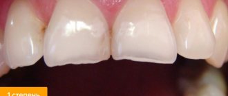
The patient, 35 years old, complained of aesthetic defects of the upper central incisors. Carious cavities are localized on the mesial surfaces of the 11th and 21st teeth. There is a significant decrease in the height of the crowns of the upper central incisors due to increased uneven abrasion of hard tissues (Fig. 1).
Rice. 1. Carious cavities on the mesial surface of teeth 1.1 and 2.1, uneven wear of hard tissues.
The use of composite material for dental restoration involves the following sequence of stages: mechanical cleaning of the tooth surface; choice of photopolymer shades; planning the size and shape of restorations; tooth preparation; use of an adhesive system; filling the defect with a composite; polymerization; coating of teeth with fluoride varnish.
Taking into account the complexity of the work and aesthetic requirements, a nanohybrid composite is used for dental restoration, representing a new class of filling materials characterized by high aesthetic properties: Grandio SO (VOCO). It combines the qualities of photopolymer with the innovation of nanotechnology. For its production, polymer resins with high molecular weight and microfillers with an average particle size of 20-30 microns were used.
Combining nanomers (zirconium silicate particles less than 100 nm in size) with glass-ceramic composite particles made it possible to increase the share of filler in the material to 87%, which reduced the content of organic resins in the matrix and, accordingly, reduced polymerization shrinkage to 1.57%. Filling the material with nanoparticles ensures high marginal stability, easy polishability and good color fastness of the restoration. Thanks to good adaptation to enamel and dentin shades, the so-called chameleon property, the stage of selecting composite shades is greatly simplified. In some cases, one shade of material from one color group (for example, red-brown, group A) of varying degrees of transparency (opaque, enamel, transparent) can be used. In more complex clinical situations, 2-3 syringes with different color characteristics will be required.
Nanocomposites are indicated for filling cavities of classes I-V according to Black, restoring teeth with traumatic injuries and hard tissue defects of non-carious origin. A large selection of shades and color stability of the nanomaterial allow it to be used for aesthetic filling of anterior teeth with correction of shape and color; restore teeth with class II cavities of significant size; used for splinting, making inlays, onlays, adhesive prostheses, restoring the tooth stump for a crown.
The first stage is the mechanical treatment of teeth with fluoride-free paste (Fig. 2). Cleaned surfaces are washed with a stream of water.
Rice. 2. Cleaning teeth from plaque.
The shades of the photopolymer are selected in natural light (the patient is in a sitting position) using the colors supplied with the material (Fig. 3).
Rice. 3. Selection of shades of tooth color.
The surfaces of the teeth are kept moist, which preserves their natural appearance. The opaque composite OA 2 is selected to match the color of the central area, this will avoid the transparency of the filling. To create a natural look, shine and transparency, enamel shades of the restoration are used, which are selected separately for the central (A 2) and proximal sections (A 2 and I), and the incisal edge (A 1 and I). Using opaque alone can create the appearance of a flat or lifeless tooth. The absence of opaque will make the restoration “transparent”. Enamel and clear shades are required to cover the main area of the restoration, the incisal edge and proximal surfaces. A transparent cutting edge with a width of more than 1 mm is assumed.
When choosing tooth shades, the phenomenon of irradiation is taken into account - a change in the size of the surface depending on color and lightness, when warm light colors (yellowish) create the illusion of convexity, and cool colors (blue) create depressions. As a result, the cutting edge of light yellow shades will be perceived as voluminous, while the bluish edge will be perceived as flat and displaced orally. In this case, it is planned to use 4 syringes of composite material: opaque OA 2, enamel A2 and A1, transparent I.
Planning the size, shape and relief is a strictly defined sequence of actions describing the specific anatomical formation of the tooth.
Teeth are measured with a micrometer. The height of the clinical crown of the central incisors is defined as the distance from the marginal level of the gums along the vertical midline to the conventional line of the cutting edge, overlapping the occlusal surface of the lower incisors by 1.5 mm.
Mesiodistal dimensions in the neck area are measured by the distance between two points at the level of the apexes of the interdental papillae, in the equator area - at the level of the middle third of the crown height, in the incisal edge area they are assessed by the distance between the protruding points of the mesial and distal edges of the crown. In this case, odontometry indicates that the width of the tooth in the equatorial region is equal to the height of the tooth and is 8.9 mm in the 11th tooth, 9.0 mm in the 21st tooth.
In the area of the cutting edge, it is not possible to measure the horizontal size of the tooth, since it is erased. The map indicates signs of belonging to a party. The sign of crown curvature is weakly expressed. Due to chipping and increased wear of teeth, the dimensions of the mesial angle are not determined. Based on visual assessment and measurement results, it is planned to change the square geometric shape of the crowns to rectangular by increasing the vertical size by lengthening the cutting edge. The planned vertical dimensions of the incisors are 9.7 mm for the right incisor and 9.8 mm for the left (the height of the teeth differs due to the higher dome of the gingival margin in tooth 21) (Fig. 4).
Rice. 4. Odontometry: tooth width in the equatorial region in 1.1 teeth is 8.9 mm, in 2.1 teeth 9.0 mm. The planned tooth height is 9.7 mm for the right incisor and 9.8 mm for the left.
It is planned to recreate the predominance of the distal angle of the crown over the mesial angle, which is a sign that the tooth belongs to a certain side. The sign of crown curvature is not pronounced: the mesial surfaces have a slight oral location compared to the distal ones, they have a penumbra, and are visually perceived as darker. It is intended to create a slight convexity of the vestibular surface closer to the mesial edge. The individual characteristics of teeth are smooth vestibular surfaces. The length of the contact points between the teeth is planned in such a way that there is enough space for the interdental papilla (from the top of the gingival papilla to the incisal edge). The cutting edge of the teeth is smooth.
To prevent chipping of the filling material, occlusal contacts and the incisal path are aligned using carbon paper.
Teeth preparation is carried out with diamond burs and begins with a thorough necrotomy of the walls and bottom of the carious cavity. In this case, diamond burs (NTI company) are used to process the enamel, first with medium (100 microns), then with fine grain (40-50 microns). The water jet is applied continuously to the drill to avoid irritation of the pulp. Dentin is prepared with carbide burs. The vestibular wall of the carious cavity is partially excised, since it is represented by a thin layer of enamel that does not have underlying dentin. As a result, class IV cavities are formed (Fig. 5).
Rice. 5. Teeth after necrectomy.
All corners are rounded with a spherical bur. On the vestibular surface of each incisor, an enamel bevel approximately 3.5-4.0 mm wide is made in order to recreate the sign of crown curvature and move the convexity of the vestibular surface closer to the mesial edge. The surface is smoothed with a fine-grained diamond bur (40-50 microns). Bevels will ensure an increase in the contact area between the tooth and the composite, as well as the aesthetics of restorations due to a smooth transition of the material at the filling-tooth interface. On the palatal surface, the enamel is ground at an angle of 45°.
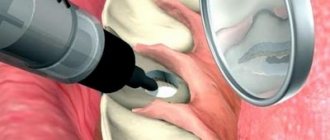
The prepared surfaces are thoroughly washed with a stream of water and dried with an air stream. The dentin is covered with glass ionomer cement. Then the enamel is covered with etching gel (30 sec.) (Fig. 6).
Rice. 6. Acid etching of enamel.
After time, the gel is thoroughly washed off, and the enamel surface is dried with a stream of air (Fig. 7). The adhesive is evenly applied to the prepared surfaces, rubbed in with a brush, left for 30 seconds to soak, distributed with a gentle stream of air and polymerized for 20 seconds.
Rice. 7. Central incisors after the acid etching stage.
Filling begins with filling the cavity. The opaque composite (OA 2) is introduced into the cavity in a layer of no more than 2.0 mm in thickness, carefully pressed against the bottom and walls, and photopolymerized for 30 seconds.
The adaptation of the material in the area of the missing vestibular wall of the cavity requires special attention: the level of the opaque layer is located 1.0-1.5 mm below the level of the tooth surface (the difference will allow the formation of the enamel layer) (Fig. 8).
Rice. 8. Adaptation of the opaque layer of the Grandio SO (VOCO) material.
After filling the carious cavity, they begin modeling the half-veneer, which is carried out in compliance with a gradual transition from recreating large details (the geometric shape of the vestibular surface) to reproducing medium ones (signs of the angle and curvature of the crown), and then to modeling smaller elements (the area of the cutting edge). Each subsequent layer is applied on top of the previous one and distributed in different directions using a wide trowel or spatula. The boundary of each layer is created in the form of buoyant waves (Fig. 9).
Rice. 9. The enamel shade of the composite is distributed.
The mesial surface is modeled with an opaque composite throughout the middle and lower tier of the tooth crown. The main landmark is the border of the transparent tooth enamel: in this case, there is about 1.0 mm of free space around the entire perimeter of the tooth, except for the cutting edge, where the width of the transparent layer will be 1.5 mm.
To recreate the sign of crown curvature, a portion of opaque photopolymer is applied in the form of a vertical roller at the border of the mesial and median sections, and then smoothed so that the maximum convexity is maintained closer to the mesial surface (Fig. 10).
Rice. 10. Scheme for modeling the sign of crown curvature.
Modeling of the sign of the crown angle is carried out in the lower tier of the tooth, taking into account the degree of its severity: the distal angle is larger in size than the mesial one. In this case, the latter is modeled in the area of the mesial lower third of the tooth crown, with the filling material distributed towards the cutting edge and mesial contour and giving the corner the desired shape (Fig. 11).
Rice. 11. Modeling the crown angle sign.
The vestibular surface is covered with a thin layer of enamel (A 2) and then transparent (I) photopolymer. Modeling of the side surfaces is also completed with enamel (A 1) and transparent composite, which is distributed taking into account the individual degree of transparency of the enamel: evenly along the entire perimeter of the restoration. At the cutting edge, the width of the transparent layer is 1.0 mm (Fig. 12).
Rice. 12. Imitation of a translucent cutting edge.
Immediately after the production of aesthetic structures, they are processed: the surface hybrid layer is removed (with bur No. H390S-018-FG, NTI) (Fig. 13), the relief is contoured, and occlusal contacts with antagonist teeth are verified.
Rice. 13. Contouring of aesthetic restorations.
Polishing is carried out with discs without significant pressure on the surface of the restoration, in the direction from the equator towards the cutting edge. To achieve shine, polishing heads containing fine aluminum oxide powder as an abrasive are used, sponges and polishing pastes are used (Fig. 14).
Rice. 14. Polishing the surface of the restoration.
At the final stage, odontometry is carried out: the vertical size of the teeth after restoration exceeds the horizontal ones and amounts to 9.7 mm and 9.8 mm for the right and left incisors, respectively, the teeth have acquired a rectangular shape (Fig. 15).
Rice. 15. Final odontometry: vertical tooth size 9.7 mm and 9.8 mm for the right and left incisors, respectively.
Finishing of proximal surfaces is carried out with strips - strips on a metal or plastic base with varying degrees of granularity of the abrasive material applied to them. The finished work is shown in Figure 16.
Rice. 16. General view of the finished restoration.
Illumination of teeth with short-wave light demonstrates the same spectral composition and fluorescence intensity of the filling material from which the half-veneers are made and the hard tissues of the tooth (Fig. 17).
Rice. 17. When illuminated with short-wavelength light, the restorations fluoresce in the same way as the hard tissues of the tooth.
The final stage of treatment is the treatment of the enamel surrounding the restoration with fluoride-containing preparations.
Quality assessment is carried out in accordance with the aesthetic quality index (EQI) [3]. The score is given separately for each of the 12 parameters presented in the table: 3 points - the result fully corresponds to the plan; 2 points - the result partially corresponds to the plan; 1 point - the result does not correspond to the planned one. Then all points are summed up. The highest possible number of points is 36.
Quality rating: 33-36 points - excellent; 29-32 points - good; 24-28 points - satisfactory; below 24 points - unsatisfactory.
Calculation of the aesthetic quality index (EQI):
EIC = n/36
where n is the total score; 36 - maximum score;
0.9-1 - excellent result;
0.7-0.8 - good result, minor correction of the restoration is needed;
<0.7 - unsatisfactory result, the structure needs to be replaced.
An assessment of the aesthetic restorations we performed showed: EIC = 1, which is interpreted as an excellent treatment result.
Table No. 1. Assessment of the quality of the manufactured structure according to EIK (aesthetic quality index)
Criteria | Intact, symmetrical tooth | Design | Score in points | |
| planned | manufactured | |||
| 1 | 2 | 3 | 4 | 5 |
| 1. Tooth dimensions: height (LCO2), mm | 8.9 mm | 9.7 mm | 9.7 mm | 3 |
| transverse size (MDCO2), mm in the cervical region | 8.9 mm | 8.9 mm | 8.9 mm | 3 |
| in the equator region | 8.9 mm | 8.9 mm | 8.9 mm | |
| in the area of the cutting edge | 8.9 mm | 8.9 mm | 8.9 mm | |
| 2. Geometric tooth shape: rectangular (square) | square | rectangular | rectangular | 3 |
| triangular | ||||
| oval | ||||
| 3. Crown angle sign: expressed not expressed | not expressed | expressed | expressed | 3 |
| 4. Sign of crown curvature (displacement of the point of greatest convexity): | 3 | |||
| mesially | ||||
| closer to the midline | ||||
| displaced distally | distally | mesially | mesially | |
| absent | ||||
| 5. Sign of root deviation: expressed not expressed | expressed | expressed | expressed | 3 |
| 6. Gingival contour: flattened | 3 | |||
| rounded | ||||
| domed | domed | domed | domed | |
| 7. Cutting edge: straight | straight | straight | 3 | |
| convex | ||||
| concave | concave | |||
| serrated | ||||
| 8. Relief of the vestibular surface: expressed | ||||
| not expressed | not expressed | not expressed | not expressed | 3 |
| 9. Degree of gloss of the enamel (presence of distortions): evenly matte | 3 | |||
| “matte” in the cervical area | ||||
| brilliant | shine | shine | shine | |
| 10. Type of tooth transparency: enamel is transparent in all areas of the tooth crown | 3 | |||
| pronounced transparency of the proximal surfaces of the tooth | ||||
| Only the cutting edge is transparent | ||||
| transparent cutting edge and proximal surfaces | + | + | + | |
| 11. Tooth color assessment using the VITA scale: in the cervical region | A 2 | A 2 | A 2 | 3 |
| in the equator region | A 2 | A 2 | A 2 | |
| at the cutting edge | A 1 + I | A 1 + I | A 1 + I | |
| on proximal surfaces | A 2 + I | A 2 + I | A 2 + I | |
| 12. The presence of individual characteristics of the tooth (stains due to hypoplasia, fluorosis, etc.) | — | — | — | |
| 13. Assessment of the quality of the manufactured structure according to EIK (aesthetic quality index) | n/36 | 36/36 = 1 | ||
Conclusion
The use of photocomposites in aesthetic dentistry makes it possible to expand the indications for dental treatment using therapeutic methods. In accordance with the basic requirements for aesthetic restorations, the main stages of treatment are carried out using light-curing materials, including the selection of optimal shades, modeling of restorations, and their processing. Efficiency is assessed in accordance with the aesthetic quality index, which reflects the main parameters of size, shape, surface topography, optical properties of the tooth and restoration.
Literature
- Lomiashvili L. M. On the way to creating the secrets of tooth shapes
/ L. M. Lomiashvili // Klin. dentistry. - 2006. - No. 2. - P. 12-15. - Lutskaya I.K. Fundamentals of aesthetic dentistry.
/ I. K. Lutskaya. - Mn.: Let's lie. school, 2005. - 332 p. - Criteria for assessing aesthetic restorations: instructions for use No. 078-0906:
approved.
Ministry of Health of the Republic of Belarus 06/26/2007 / I.K. Lutskaya, N.V. Novak, T.A. Zapashnik, V.P. Kavetsky // Modern methods of diagnosis, treatment and prevention of diseases
: collection. instruction—method. doc. - Minsk: RNMB, 2007. - T. 5. - Issue. 8. - pp. 75-79. - Nikolaev A.I. Physico-mechanical properties of modern filling materials: significance for practical dentistry.
III. Dry shine and strength of composites / A. I. Nikolaev, L. M. Tsepov, P. G. Adamov // Maestro of Dentistry. - 2003. - No. 3. - P. 28-32 - Pogorovskaya I. Ya. Comparative assessment in vitro of color indicators and color stability of composite and glass ionomer materials for aesthetic restoration of teeth
/ I. Ya. Pogorovskaya, I. M. Makeeva, E. A. Emilenko. - M., 2001. - P. 83-84.