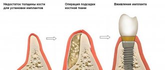
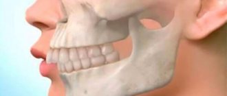
Difference between molars and premolars
The main difference between molar teeth and premolars is their location on the jaw.
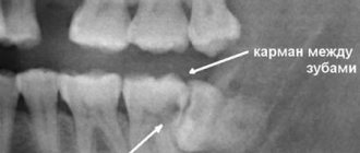
Premolars are located closer to the front of the dentition, and molars are slightly moved inward. Their quantity also varies. There are always two premolars on each side of the upper and lower jaws. There can be either two or three molars. Still others are also called “wisdom teeth.” They grow later than others and often appear only at 20-25 years of age. This process is often accompanied by malaise and fever. However, not all people on earth have third ones. A certain genetic predisposition can cause the absence of wisdom teeth in the oral cavity.
Subsequence
The order in which teeth appear may vary, which is rare and is not considered pathological. For some boys and girls, the upper incisors appear first, rather than the lower incisors as usual. But basically the sequence of teething in children is as follows (teeth appear in pairs):
- lower central incisors;
- upper central incisors;
- upper lateral incisors;
- lower lateral incisors;
- first upper molars (molars);
- first lower molars;
- upper canines;
- lower canines;
- second lower molars;
- second upper molars.
By the age of 2.5–3 years, the child has a full set of baby teeth: 10 on each jaw. After about 4–6 years, the roots begin to dissolve, the teeth lose support and fall out in the same order in which they erupted.
As the jaw and facial bones grow, spaces form between the primary teeth. This provides space for permanent, larger teeth to emerge, which erupt from 5 to 15 years of age.
Features of the structure of molars
Molars have a characteristic structure. On the upper jaw they have three roots and four canals. The lower jaw has two roots and three canals. Moreover, the number of canals also differs depending on the location of the individual tooth. Thus, the first of them often have one more channel than the next two.
The main characteristic feature of this type of teeth is the area of their chewing surface. They bear the heaviest load when chewing food particles. The molars themselves also have differences among themselves, which are associated with the structure of the jaw.
Upper molars
For the upper row of the jaw, the molars have the following anatomical surfaces:
- The buccal part is convex, with a prominent groove that is most susceptible to caries. When performing hygienic cleaning, it is recommended to pay special attention to this area.
- The chewing side has some differences and a large number of tubercles, which increases the role of this group in performing the chewing function. One of these features is the triangular trigon, which unites the following types of tubercles - protocone, metacone, paracone.
- The palatal part is distinguished by such a characteristic feature as the Carabelli tubercle, located on the border of the surface from the side of the palate and the medial area. In rare cases it has a separate root.
- The mesial part has a convex or straight line between the cementum and enamel. The root system is complex and can be barrel-shaped, divergent, or cylindrical.
- The distal one resembles the medial one and requires special attention when cleaning (dental floss is recommended to completely remove any remaining food).
Molar Differences
Often the surface of each molar tooth is shaped like a triangle. There is a certain number of tubercles on it, which take an active part in chewing food. The number of such tubercles may vary. Usually there are three, but sometimes there are more.
Such mounds are connected to each other by special ridges. On the upper and lower jaws the structure of these elements is different. In the upper dentition, the apex of the surface triangle is directed towards the tongue. This form is called a trigon. On the lower jaw, the apex of such a triangle is directed towards the cheek, which is called the trigonid. The size of the first and second types of teeth is practically the same.
Molar placement
The molar large units are located on the back of the jaw. Each jaw has six of them. They decrease in size from front to back. That is:
- the first rudiment is the largest;
- the second one is smaller;
- the third tooth is small;
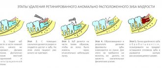
The molar units are located most posteriorly in the arc. They are also called extreme. The number eight, that is, the wise tooth, grows in the period from 18 to 35 years. It happens that he does not appear at all. Row anomalies and bite defects can form in the absence of individual units or groups of rudiments. Often this concerns the end organs. Remember that normally there should be 28 units without eights, and with them 32 organs. The primary dentition has eight outermost units, the permanent dentition has twelve. In the molar group, the sixth units are key, and the 7s are variable, the most variable being the eights.
If caries treatment is carried out in a timely manner, the chewing organs will last a long time. The health of the whole body, the functioning of the gastrointestinal tract, liver and kidneys depends on this. Therapy should begin at the white spot stage, while the cavity has not yet formed. Prevention of caries is of great importance. To do this, you need to visit the dentist at least once every 6 months, carefully care for your oral cavity, and follow the dentist’s recommendations. Modern medicine has technologies the use of which can save even a dilapidated organ. Therefore, the key to health is timely treatment after a complete diagnosis and correct diagnosis.
It is important from childhood to teach a child to brush his teeth and treat the oral cavity. Children should understand that the main cause of caries is pathogenic bacteria living in dirty plaque. Therefore, trips to the hygienist will not allow the active proliferation of pathogenic microbes, subject to regular preventive cleanings.
Structural features
Those who looked at their teeth in the mirror noticed that they all had a different design. The structure of the crowns depends on:
- from the arrangement of organs in a row;
- depending on the shape of the unit;
- depending on the size of the rudiment;
The anatomical structure of the mammary organs is basically the same as the permanent units. True, temporary rudiments have distinctive features.
- They are smaller in size;
- The width of their crown prevails over the height;
- In the cervical area, the enamel layer is thickened;
- The shade of the enamel is bluish;
- The roots are widely spaced and short;
In addition, in the alveolar arch the temporal units are arranged steeply vertically. This is influenced by the fact that behind the roots there are already rudiments of permanent teeth. The upper units have 3 roots, one of them is lingual and 2 are buccal. The crowns have a cuboid shape, on the chewing surface of which there are 3 or more tubercles. The groove that separates the tubercles has two parallel lines and one perpendicular. The crowns of the rudiments on the lower jaw are somewhat elongated, and the fissures are located almost cross-shaped. These chewing organs have 2 roots. In eights, the roots often merge, the shape of such a root is conical. It happens that wise teeth grow with 4-5 roots. Since the upper and lower eights are close to each other, occasionally they can grow together.
To keep the chewing organs healthy, dentists may prescribe dental remineralization. This procedure restores the enamel layer well due to its saturation with deficient elements. The enamel ceases to be porous, acids do not penetrate its pores and do not contribute to the leaching of useful minerals. This event prevents and eliminates caries at an early stage, improves the aesthetics of crowns with fluorosis, reduces the sensitivity of units, brightens crowns and normalizes the microflora of the oral cavity.
Possible damage to small molars
Carious lesions of primary and molar teeth in children are observed quite often. This is due to the bumpiness of their surface, which can trap food particles and bacteria. Since children at an early age do not always actively and correctly brush their teeth, these residues can lead to damage to the enamel and deeper spread of caries.
Depending on the degree of damage, caries can penetrate either exclusively into the enamel, or into dentin or even cement. In the latter case, we are talking about deep caries, which often requires serious treatment or even tooth extraction.
Another variant of damage is a change in tissue structure, which may be associated with metabolic disorders in the human body or other diseases. Such a disease can also be the result of poor nutrition or constant diets, in which the body does not receive enough nutrients.
At any age, a person can experience excessive tooth wear, which leads to the destruction of enamel and can cause other serious damage. Teeth can wear down in different situations, such as:
- if a person has bad habits of grinding his teeth, etc.;
- in case of improper or insufficient nutrition;
- in the presence of diseases of the endocrine system;
- due to a genetic predisposition to enamel depletion.
All of these lesions can be the result of both an incorrect lifestyle and various diseases or heredity. Regardless of the reasons, it is necessary to begin treatment of emerging injuries immediately so as not to aggravate the process.
3D diagnostics and principles of visualization of 3 molars of the lower jaw
Rogatskin D. V. Radiologist, Ortos LLC (Smolensk)
A fragment of a chapter from the forthcoming book “Radiodiagnosis in Dentistry. 2D/3D".
Teeth 48, 38, often called “lower wisdom teeth,” usually have a two- or three-root structure, but the length and configuration of the roots are extremely variable. These can be either short fused roots or excessively long curved ones with pronounced divergence (Fig. 1).
Rice. 1. Fragment of a panoramic tomogram, the area of the molars of the lower jaw on the left. The anterior root of tooth 38 is enlarged and curved. The shadow of the root crosses the lumen of the mandibular canal.
There are a number of clinical indications for the removal of third molars of the mandible, however, surgical intervention is often complex and complicated, so at the planning stage there is a need for careful preoperative diagnosis. This is due to the fact that these teeth occupy a distal position in the dentition and are often in a state of retention. According to modern studies, complications associated with damage to the mandibular nerve occur on average in 13% of cases of the total number of removals (Wang et. al., 2016). Traditional diagnosis of the position of the mandibular canal relative to the roots of the third molar using a panoramic image can no longer be considered sufficient and objective. In some cases, a three-dimensional study performed for clarification forces the surgeon to change the intervention tactics (Matzen et. al, 2013).
Impacted teeth are teeth that have not fully erupted due to difficult conditions for eruption, or have formed impacted, that is, completely immersed in bone tissue.
To describe the position of the third molar, we can recommend the principles laid down in the Winter and Pall-Gregory classifications. Winter (1926) assessed the position of the vertical axis of the wisdom tooth relative to the 2nd molar and identified 5 position options - vertical (V - in Fig. 2), mesial inclination (M - in Fig. 2), horizontal position (H - in Fig. 2), transversal position, distal tilt and inversion. To this it should be added that the wisdom tooth can also be in an ectopic position - that is, outside the dentition, for example, in the jaw branch. The Pell-Gregory classification (Pell&Gregory, 1933) is based on determining the degree of immersion of the 8th tooth relative to the crown of the 7th and the degree of overlap of the crown of the 8th by the bone tissue of the anterior edge of the jaw branch. The authors distinguish 3 classes of overlap (Fig. 2):
- 1 – the crown is not covered by bone tissue
- 2 – bone tissue covers up to half of the crown surface
- 3 – the crown is completely immersed vertically in the bone tissue
and 3 levels of immersion:
- A – the crown of the 3rd molar is in contact with the crown of the 2nd molar
- B - the crown of the 3rd molar is in contact with the cervical region of the 2nd molar
- C - the crown of the 3rd molar is in contact with the root of the 2nd molar
Depending on the class, level and relationship of the root with the mandibular nerve, the surgeon chooses the method of tooth fragmentation and determines the tactics of surgical intervention (Maegawa et al., 2003).
Rice. 2. Illustration for the classification of the position of the 3rd molars of the lower jaw according to Pell&Gregory, Winter. Visualization based on panoramic tomograms. Vertical columns: V – vertical position, M – mesial slope, H – horizontal position. Horizontal rows: A, B, C - immersion levels (explanation in the text). The numbers in the table cells indicate the degree of overlap of the crown with bone tissue.
In 2015, Italian researchers (Maglione et al.) proposed a detailed and rather complex classification of the relationship between the roots of impacted wisdom teeth and the mandibular nerve. The authors identified 7 classes and a number of subclasses. It is likely that the proposed gradation has important clinical significance, but from the point of view of primary diagnosis and description of the tomogram, such detail may be regarded as unnecessary. For diagnostic purposes, Chinese experts only propose to state that the nerve passes below the tooth, vestibular from the root, lingually or between the roots (Wang et. al., 2016). In this context, this approach is the simplest and most logical. However, there are a number of nuances associated with the features of the virtual positioning of the software coordinate system.
First of all, it is quite difficult to adequately assess the relationship between the root and the mandibular canal with a minimum of options and using a non-interactive coordinate system fixed in the anthropometric position. In this case, the area under study is visualized in an oblique section without taking into account the individual structure and position of the tooth; the shape, size and relationship of the structures are conveyed with distortion, and there is no information about the neighboring tooth (Fig. 3). To obtain the most informative image, it is necessary to orient the axial and sagittal planes according to the mesio-distal length of the canal, taking into account its position and configuration, and fix the coronal plane at the point of greatest contact of the root with the nerve (Fig. 4, 11). Optimal visualization is carried out in three dimensions, indicating the location through which the main section passes. The working depth of the layer for the axial and coronal sections is recommended to be minimal, for the sagittal (mesio-distal section) - a zonogram from 3 to 10 mm. In addition, for each case, standardized polypositional visualization of objects is necessary - separately the tooth, in order to assess its structure and configuration (Fig. 5), and separately the relationship of the root with the mandibular nerve if their contact is visually determined (Fig. 6). For this purpose, the following formulations can be recommended:
- I – inferior – the nerve is in a lower position relative to the tooth. Periapical (apical) in a vertical position and mesial tilt - IA (Fig. 7), periradicular in a horizontal position - IR (Fig. 6). In the case of simultaneous periradicular inferior fit and periapical distal, in a horizontal position or mesial tilt - IRD (Fig. 8).
- L – lateralis – the nerve is located on the side of the tooth. LL – lingual or LB – buccal (Fig. 4).
- IR – inter-radicularis – the nerve is located between the roots in the furcation area. In this case, there can be two options: IR1 - the apices of the roots do not close under the nerve (Fig. 9), IR2 - the apices of the roots close under the nerve, enveloping it in a ring (Fig. 10).
Rice. 3. An example of incorrect visualization of tooth 48 and its relationship with the mandibular nerve.
Rice. 4. The same tooth (Fig. 3), correct MPR visualization using an interactive coordinate system and adjustable depth of the selected layer. Targeted visualization of tooth 48 and the mandibular canal. The depth of the selected layer is 5 mm. Immersion level B, degree of crown overlap 3, position M, relationship with nerve LL – B3, M, LL (explanation in the text).
Rice. 5. Targeted imaging of tooth 46, MPR. The axial section is longitudinal along the distal (“upper”) root. A two-root structure, a pericoronal decrease in the density of bone tissue of a hemispherical shape is determined, corresponding to the initial signs of the development of an odontogenic cyst. C3, H, IRD.
Rice. 6. The same tooth (Fig. 5), transverse and longitudinal section (coronal and sagittal reformats), targeted visualization of the relationship of the root of tooth 48 with the mandibular nerve. The correct location of coordinates to determine the lower contact with the root along the length.
Rice. 7. Correct position of coordinates for the purpose of visualizing the relationship of the anterior root of tooth 48 with the mandibular nerve at B2, V, IA.
Rice. 8. Three-dimensional visualization of the lower and distal relationship of the root of tooth 48 with the mandibular nerve along its length. C3, H, IRD, length 10 mm.
Rice. 9. MPR, position of the nerve between the roots of tooth 38 without closure of the apexes. B2, M, IR1.
Fig. 10. MPR, position of the nerve between the roots of tooth 38 with closed apexes. C3, M, IR2.
The above abbreviations and designations characterizing the position of the third molars and the relationship of their roots with the mandibular canal can be used as indices in statistical studies. For example, in Fig. 4, the position of the tooth corresponds to the index B3, M, LL, where B is the level of immersion, 3 is the degree of crown overlap, M is the position of the tooth relative to the vertical axis and LL is the relationship with the nerve (see Fig. 2).
In some cases, the mandibular canal is defined as being located at some distance from the root of the impacted tooth (Fig. 11). In addition, the bone marrow space present in the lower position can simulate the lumen of the mandibular canal (Fig. 12).
Rice. 11. Transverse and longitudinal section through the mesial root of tooth 38. The root is curved in the vesibulo-oral direction, the apical part is located at a distance from the canal of the mandibular nerve.
Rice. 12. MPR. Visualization of the distal root of tooth 38. B3, M, LL. Lateral-lingual position of the nerve relative to the root, periapical decrease in bone density due to the presence of bone marrow space.
The contact of the tooth with the nerve can be either point-like or over a certain extent relative to the surface of the tooth, for example, lower and distal contact with mesial inclination and horizontal position (Fig. 5, 6, 8). In such cases, the location and extent of the intended contact should be indicated.
Thus, in the process of initial diagnosis of the condition and position of the third molars of the mandible, the following aspects should be assessed and described:
- The shape, configuration and degree of preservation of the tooth itself;
- Degree of immersion relative to the 2nd molar;
- The degree of overlap of the crown with bone tissue;
The presence or absence of an inclination of the vertical axis of the 3rd molar relative to the 2nd, the presence of inversion, transverse position or ectopia;
- Relationship between the root and the mandibular canal;
- The presence of pathological changes in the surrounding bone tissue.
Fully erupted third molars can be partially blocked by soft tissues of the retromolar region, which contributes to the development of pericoronitis - inflammation of the overhanging gingival hood, which in turn creates conditions for the formation of a paradental cyst - Craig's cyst, retromolar cyst (Fig. 13). This cyst, like a radicular one, is inflammatory, that is, it arises as a result of a chronic inflammatory process. It was described as an independent pathological condition in 1976 (Craig). It develops in the cervical region of wisdom teeth standing in the dentition or slightly immersed vertically, most often distally, but can also form from the lingual or vestibular surface (Fig. 14). In the process of its development, it destroys bone tissue in the retromolar area, easily suppurates, complicates the removal of the causative tooth, and in rare cases, spontaneous regression of the paradental cyst is observed.
Rice. 13. MPR. Paradental cyst in the retromolar region associated with tooth 38.
Rice. 14. MPR. Paradental cyst with vestibular growth associated with tooth 38.
The most common pathological condition associated with the presence of an impacted wisdom tooth is an odontogenic cyst, which is traditionally called a follicular cyst. Unlike Craig's periodontal cyst, it is not inflammatory and can develop around the crown of any impacted tooth. The definition of “follicular” is no longer used in modern classifications or is referred to as a synonym, since this term was considered incorrect (Browne & Smith, 1991). Firstly, the definition of “follicular cyst” was used in gynecology and dermatology much earlier than in dentistry. Secondly, the process does not originate from the tooth follicle itself, but from the preserved enamel epithelium of the formed tooth crown. In the English-language literature, the definition used for this pathological condition is dentigeros cyst - “dental cyst”, “cyst from the tooth”, that is, in fact, the translation of the term cysta odontogenica - odontogenic cyst.
The definition of “tooth-bearing cyst” is incorrect, since in the lumen of the cyst in this pathological condition there is only the crown of the tooth, the shell is fixed in the cervical region, and the root remains in intact bone tissue.
Radiologically, two-dimensional images usually distinguish between central, lateral and circumferential types of growth of dental cysts (Shear & Speight, 2007). In the central type, the clearing evenly surrounds the crown along the perimeter; in the lateral type, the lumen of the cyst extends to one side of the crown—mesially or distally. With the circumferential type of growth, the shadow of the tooth is completely projected onto the lumen of the cyst, creating the impression that it “envelops” most of the root. However, this effect should rather be considered as a two-dimensional projection summation of the cyst lumen and the shadow of the tooth. When working with a three-dimensional image, the determination of the central (Fig. 15) and lateral type of growth of the odontogenic whale remains relevant. In the lateral type, mesial growth is usually observed from teeth with a mesial inclination or a horizontal position of the tooth axis (Fig. 16) and the caudal body of the mandible itself is subject to destruction. Such cysts quite quickly cause symptoms associated with compression of the mandibular nerve or suppuration. With the distal type of growth, the angle and branch of the jaw are destroyed (Fig. 17). Such cysts are rare, can reach large sizes and do not cause any symptoms for a long time. In both cases, the crown is defined as being completely located in the lumen of destruction, and the root as being located mostly in the bone tissue.
Rice. 15. MPR. Odontogenic (follicular) cyst from tooth 48 with a central type of growth.
Rice. 16. MPR. Odontogenic cyst from tooth 38 with a lateral mesial type of growth.
Rice. 17. MPR. Odontogenic cyst from tooth 38 with a lateral distal type of growth.
Simple odontogenic cysts should be differentiated from tooth-related odontogenic keratocysts (Fig. 18), inflammatory paradental cysts (Fig. 13, 14), intraluminal (unicistic, single-chamber) ameloblastomas (Fig. 19) and odontogenic ameloblastic fibromas (Fig. 20).
Rice. 18. Panoramic reformat. Keratocyst associated with tooth 48.
Rice. 19. MPR. Intraluminal ameloblastoma associated with tooth 37.
Rice. 20. MPR. Ameloblastic fibroma associated with tooth 37.
The lower third molars sometimes develop abnormally. The most common anomaly in the development of wisdom teeth is radicular dilaceration - curvature of the root at a large angle (Fig. 1, 21) and the presence of an additional lingual root (Fig. 22). In addition, the third molars themselves can be formed as a simple or compound odontoma (Fig. 23).
Rice. 21. Sagittal reformat mesio-distal section of the area of teeth 37, 38. Dilaceration of the root of tooth 38.
Rice. 22. MPR. Focused visualization of the additional lingual root (radix entomolaris) of tooth 38. The root is curved at a right angle relative to the axis of the tooth.
Rice. 23. MPR. Composite odontoma developed instead of tooth 48, impaction of tooth 37, 4-root structure of tooth 37, position of the mandibular nerve between the roots of tooth 37.
Information about the author/literature Dmitry Vasilievich Rogatskin, radiologist at Ortos LLC, Russia, Smolensk
Rogatskin DV, radiologist, LLC Ortos, Russia, Smolensk
3D diagnostics and visualization principles of 3 molars of the lower jaw
Annotation. According to modern research, complications associated with damage to the mandibular nerve occur on average in 13% of cases of the total number of removals. Traditional diagnosis of the position of the mandibular canal relative to the roots of the third molar using a panoramic image can no longer be considered sufficient and objective. In this article, which is an excerpt from the book “Radiodiagnosis in Dentistry. 2D/3D”, describes the principles and methods of visualization of the 3rd molars of the lower jaw. The descriptions are supplemented with detailed illustrations from medical practice.
Annotation. According to modern research, complications associated with damage to the mandibular nerve occur on average in 13% of the total number of removals. The traditional diagnosis of the position of the mandibular canal relative to the roots of the third molar using a panoramic image can no longer be considered sufficient and objective. In this article, which is a fragment from the book “Radio Diagnostics in Dentistry. 2D / 3D ”, describes the principles and methods of visualization of 3 molars of the lower jaw. Descriptions are supplemented by detailed illustrations from medical practice.
Key words: lower wisdom teeth; damage to the mandibular nerve; impacted teeth; follicular cyst; three-dimensional images.
Key words: lower teeth of wisdom; damage to the mandibular nerve; retarded teeth; follicular cyst; three-dimensional images.
Literature
- Maglione M, Costantinides F, Bazzocchi G. Classification of impacted mandibular third molars on cone-beam CT images. J Clin Exp Dent. 2015;7(2):e224-31.
- Winter GB Impacted mandibular third molars. St. Louis: American Medical Book Co.; 1926. p. 241–79.
- Pell GJ, Gregory BT. Impacted mandibular third molars: classification and modified techniques for removal. Dent Digest 1933;39:330–338.
- Dongmiao Wang, Tangyi Lin, Yanling Wang, Chao Sun, Lianfeng Yang, Hongbing Jiang, Jie Cheng. Radiographic features of anatomic relationship between impacted third molar and inferior alveolar canal on coronal CBCT images: risk factors for nerve injury after tooth extraction. Arch Med Sci. 2022 Apr;14(3):532-540.
- Matzen LH, Christensen J, Hintze H, Schou S, Wenzel A. Influence of cone beam CT on treatment plan before surgical intervention of mandibular third molars and impact of radiographic factors on deciding on coronectomy vs surgical removal. Dentomaxillofac Radiol. 2013;42(1):98870341.Epub 2012 Aug 29.
- Hidenobu Maegawa, Kazuo Sano, Yoshimasa Kitagawa, Toshiyuki Ogasawara, Kazuki Miyauchi, Joji Sekine, Tsugio Inokuchi, Preoperative assessment of the relationship between the mandibular third molar and the mandibular canal by axial computed tomography with coronal and sagittal reconstruction Oral Surg Oral Med Oral Pathol Oral Radiol Endod 2003;96:639-46
- Craig G.T. The paradental cyst. A specifi c infl ammatory odontogenic cyst. Br Dent J 1976;141:9-14.
- Browne RM, Smith AJ. Pathogenesis of odontogenic cysts. In: Browne RM, editor. Investigative Pathology of the Odontogenic Cyst. Boca Ratonl: CRC Press; 1991. pp. 88–10
- Shear M, Speight P: Cysts of the oral and maxillofacial regions, ed 4, Oxford, 2007, Blackwell.
Replacement of primary molars with permanent permanent molars
Molars are the first permanent teeth to appear in a child's mouth. This begins around the age of five. Growth begins with the first molar tooth, which appears in a free space in the depths of the jaw, closer to the milk one that has not yet fallen out.
The second molar usually grows in at the age of 12-13 years. It also takes up free space and does not replace a baby tooth. The latter, or “wisdom teeth,” may take up to 25 years to grow or may not appear at all.
As for dairy products, they begin to fall out from the age of 9. In their place, permanent teeth grow, which occurs at approximately 10-12 years of age. Usually these are the last molars, the growth of which must wait to fill the adult dentition (not counting “wisdom teeth”).
Is it possible to loosen baby molars?
The process of tooth loss always begins with softening of its root. This occurs as the jaw grows, freeing up more space for a permanent tooth. Thus, while the baby tooth is still in the hole, the molar is already beginning to take its correct place.
In this regard, dentists categorically do not recommend deliberately loosening baby molars. If they fall out prematurely, jaw growth may be stunted. As a result, there will not be enough space for permanent teeth, and they will begin to grow crookedly, getting out of the general dentition.
Signs of imminent appearance of molars
The first signs that molars are about to appear are noticeable even before the baby teeth fall out. These include the following:
- widening of the jaw, which can be seen when gaps appear between other baby teeth;
- the appearance of sufficient free space behind the outer lateral milk teeth;
- swelling of the gums.
In this case, the temperature does not necessarily have to rise or the child’s well-being deteriorates, as was the case with the growth of baby teeth. This is why the appearance of the first molars often goes unnoticed.
Helping your child replace teeth
The process of replacing teeth often takes place without pain or discomfort. The roots of baby teeth dissolve on their own, and the dental crown falls out freely. However, there are a number of recommendations that should be followed during this period.
The main one is systematic rinsing of teeth for disinfection purposes. This is necessary in order to avoid bacteria from entering the hole that will form at the site of the falling tooth. In addition, the sharp edges of a crown that has separated from the gum may cause minor damage to soft tissue. To prevent them from becoming inflamed, it is important to exclude any infection.
If your child experiences any pain during the tooth replacement stage, you should immediately consult a doctor. Under no circumstances should pain be eliminated using improvised methods.
Molars and prevention of their loss
Molars are stronger than baby teeth. However, they need proper care to prevent their loss, because new ones will not grow in place of a lost tooth.
Prevention of molar tooth loss involves proper oral hygiene. It includes systematic brushing of teeth, use of dental floss and mouthwash. In addition, you should carefully monitor the condition of your teeth and consult a doctor if even minor damage is detected.
Proper balanced nutrition is also mandatory. Especially in childhood and adolescence, you need to ensure that your body gets enough calcium and vitamin D.
When does teeth change?
In order to monitor the order of changing teeth and be able to identify pathology if necessary, you should remember the time frame:
- Children's incisors change at the age of 6-8 years.
- Fangs fall out and new ones appear at 9-12 years of age.
- Molars begin to change at 9-10, and can change up to 12 years.
- Large molars erupt from the age of 6 to 11-13 years, and the last ones (“eights”, so-called “wisdom teeth”) can appear at 20-30 years.
And if you notice a change in incisors and fangs immediately - the child is happy to show everyone his charming toothless smile, then with molars everything is not so simple.
How to distinguish a permanent molar from a baby tooth
You can distinguish between temporary and permanent teeth, first of all, by color. Milk enamel is white and even has a bluish tint.
These are the teeth that are called snow-white. But the permanent ones are always a little darker and have more of a yellowish or grayish tint.
Also, temporary teeth are smaller, and permanent teeth
are larger
; molars and premolars have a large chewing surface and several large tubercles on it. The teeth give the impression of being impressive and strong.
The formula of the dentition in children whose teeth have already been replaced will be similar to that of an adult - 8 incisors, 4 canines, 8 premolars and 8 molars. That is, on one side of the jaw there will be 2 small molars and 2 large molars. To understand this formula and learn to distinguish a permanent molar from a baby tooth, we recommend comparing them in the photo.
Maxillary premolars
The crown of the premolars on the upper dentition has a prismatic shape. The buccal and palatal bones often have a convex surface. The first and second premolars in the row also have their differences.
First premolar
The difference between the first premolar of the upper jaw is the predominance of the vestibular surface over the palatal surface. Its contact surfaces are rectangular in shape. The buccal tubercle has two distinct slopes. The root of such a tooth often has a bifurcation.
Second premolar
In such a tooth, the buccal surface usually predominates over the palatal surface, which is more rounded. In most cases, this premolar tooth has a single cone-shaped root. However, there are also cases when it has a split.