Dear friends, last time we talked about what wisdom teeth are like, when they need to be removed and when not. And today I will tell you in detail and in every detail how the removal of “sentenced” teeth actually takes place. With pictures. Therefore, I recommend that especially impressionable people and pregnant women press the “Ctrl +” key combination. Joke.
Where does the removal of the 8th, and, in principle, any other tooth begin?
With anesthesia.
So:
Anesthesia (pain relief)
To minimize discomfort during an injection, you need to treat the injection site with a special anesthetic gel. This is the so-called topical anesthesia. It is very often used in pediatric dentistry, but we also use it in working with adults. As practice shows, there are fewer unpleasant sensations, and the taste is pleasant... at least some kind of joy.
When removing teeth from the upper jaw, as a rule, a simple infiltration of anesthetic into the area of the tooth being removed is sufficient. It is carried out using a special syringe with specially selected anesthetics and is called infiltration .
When removing teeth on the lower jaw, infiltration anesthesia is usually not enough (with the exception of the frontal group of teeth, from canine to canine). Therefore, the anesthesia technique changes somewhat - the anesthetic, using a long but very thin needle, is applied directly to the nerve bundle responsible for the innervation of the desired areas. This anesthesia allows you to “turn off” sensitivity not only in the area of the tooth being removed, but also in the lip, chin, part of the tongue, etc.
It should be noted that during and immediately after anesthesia, a number of interesting phenomena may be observed - increased heart rate, trembling of limbs, an inexplicable feeling of anxiety. Many patients begin to panic about this. But there is no need to panic! These are side effects of most modern anesthetics and go away on their own within 10-15 minutes.
Well, the anesthesia is done! Now you need to make sure that it was carried out successfully?
Those places that should ideally be numb are listed above. Also, using a special instrument and pressing on the gum in the area of the operated tooth, we determine whether the pain still remains or no longer exists. The only thing that should be felt is the sensation of “something” touching the gum. That is, tactile sensations are still preserved, but pain is no longer present.
And then our actions differ depending on what type of wisdom tooth we are dealing with.
What instruments are used to remove teeth?
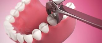
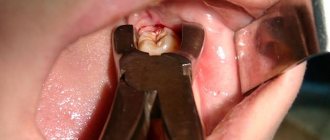
Dental forceps have been and remain tools for removing teeth. For certain groups of teeth, a different type of forceps is used, since our teeth have different structures and are located differently in the dentition. For example, to remove the upper anterior tooth and the maxillary canine, there are straight forceps, and the remaining upper teeth are removed using S-shaped ones. The incisors of the lower jaw are pulled out using forceps curved at 90º with narrow cheeks (the part of the forceps that grasps the crown or root of the tooth being removed). The fangs and the two teeth following them are pulled with forceps, on the contrary, with wide cheeks. To remove large molars of the lower jaw, forceps with spikes that go between the roots are used.
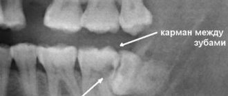
Impacted wisdom tooth
These are usually the hardest eights to remove out of all the others. We have already numbed the surgical field. What's next?
It's under the gum! So, we take a scalpel in our hands and make a delicate incision in the area of the tooth being removed. This creates access to the wisdom tooth being removed. It is isolated from the surrounding tissues using special instruments, and now we can visually assess its position and choose a removal technique.
If the tooth does not erupt, it means that something is preventing it. This “something” will also interfere with its removal, and this “something” could be a neighboring tooth, a bony protrusion, etc. However, you won’t also remove the seven to get to the wisdom tooth, right?
Special surgical tip for removing wisdom teeth. Rotates at the right frequency, provides the right torque, does not burn tissue or inflate emphysema. In surgery, we use only such devices.
Therefore, we divide the tooth into parts. Using a special tip with a cutter speed of 150,000 rpm - this is no longer a simple angle cutter, but not yet a turbine cutter. The latter, by the way, is highly undesirable to use for removing teeth, because at 500,000 rpm it is easy to burn everything with a hellish flame, and with air from the cooling nozzle you can also inflate emphysema over half your face. In general, for removal you need to choose the right tools; there are no trifles or compromises here and cannot be. And you should think a hundred times before removing such problematic teeth in a one-chair dental office at a rural club on the “Half-Empty Bins” collective farm.
Impacted teeth are removed mainly with an elevator, and not with forceps, as many are accustomed to thinking
So, we divide the tooth into 2-3 parts in order to remove it carefully and with little trauma to the surrounding tissues. And teeth are usually removed using an “elevator” (in the picture on the left). Forceps, which everyone associates with removal, are actually used extremely rarely.
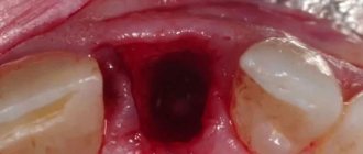
Well, the tooth has been removed. Next, we clean the tooth socket from “sawdust” and small tooth fragments that might remain. Using a curette.
When removing wisdom teeth, no biomaterials are used; the hole is filled with a blood clot on its own, this is quite enough for normal healing.
Moreover, “pushing” biomaterials into the hole can complicate the healing process, so let the regeneration process take place naturally and simply, and not fancy, as some doctors suggest.
After removal, resorbable (absorbable) sutures are placed on the hole; most often they do not need to be removed.
The clot is in place. Next, we bring the edges of the wound together and put stitches so that food doesn’t get stuck in the wound, it doesn’t bleed too much, and it heals faster. But at the same time, the sutures should not be tight, because the wound may bleed significantly during the first 24 hours. And if you don’t create an outflow, edema often develops.
Main indications and contraindications
Complex removal is required in a number of complex clinical cases when it is not possible to perform surgical intervention using standard techniques and instruments. Very often it is carried out in relation to the last painters (“eights”). As a rule, they do not have enough space, against which they begin to grow incorrectly (for example, erupt towards the cheek, injuring the mucous membrane), slowly erupt, accompanied by painful sensations. The last molars do not participate in the chewing process, so most often the dental surgeon decides to remove them in such situations.
Complex surgical procedures are resorted to in the presence of a chronic inflammatory process, against the background of which the bone tissue and the molar or premolar itself have fused. A complex operation cannot be avoided if the molar has several roots or its area is severely curved. Benign neoplasms (for example, granulomas or cysts) are often an indication for removal if the tooth is destroyed and there is no point in trying to restore it. Sometimes a complex extraction of a previously treated tooth is performed several times. This is due to the fact that it is not durable, its density is weakened, and it is capable of cracking.
As with any surgical intervention, there are a number of absolute and temporary contraindications to complex removal of molars or premolars. Among them:
- diseases of viral etiology;
- serious deviations from the cardiovascular system;
- pregnancy and lactation;
- blood diseases (anemia, poor clotting, leukemia);
- chronic pathologies during exacerbation;
- recent strokes and heart attacks;
- serious mental disorders (schizophrenia);
- long-term use of medications that affect blood clotting.
Semi-retinated tooth
In principle, the method of removing such a tooth is no different from removing a completely impacted tooth. But, as a rule, it is a little easier, because the tooth is not so deep. The main stages are essentially the same: anesthesia, creating access to the tooth (and sometimes you can do without incisions), fragmentation (dividing the tooth into parts) and, in fact, removing the teeth in parts.
After removing the lower semi-impacted tooth, sutures are placed on the socket; in the area of the upper wisdom teeth, sutures are not necessary.
When is complex tooth extraction indicated?
Tooth extraction is considered difficult due to tumor or edema, periodontitis, periodontitis, abscess and gumboil. The presence of a cyst and a fistulous tract in the tooth also complicates the removal procedure. Impacted (unerupted) teeth are also indications for surgical tooth extraction. Complex cases include the removal of a dystopic wisdom tooth located outside the dentition; removal of 4 teeth to correct malocclusion; removal of baby teeth in children at an early age. Severe curvature of the roots and fracture of the apical part of the root are also indications for surgery. Please note that complex tooth extraction is not performed during pregnancy.
The method in which your tooth will be removed depends on the individual case. Only a specialist can determine the removal strategy. In any case, you should not be afraid of this procedure. A competent doctor will perform the removal correctly, and all you have to do is say “thank you”
Dystopic tooth
Removing such teeth can be called a simpler case compared to others, but only if the tooth has one straight root. Then the removal can take place quite quickly. But such clinical cases are extremely rare. And, looking at the picture, we see hooks, not roots, which, with proper pressure, can simply break. There are usually 2 roots, and in this case we just need to separate one root from the other using the same tool - the “raising” tip. And carefully remove each of the roots separately. The beginning and completion of the removal of such teeth is the same as for all others.
Tooth root removal using an elevator
In cases where the tooth root is located very deep and is difficult to reach with forceps, an elevator is used to remove it. The doctor inserts this instrument into the narrow gap between the edge of the hole and the root, and then rotates it while simultaneously pressing lightly. With these movements, the periodontal ligaments holding the root are torn. Using the elevator as a kind of lever, the doctor seems to push the root out of the hole. Even if it does not come out of the gum completely, the exposed part is enough to pull it out with forceps.
And it also happens...
... that wisdom teeth block the neighboring teeth and prevent them from erupting normally. In such cases, patients are referred to a surgeon by an orthodontist.
Of course, the germ of the eighth tooth needs to be removed. This is a fairly simple and relatively comfortable operation.
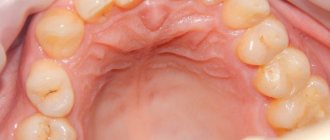
Look at the pictures on the right. There is a difference of three weeks between the top and bottom. It is clearly visible from them that after removing the rudiments of the eights and “unblocking”, the seventh teeth immediately began to grow.
Wisdom tooth removed. The patient is satisfied. But the fun is yet to come. Namely, the postoperative period.
Removing a tooth root using forceps
First, the doctor separates the periodontal tissue from the root so that the exposed part of the root can be easily grabbed with forceps. If this is difficult to do, the doctor separates the periosteum and the edge of the mucosa from the socket. It is much more difficult to remove upper teeth than lower teeth, so bayonet-shaped and S-shaped forceps are used for this. The doctor places the instrument as tightly as possible and makes rotational movements with it. If the tissues surrounding the tooth are affected by an inflammatory process, then the problematic root can be extracted quite quickly. The most difficult teeth in this regard are the canines and lower molars, since they have large alveolar processes.
Elimination Features
To eliminate a damaged tooth, an elevator is inserted between the wall and the root; for multi-rooted units, between the individual elements of the root. The concave side faces the tooth, the convex side faces the socket. The doctor presses on the handle and forgives the instrument, moving its working part deeper. The success of removal depends on how correctly the dentist chooses the type of instrument and performs the operation. Before starting work, you need to carefully examine the damaged and removed tooth and evaluate the strength of the crown part. If the external crown is in the way, its preliminary removal is indicated.
Elevator usage options:
- With slight bending, the root part is deflected, the socket is enlarged, and the unit being removed is dislocated.
- For third molars, sectioning and insertion of the cheek from the distal side are necessary. The dental crown is displaced and the units are removed.
- If the roots are sufficiently bent, it is necessary to rupture the periodontal ligaments. To do this, the tooth is divided into three separate parts using special tools, which are removed one by one.
- In case of buccal as well as lingual curvature, the elevator widens the periodontal gap, after which removal is carried out.
- If the alveolar process is of significant thickness or the crown is significantly destroyed, the cheek is inserted between the root part and adjacent units.
- The straight type of instrument is used to extract figure eights; the working part is carefully inserted along the surface of the tooth, after which it is removed.
Before starting the procedure, you must thoroughly sterilize the elevator and other instruments. First, they are soaked in an antiseptic solution and washed in clean running water. The next step is immersion in the biolot, heated to 40 degrees. The instruments are dried before use.
Complications
Deterioration during removal rarely develops. But such a situation is still possible; in some cases, the Patient’s condition seriously worsens, especially if the doctor violates the removal technology. In practice, the following complications are noted:
- damage to blood vessels, which causes severe bleeding;
- removal, serious damage to the primordia of permanent units when removing milk;
- puncture wounds of tissue due to puncture of instruments, in this situation there is a high risk of damage to the carotid artery and other large vessels;
- development of an infectious lesion, alveolitis (if the instrument is not properly sterilized);
- ruptures, dislocations of ligaments;
- breakage, part of the elevator getting stuck in the wound;
- fractures of hard tissues when the load is exceeded towards the tips of the root part.
Rare cases include the formation of a granuloma in the area where the unit was removed. After eliminating the cyst, part of the nail of the doctor who performed the extraction was found in the tissue. Apparently it was cut off with an instrument during surgery.
Elevators are indicated for use if the coronal part is destroyed or for other reasons it is not possible to use forceps. For the operation, local anesthesia is used, which allows the Patient not to feel pain or discomfort.
Is it necessary to remove tooth roots?
The issue of tooth root removal can only be decided by your attending dentist based on the examination and the X-ray obtained. If it is not a source of infection and the tissues surrounding it are healthy, then the root can be used in the future as a support for an artificial crown, having previously “extended” it using a stump tab. Tooth root extraction is highly recommended in the following situations:
- a cyst has formed on the root of the tooth;
- the tooth was severely damaged by caries;
- root fracture;
- one of the walls of the tooth has broken off, and the chip goes deep into the gums;
- There is severe tooth mobility caused by periodontal diseases.

