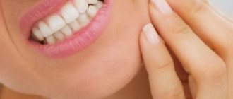
What are blockades
Therapeutic blockades are the relief of acute or chronic pain and spasms. It is performed by injection into the area where the nerve trunks and plexuses pass. Severe pain causes physical and psychological suffering, incapacitates the patient and can cause disability. Depression that occurs against this background often leads to suicide attempts.
Therapeutic blockades quickly relieve acute pain. After some time, swelling, inflammation, spasm of muscles and blood vessels decrease. This leads to a lasting improvement in the condition. At the 100med medical center, blockades are performed by neurologists and anesthesiologists. Several groups of drugs are used for the procedure:
- local anesthetics;
- corticosteroid hormones;
- chondroprotectors.
The effect of drug administration lasts from several days to several weeks. After the medicine stops working, the pain may return. However, its severity will be less by about 50%.
During the procedure, the patient may feel some discomfort associated with the injection. However, the discomfort quickly passes. There is no need to be afraid of blockades, as this method is one of the safest. Our specialists strictly follow all the rules for performing the procedure, as well as asepsis and antiseptics. This guarantees the effectiveness and safety of blockades in our clinic.
Drugs used for therapeutic blockades
First, let's look at the most commonly used local anesthetics.
"Novocaine"
Can be injected into nerves and tissues, relieves pain and relieves spasms. In the pathological focus, strong irritation occurs and peripheral innervation is switched off. The drug improves tissue trophism, as well as:
- reduces the permeability of vascular walls;
- acts as an antiseptic and bacteriostatic;
- increases resistance to allergens;
- evens out vascular tone;
- improves nerve trophism.
It is considered the safest means of pain suppression, characterized by a minimum of side effects. Has a certain degree of toxicity.
"Lidocaine"
A local anesthetic that provides a more intense and long-lasting effect compared to Novocain. It is used in the form of hydrochloride. Blocks voltage-gated sodium channels, which stops the generation of impulses in nerve endings and blocks the conduction of impulses along nerves. Blocks not only pain impulses, but also others. Dilates blood vessels and has no local irritating effect. A few minutes after the administration of Lidocaine, the pain disappears and the muscles relax. Toxicity is lower than that of Novocain.
"Bupivacaine"
Amide drug. Its effect develops slowly, but lasts for a long time. This is a drug approximately 16 times more powerful than Novocaine. The entire group of amide anesthetics is more stable compared to ester anesthetics (for example, Novocain). Their long-lasting effect is due to the fact that they have a longer half-life. This is due to the fact that drugs are processed not by plasma, but by the liver. Bupivacaine prevents the pain impulse from spreading along the nerves by blocking sodium channels in the neuron membrane. The blockade is sequential, depending on the size of the nerve fibers (small ones are more sensitive compared to large ones), the degree of activation (a higher frequency of impulses opens more channels for blocking), and the degree of myelination (fibers with a myelin sheath are blocked better). The effect of the procedure occurs within 5-10 minutes.
Now let's move on to the most popular glucocorticosteroids. Drugs in this group are designed to enhance the effectiveness of the blockade, relieve inflammation and allergic reactions. Can also be used independently.
"Hydrocortisone"
Belongs to a group of drugs with a short duration of action. Contains artificial hormones that are similar to those produced by the adrenal glands. It is used to treat heel spurs with a course of 3-5 injections. The active substance of the drug accumulates in tissues, relieving inflammation. The main advantage is low price. However, it loses to Diprospan in terms of effectiveness.
When injected into joints together with Lidocaine, the effect develops within 24 hours and lasts up to several weeks. Able to increase blood pressure while simultaneously reducing the concentration of lymphocytes. Thanks to the first, the volume of blood circulation increases, and the second, the intensity of the immune response decreases. The drug is suitable for neurological blockades. For peri- or intra-articular blockades, it is necessary to mix the microcrystalline suspension with a local anesthetic to prevent necrosis.
"Dexamethasone"
Long-acting glucocorticosteroid. One of the most powerful drugs. It has a pronounced anti-inflammatory, antiallergic, immunosuppressive and desensitizing effect. It works especially well when used together with an anesthetic for degenerative pathologies. Reduces inadequate response to anesthetic drugs. Acts quickly, suitable for blockades of soft tissues and joints. Compared to Hydrocortisone, Dexamethasone is 25-30 times more active. There are no known cases of necrosis with its use; it has little effect on electrolyte metabolism.
"Depo-Medrol"
Methylprednisolone acetate is a long-acting form of methylprednisolone. This is due to poorer solubility and lower metabolization activity. Used for blockades of soft tissues and joints. For epidural blocks - with caution, because it can provoke inflammation of the arachnoid membrane.
Used to treat all joints, with the exception of those that are difficult to reach and do not have a synovial cavity. When injecting into the hip joint, it is necessary to avoid entering large vessels. Injections into the tissue surrounding the joint may have little or no effect. If there is no positive effect from the first injection, the procedure is not repeated.
"Diprospan"
A drug based on betamethasone. Used to treat heel spurs. Provides rapid analgesic and anti-inflammatory effect. Relieves allergic reactions. Usually it allows you to cure a spur in 1-2 sessions. A significant drawback is the destructive effect on the adrenal glands and pituitary gland. However, the likelihood of complications in this drug is sharply reduced compared to analogues. It differs from other similar drugs in its microcrystalline structure. Can be used for epidural blockades, as well as blockades of spinal nerves, muscles, etc. It is effective for destructive-dystrophic diseases of the spine, in particular the lumbar region.
Nerve block
A nerve block is a treatment in which a local anesthetic is injected into the soft tissue where the peripheral nerve passes. The procedure allows you to relieve pain, spasm of muscles and blood vessels, swelling and inflammation. Indications for manipulation:
- neuritis;
- neuralgia;
- tunnel syndrome;
- muscular-tonic syndrome;
- diseases of the spine;
- pain after injury or surgery;
- chronic pain syndrome;
- oncological diseases.
Nerve blocks do not require special preparation. After a thorough examination and examination of the patient, the doctor determines the injection site and treats the skin with an antiseptic. An injection of pain medication is given into the soft tissue. After a few minutes, numbness, warmth and a feeling of heaviness appear in this place. The course of injections (up to 3 sessions) can be repeated every year.
Joint blockade
Joint blockade is a method of introducing a drug into the joint cavity or adjacent soft tissues (articular capsule). The method not only relieves pain, but eliminates inflammation, relieves muscle and vascular spasm, and restores range of motion in the joint. The procedure is indicated for the following diseases:
- arthritis of joints of various sizes;
- arthrosis;
- arthralgia;
- synovitis;
- bursitis;
- tendovaginitis;
- periarthritis;
- facet syndrome;
- Reiter's disease;
- Bekhterev's disease.
The injection is performed without additional preparation after a thorough examination. The patient is seated or laid on a couch and the skin over the joint is treated. The injection is made into the cavity of the joint capsule, into the area of the periarticular ligaments or bursa. The effect occurs within a few minutes and lasts up to 3 weeks. The duration of the course depends on the severity of the lesion and can be up to 15 procedures.
Traumatic neuropathy. Etiology.
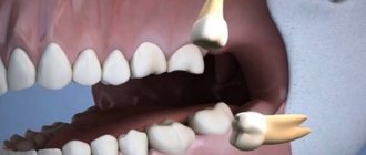
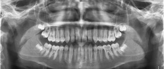
Traumatic neuropathy most often occurs in the case of surgical interventions on teeth (traumatic extraction of teeth, removal of filling material beyond the apex of the tooth root, anesthesia with injury to the nerve trunks, removal of bone or tumor of the jaws), as well as in cases of surgical interventions on the paranasal sinuses and infraorbital canal.
Damage to the first branch of the trigeminal nerve, as a rule, is practically not observed. Most often, the third branch of the trigeminal nerve is affected, which is apparently due to the anatomical location of the inferior alveolar nerve, which makes it easily accessible during a variety of traumatic dental procedures. This is especially true for dental interventions on third molars. The cause of traumatic neuropathy of the inferior alveolar nerve can also be filling the mental canal during the treatment of pulpitis of the 4th and 5th teeth of the lower jaw.
Combined damage to the I and II branches of the trigeminal nerve can occur after inflammatory diseases of the brain with the development of adhesions or in the case of sinusitis, when the maxillary and frontal sinuses are simultaneously involved in the inflammatory process.
Clinic.
Patients complain of constant aching, sometimes pulsating pain in the area of innervation of the injured nerve, a feeling of numbness and “crawling goosebumps”. In case of injury to the mandibular nerve, tooth alignment occurs, which is associated with damage to the motor part of the nerve; patients cannot eat or talk. Trigger zones on the face and in the oral cavity are not identified.
During an objective examination, hypoesthesia or anesthesia (hyperpathy is also possible) of the skin and mucous membrane in the area of nerve innervation is detected. On palpation, pain is noted at the exit points of the II and III branches of the trigeminal nerve, as well as in the case of vertical percussion of the teeth and deep palpation of the lower jaw.
Diagnostics.
The main diagnostic criterion is the occurrence of pain after interventions on the dental system. The disease is characterized by clinical polymorphism and significant duration. During weather changes, stressful situations and in the presence of somatic diseases, exacerbation of the pain syndrome may occur.
In the event of scar changes in the nerves or retraction of the nerve into the scar of soft tissues (after gunshot wounds, in the case of defects of soft and bone tissues after resection of the jaws), constant aching pain of unexpressed intensity with persistent sensory disturbances is observed.
Celiac plexus block
With this type of blockade, the medicine is injected into the solar plexus area. The injection is done under the control of an X-ray machine or ultrasound.
The procedure is effective for:
- chronic pancreatitis, pancreatic necrosis;
- abdominal adhesions;
- Crohn's disease;
- tumors of the abdominal organs;
- ineffectiveness of painkillers.
You should prepare for the blockade in advance. A few days before the procedure, the doctor at our clinic recommends stopping taking anticoagulants and diuretics and adjusting the dose of glucose-lowering medications. The patient is placed on his stomach with a bolster or pillow. First, the doctor anesthetizes the soft tissue at the injection site. Then, under X-ray control, a special long needle is inserted. Through it, a local anesthetic or a drug that destroys nerve cells is supplied to the solar plexus. The patient feels warmth in the abdomen and a decrease in pain. After the procedure, observation is required in our hospital for 24 hours.
Anesthesia of the anterior and middle superior alveolar nerves with a single injection into the palate
Traditionally, anesthesia of teeth in the upper jaw is carried out by injection into the transitional fold in the projection of the apexes of the roots. It is a simple, safe and effective way to achieve anesthesia of the pulp and associated tissues. The main disadvantage of this procedure is the need for multiple injections to anesthetize more than one tooth, as well as excessive anesthesia for the lip and facial muscles.
However, the sensitivity of the teeth in the upper jaw can also be turned off using conduction anesthesia (infraorbital, torusal) or intraosseous and intraligamentary. The literature does not contain information about performing anesthesia with one injection for several teeth, which would not turn off facial sensitivity. Therefore, Friedman and Hochman (1998) proposed a maxillary anesthetic technique to block the anterior and middle superior alveolar rami. The authors describe effective anesthesia from the central incisor to the second premolar with a single injection into the palate. Anesthesia is expected to last 45 minutes to 1 hour (Figure 1), and the injection is performed using a computerized Wand Plus system (Milestone Scientific, Deerfield). In addition, the authors claim that numbness of the lips and facial muscles does not occur during anesthesia.
Photo 1. Anesthesia zone for blocking the anterior and middle alveolar nerves: gingiva of the vestibule of the oral cavity, teeth (central, lateral incisors, canine, first premolar, second premolar and mesial buccal root of the first molar), gums from the palatal side and oral mucosa to midline.
The essence of anesthesia is the supply of anesthetic to the anterior and middle superior alveolar branches by diffusion through many nutrient foramina on the palatine process of the maxilla (Photo 2). Both nerves are collaterals of the infraorbital nerve in the canal of the same name, which is a branch of the maxillary nerve. The anterior superior alveolar nerve departs from the infraorbital branch not reaching 5-8 mm of the infraorbital foramen. It is responsible for the innervation of the pulp of the central, lateral incisors and canines. The middle superior alveolar nerve arises from the infraorbital branch approximately 10 mm before the infraorbital foramen. This nerve supplies the pulp of the premolars and the mesial buccal root of the first molar. However, the middle branches are not present in all people. Research reports that they are found in 30-72% of individuals. When these branches are absent, the innervation of the corresponding zone is provided by plexuses between the posterior and anterior branches.
Photo 2. Arrows indicate nutrient openings
The zone of anesthesia for an anterior and middle rami block extends from the palatal side, detaching the premolars and reaching the midpalatal suture, while affecting the free gingiva (Photo 3). Theoretically, this technique is advantageous because the bilateral block of the anterior and middle branches provides simultaneous anesthesia of 10 upper teeth without numbness of soft tissues and facial muscles, which is especially convenient for restorative manipulations.
Photo 3. The injection site is indicated by an arrow, the middle of an imaginary line (a) connecting the midpalatal suture (b) and the free gingival margin (c).
Fukayama and Lee found high efficiency when using this technique using a computer system (Wand), but the resulting pulpal anesthesia percentage remained controversial. Other studies have reported the use of the technique in periodontal surgery, but only using a conventional syringe. The advantages include excellent hemostasis, no facial anesthesia and fewer injections.
The purpose of this study was to study the level of successful anesthesia of the anterior and middle superior alveolar nerves using a conventional syringe (carpule type).
Materials and methods
This clinical controlled study involved 30 Caucasian patients (16 men and 14 women) with a mean age of 22 years (range 21-25 years). During the selection process, exclusion criteria from the study were established: the presence of systemic pathology that prevents the use of an anesthetic with a vasoconstrictor, pregnancy, taking medications that affect pain perception, active orthodontic treatment, condition after tooth extraction, patients with fixed dentures, a large number of filled and endodontically treated teeth in the examined area (upper central incisor, lateral incisor, canine, first and second premolars). All information was obtained through a written survey and clinical examination. The study was approved by the ethics committee of the school of dentistry, and written informed consent was obtained from each patient.
30 patients underwent anesthesia of the anterior and middle alveolar branches on a randomly selected one half of the jaw using a carpule syringe (Hu-Friedly), 1 ml of 2% lidocaine with epinephrine 1:100,000. All injections were performed by a single specialist.
Then the upper central incisor, lateral incisor, canine, and first and second premolars of the selected side were examined. The lower canine is selected as a control tooth to monitor the performance of the pulp tester. At the very beginning, before the injection, the experimental tooth and the control canine were examined for vitality using a pulp tester (Sybron Endo). The pulse level on the pulp tester is adjusted from 0 to a maximum of 80.
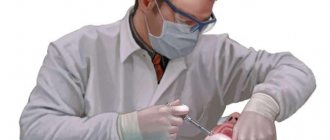
The injection was carried out according to the landmarks described in the original technique of Friedman and Hochman. The injection site is located midway between the midpalatal suture and the gingival margin of the first and second premolars (Figure 3). An application of local anesthetic (20% benzocaine) is applied for 1 minute at the injection site.
The injection was performed with a 30G 22mm needle (Teruno). The bevel of the needle to the bone was oriented at an angle of 45 degrees. The needle was then slowly penetrated into the palatal mucosa until it contacted the bone and 1 ml of anesthetic solution (lidocaine 2% with epinephrine 1:100,000) was injected over three minutes (Figure 4).
Photo 4. Implementation of the technique.
The success of anesthesia was monitored by an electronic pulp tester (Sybron Endo). During the study, a protocol was drawn up. At 1 minute after injection, pulp test data were obtained for the first and second premolars. After 2 minutes the canine was tested, 3 minutes - the lateral and central incisor. At the fourth minute, the control canine was tested. The control continued with a 4-minute cycle for an hour. The criterion for pulpal anesthesia was a negative response at the maximum impulse (80). Anesthesia was considered successful when at least two consecutive negative responses were obtained at pulse 80 within one hour.
The time of onset of anesthesia and duration of effect were also taken into account.
results
The study involved 30 adult patients, 16 of whom were men and 14 women. The average age of the group was 22 years (with a range of 21-25 years). All patients received an injection to anesthetize the anterior and middle superior branches of the maxillary nerve on one side of the jaw. The injection consisted of 1 ml of 2% lidocaine with epinephrine 1:100,000. Successful pulpal anesthesia was 16-66% in the teeth examined. The onset of anesthesia is from 6 to 12 minutes and the duration is 23-40 minutes.
All subjects experienced anesthesia of the palatal tissue from the central incisor to the mesial buccal cusp of the first molar, without crossing the midline. No patient experienced pulpal anesthesia from the second premolar to the central incisor and facial tissues. While 8 patients (26.7%) did not experience anesthesia at all in any of the experimental teeth. No severe pain was experienced when the anesthetic was injected into the tissue of the palate. Also, no complications or adverse reactions were observed during and after the technique.
Discussion
The main advantage of the technique of one-stage block of the anterior and middle superior alveolar branches is the reduction in the number of injections and the amount of anesthetic solution compared to conventional infiltration anesthesia for each tooth. In addition, this technique can be ideal in cosmetic dentistry since it does not cause numbness to the lip and face.
However, the study showed the success of anesthesia in only 16.7-66.6% of cases. The criterion was a device impulse block with a value of 80. Dreven and Certosimo proved that transmission disruption at any device reading below 80 would indicate the presence of pain during the intervention.
In our study, blockage of all five experimental teeth did not occur in any of the cases, as was described by Friedman and Hochman. The effect of the anesthetic gradually decreased over 60 minutes; our study could not confirm the data of other authors who reported the effect of anesthesia within an hour. On the other hand, the absence of numbness of the lips and facial muscles is completely confirmed.
Tests of this technique using a computer system (Fukayama, Lee) showed a range of 42-86% and 35-58% successful anesthesia. In our study, when using a conventional syringe, the rates were significantly lower (17-66%). The low percentage when using a conventional syringe may be due to the less perfection of the method compared to the computer-controlled one. Automated injections are characterized by a controlled constant flow of solution. With the traditional technique, the solution flow and pressure depend on the specialist, although all injections in the study were carried out by one doctor. Lee suggests that computer-controlled injections create a special pressure gradient that allows the anesthetic to better penetrate the many feeding holes on the hard palate.
In cases of ineffective anesthesia of the central and lateral incisors, it can be assumed that the primary innervation is from the middle upper branches. While the middle branches are well anesthetized in such cases, the anterior branches remain uninvolved due to their distant location to the injection site. The study of cadaveric materials has shown that the middle superior alveolar branches are present in only 30-72% of people, and in cases of their absence, the innervation of the corresponding area is carried out by a plexus formed by the anterior and posterior alveolar nerves. The exact role of the effect of the absence of middle branches on the success of the block remains unclear.
The technique is described by the authors as a nerve block, while Malamed calls a nerve block only the process of bringing the anesthetic solution very close to the main nerve trunk, in this particular case directly to the middle and anterior superior alveolar nerves, which will ensure high efficiency of anesthesia. However, we do not observe this in the described study, since the anesthetic is deposited at the palatine process of the maxilla.
On the other hand, reducing the number of injections allows for the introduction of a smaller amount of vasoconstrictor, which is especially important in patients with cardiovascular pathology. For interventions on the mucosa and gums, anesthesia of the anterior and middle branches provides excellent hemostasis, which does not require reinforcement with subsequent injections.
One of the main disadvantages of this technique is the severe pain of the injection into the palate. Wahl states that palate injections are the most painful of all oral injections, possibly due to the pressure created in the tissue. However, in our study, patients did not experience significant discomfort due to pre-application of local anesthetic, as well as slow and controlled administration of the drug. Another disadvantage is that the technique is limited to anesthetizing only five teeth, so periodontal surgery requires a different, more complete method of anesthesia.
Another option for anesthesia with one injection from the central incisor to the second premolar is an infraorbital block. This injection is less painful, since it is not performed on the palate, however, the inconvenience of performing it causes some side effects, including numbness of the face. Karkut found that infraorbital nerve blocks are ineffective for complete pulpal anesthesia of the upper central incisor (15% success) and lateral incisor (22% success). The effectiveness of anesthesia for the canine is 92% and for premolars is 80%. Moreover, anesthesia lasts much less than an hour in all of these teeth. According to Berbeich, who studied 40 patients, the ineffectiveness of the technique in relation to incisors is also confirmed. Successful pulpal anesthesia of the canine and premolar teeth is estimated to range from 75-92%.
Corbett conducted a study comparing anterior and middle alveolar branch blocks with infraorbital foramen blocks. The success of anesthesia in the incisors was much more pronounced when performing blockade of the middle and anterior branches (42.9%). However, anesthesia of canines and premolars occurred better with blockade at the infraorbital foramen. Lip numbness was observed in 100% of cases with the infraorbital technique and in 14.3% with the palatal technique. There was no significant difference in discomfort between both techniques. It has been determined that the infraorbital technique causes more pronounced pulpal anesthesia, while the palatal one is more widespread and does not cause numbness of the face.
The shape of the palate was not taken into account in the study, but we believe that the distribution of the anesthetic between different parts of the palate was in any case homogeneous. Since the technique was used in a group of young people, the results may not be applicable to children or the elderly.
Conclusion
Since the described technique is unpredictable and not always effective, it cannot be considered the technique of choice in clinical practice. However, it is very useful when carrying out restorative manipulations, while it does not distort the smile and does not affect the facial muscles. The technique is also applicable in periodontal surgery due to excellent hemostasis in the soft tissues of the palate.
Authors: Ignacio Velasco, Reinaldo Soto Department of Oral and Maxillofacial Surgery, Los Andes University, School of Dentistry, Santiago, Chile
Paravertebral blockade
Paravertebral blockade is performed to reduce pain in the spine. The effect is achieved by blocking the ganglia or spinal nerves on one or both sides. Indications for this are:
- osteochondrosis;
- radiculitis, lumbago;
- neuralgia;
- myositis;
- hernias and protrusions of intervertebral discs;
- spinal injuries.
To carry out the manipulation, the patient is placed on a flat couch. Local soft tissue anesthesia is first performed. The medicine is injected into the segment of the spine where the pain is felt the most. The injection is given on one or both sides depending on the patient’s condition.
Epidural block
An epidural block is the injection of medication into the space between the dura mater and the periosteum of the spinal canal. Depending on the injection site, caudal, translumbar, and transforaminal blockades are distinguished. A good effect of blockades is observed when:
- damage to the radicular nerves;
- periduritis;
- intervertebral hernias;
- spondylolisthesis;
- narrowing of the spinal canal;
- back pain after injury or surgery.
The procedure must be carried out on an empty stomach. The patient is placed on a couch, and the doctor applies local anesthesia to the desired area. Then, under the control of an X-ray machine, a needle is inserted through which the medicine is administered. When performed correctly, the patient feels a slight burst in the back.