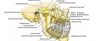
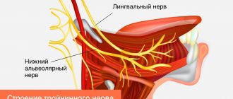
What is mandibular nerve injury?
By this concept, dentists mean injury to one of the nerves:
- chin;
- lingual;
- alveolar.
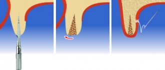
Types of injuries include sprain, compression, crushing and rupture - partial or complete. The cause of the stretching is the long-term retraction of the mucoperiosteal flap, which is created by an implant of greater length than necessary. Crush injuries and compression are caused by needle injuries during the administration of anesthesia. Rupture occurs in two cases: when cutting the mucosa or during preparation of the hole for the implant.
Basic techniques
On the lower jaw:
• Mandibular
• Buccal nerve anesthesia
• Mental
• according to Go-Gates
• according to Akinosi
On the upper jaw:
• Posterior superior alveolar nerve anesthesia
• Incisal
• Palatal anesthesia
• Infraorbital
• Maxillary nerve
Causes and prevention of mandibular nerve injuries
The only cause of such damage is considered to be medical errors. Since in preparation for implantation, X-rays of the jaw are taken, which the doctor must carefully study so that when choosing an implant and a place for it, he does not injure the nerve, the injuries are caused by his unprofessionalism or negligence.
Damage to the mandibular nerve most often occurs when:
- improper administration of anesthesia - needle injury;
- choosing an implant that is too long;
- damage by an instrument - when preparing the site for the implant.
The only way to avoid such an injury is for a doctor to responsibly approach the stage of preparation for surgery, carefully studying the condition and structure of his patient’s jaw. The only way of prevention for the patient is to choose a trusted clinic and a highly qualified doctor. Specialists at the Implantmaster clinic have been able to reduce the number of injuries of this kind to 2%, since they carefully study three-dimensional photographs of a person’s jaw before implantation, and can correctly assess the condition of the bone tissue, the location of nerves and blood vessels, and select the optimal size of the implant.
Diagnostics
The diagnosis of trigeminal neuropathy is based primarily on the patient's medical history, symptoms, and physical and neurological examination findings. To make a diagnosis of NTN, it is necessary to exclude other diseases that may manifest as pain in the facial area. Some conditions that cause facial pain include: post-herpetic neuralgia, headaches, and temporomandibular joint disorders.
Due to the commonality of symptoms and the large number of conditions that can lead to facial pain, making a correct diagnosis is often difficult, but finding out the exact cause of the pain is very important, since treatment tactics for different types of pain may differ.
Most NTN patients will eventually need to undergo a magnetic resonance imaging (MRI) scan to rule out a tumor or multiple sclerosis as the cause of the pain. This test method can clearly show the compression of a nerve by a blood vessel. Special MRI techniques can reveal the presence and degree of compression of a nerve by a blood vessel.
The diagnosis of classic trigeminal neuropathy can also be confirmed by the positive effect of taking anticonvulsant medications for a short period of time. Diagnosis of T2 is more complex and difficult, but is usually confirmed by a positive response to low-dose tricyclic antidepressants in the same way as other neuropathic pain.
Our team of doctors
Maxillofacial surgeon, Implantologist
Bocharov Maxim Viktorovich
Experience: 11 years
Dental surgeon, Implantologist
Chernov Dmitry Anatolievich
Experience: 29 years
Orthopedist, Neuromuscular dentist
Stepanov Andrey Vasilievich
Experience: 22 years
Endodontist, Therapist
Skalet Yana Alexandrovna
Experience: 22 years
Orthopedic dentist
Tsoi Sergey Konstantinovich
Experience: 19 years
Dentist-orthodontist
Enikeeva Anna Stanislavovna
Experience: 3 years
Symptoms and stages of damage
The symptoms by which this complication can be recognized are as follows:
- numbness of parts of the head - tongue, lips, chin, cheeks, etc.;
- biting lips and tongue;
- choking while eating or drinking;
- profuse salivation.
All this creates a number of inconveniences for the patient: it makes it difficult to eat and talk, disrupts facial expressions, and also prevents men from shaving and women from applying makeup. The severity of this injury is determined by its degree: a minor one goes away on its own or with the help of drug treatment, a severe one leads to irreversible processes of nerve degeneration and is not curable. Damage to the mandibular nerve, the symptoms of which the patient observes, requires immediate consultation with a doctor - only a specialist will be able to determine its extent and provide timely assistance.
Dentists distinguish the following stages of this implantation complication:
- minor - neuropraxia;
- more severe, but partial damage - axonotmesis;
- a serious injury that leads to complete loss of sensitivity - neurotmesis.
Clinical algorithm for determining post-traumatic neuropathy
Trigeminal Neuropathy Assessment Questionnaire
Important! Severe pain experienced during treatment may indicate a possible nerve injury.
- History of the initial onset of pain.
- Development of pain.
- Duration of pain.
- History of regular pain SOCRATES (Site, Onset, Character, Radiation, Associated signs, Timing, Exacerbating and relieving factors, Severity) - Localization, Occurrence (frequency of attacks), Character, Irradiation, Associated symptoms, Duration, Exacerbation and relief factors (what intensifies and relieves pain), severity of pain.
- Psychological screening.
- Functional screening (impact on daily life).
Mechanosensory tests (mapping the affected area)
A protocol for examining the dermatome to assess the extra-oral mechanosensory function of the alveolar branch of the trigeminal nerve.
Dermatomes are segments of skin into which the entire surface of the human body is divided in connection with its innervation by various roots of the spinal cord, in this case the trigeminal nerve.
Figure 7. Dermatomes of the branches of the trigeminal nerve
1. Logging the affected area.
Using surgical forceps, move from the normal to the neuropathic (changed) area, warning the patient that there may be increased sensitivity and/or decreased sensitivity. Mark an area on the patient's face with marker marks and take a photo. Assess the % or area of the extra-oral dermatome that is affected by neuropathy.
2. Recording the assessment of subjective sensation.
Press the surgical forceps or probe firmly (but not painfully) against the patient's arm several times at intervals (5 times per minute), explaining that this is a "normal" subjective rating of function on a scale of 10 out of 10. Press with the same pressure on the unaffected side of the face or tongue and repeat the stimulation, explaining that it should be 10 out of 10. Then remove the forceps or probe and explain that the missing stimulation is 0 out of 10. Only then repeat the same actions in the area of neuropathy that you have already confirmed and marked with markers labels, and ask the patient to report the stimulus level out of a possible 10 (if >10 = hyperesthesia and <10 = hypoesthesia). This test should be repeated in different areas of the neuropathy (lip border, lip skin, chin, tongue, etc.)
3. Recording the light touch assessment
To assess light touch thresholds, it is recommended to use a frayed cotton swab, repeating touches at intervals of 5 times per minute. First on the unaffected side and then repeating on the affected side, ask the patient to report the differences. If the patient is experiencing numbness, then the stimulation will have a reduced threshold for detecting light touch, however, if the patient suffers from hyperesthesia and possible allodynia (pain to touch), then this test can be very uncomfortable and irritating.
Dermatomal Involvement Map Interpretation and Neuropathy Assessment —Does the area of neuropathy correspond to the dermatomal area in which surgery was performed? — Dynamics of the area of the involved extraoral and intraoral area when mapping neuropathic areas (criterion reliability is low)
Important! Localized sensory neuropathy is not always present in patients, but there is almost always an area of abnormal sensation, and the patient's maximum pain is associated with the area of sensory deficit, that is, suffering from a combination of pain, numbness and altered sensation. This is an important diagnostic distinction for sensory neuropathy.
Subjective function
- Is neuropathy hyperesthesia or hypoesthesia?
- Thermal allodynia test is the occurrence of pain when exposed to heat or cold. Thermal allodynia, especially cold allodynia, is a feature in patients with trigeminal nerve damage.
- Thermal hyperalgesia test. Increased pain that occurs after a weak stimulus.
- Test "Direction of movement". The patient closes his eyes, the doctor uses a soft brush to determine the patient's ability to detect both the sensation and the direction of movement of the brush.
- Test for sensitivity to temperature stimuli. A cotton swab with cold test spray and a dental mirror handle heated to 43 - 45 ° C are used to determine the patient's ability to feel cold and heat. Alternatively, test tubes can be filled with hot (43 -45 °C) water and cold water.
There are WHO recommendations regarding which parameters of peripheral sensory consequences should be taken into account to predict the results of microsurgical restoration of a damaged sensory nerve. Zuniga JR and Yates DM adapted the recommendations to trigeminal nerve lesions. More details here: Zuniga JR and Yates DM Factors Determining Outcome After Trigeminal Nerve Surgery for Neuropathic Pain. J Oral Maxillofac Surg 2016 Jul;74(7):1323-9.
Today, a guideline for mandatory X-ray monitoring after endodontic treatment and dental implantation surgery has already been adopted in order to indicate the relationship between the root and the root filling, as well as the proximity of the implant bed and/or the implant itself to the canal of the inferior alveolar nerve. Intraoral dental radiography is considered to be sufficient to detect iatrogenicity, although post-traumatic neuropathy is primarily a clinical diagnosis.
Renton T, Yilmaz Z. Managing iatrogenic trigeminal nerve injury: a case series and review of the literature. Int J Oral Maxillofac Surg 2012 May;41(5):629-637.
Recovery and treatment
In the first case, self-recovery takes approximately 1 month; the help of doctors is not needed, since there is no anatomical damage. Symptoms of the second appear after a while - usually 6-8 weeks, so recovery can be painful and incomplete: it will take more than 2 months. In the third stage of damage to the mandibular nerve, treatment gives results only at the beginning and is performed surgically, since we are talking about degeneration with a violation of integrity. Loss of sensitivity, which is observed in a patient for more than 3 months, indicates a high probability of losing it forever. Damage to the mandibular nerve, the consequences of which is the lack of sensitivity of the nerve for a year, leads to irreversible changes. Only the professionalism and responsibility of the doctor, which is guaranteed by the specialists of our Implantmaster clinic, can protect the patient from such unpleasant injuries.
Author:
When performing dental implantation in the lower jaw, there is a potential risk of damage to the inferior alveolar nerve (IAN) and, as a result, disruption of its function [6, 8]. According to a number of authors [1, 3, 5], the incidence of this kind of complications during dental implantation ranges from 8.5 to 33% of the total number.
Perforation of the mandibular canal and nerve injury during the formation of the implantation bed, as well as compression of the nerve directly by the dental implant or due to postoperative swelling lead to peripheral neuropathy. This complication manifests itself in the form of absence and/or long-term changes in the sensitivity of tissues in the innervation zone, the development of pain of varying intensity, and is also accompanied by emotional and stress disorders and significantly worsens the patient’s quality of life [6, 8, 10].
After injury, complete regeneration of the nerve is possible provided there is no prolonged influence of the traumatic agent, as well as obstacles to axon growth and restoration of blood flow in the damaged area. Otherwise, already 2 months after the injury, atrophy and sclerosis of the nerve distal to the damaged area begin [4]. This can cause long-term sensory disturbances in the innervation zone. Consequently, there is a need for the earliest possible diagnosis of complications and treatment, which is aimed at restoring the anatomical integrity and functional viability of the NAS.
The purpose of the study is a clinical and physiological study of dysfunction of the NAS as a complication of dental implantation, determination of tactics and treatment strategies.
Material and methods
The main criterion for including patients in this study was the presence of signs of dysfunction of the NAS after dental implantation performed in the lateral areas of the lower jaw. In the specialized department of the Department of Faculty Surgical Dentistry and Implantology of the Moscow State Medical and Dental University, 32 patients (24 women and 8 men) aged from 26 to 60 years with installed screw implants of various systems in the amount of 1 to 4 on the side of the nerve injury were observed . After a thorough interview and history taking, all patients underwent general examination methods, as well as an assessment of local neurological status. Pain intensity was assessed using a 10-point visual analogue scale (VAS). The sensitivity of the tissues of the chin area, lower lip on the side of the injury and the symmetrical side was studied using sensory tests. The response to tactile (touching the skin with a cotton ball), pain (tingling with a needle) and temperature (metal surface, test tube with hot water) stimuli was assessed. The area of sensitivity impairment was calculated in centimeters using the formula for calculating the area of a trapezoid or rectangle and was necessarily photographed for comparison in dynamics (Fig. 1).
Figure 1. Area of sensory impairment based on sensory tests.
The degree of sensory impairment after dental implantation was assessed by measuring sensitivity thresholds in the zone of innervation of the inferior alveolar nerve. Single pulses of electric current with a duration of 0.1 ms and a frequency of 0.5 pulses/s with a gradually increasing amplitude from 0.5 to 99 mA were used as a stimulus. The electrode was installed on the skin in the projection of the lower lip and chin area both on the side of the nerve damage and on the symmetrical side, comparing the results with the average values [2]. When conducting research, the following criteria were used: sensation threshold, pain threshold, pain tolerance level. The selected criteria correspond to the definition of these concepts, which was adopted by the International Association for the Study of Pain [9].
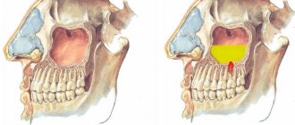
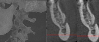
Many scientific studies have established a high data error in orthopantomography, therefore, to determine the anatomical features of the mandibular canal and clarify the localization of the dental implant, all patients underwent computed tomography of the mandible [7, 10]. Based on the results of computed tomography, the patients were divided into two groups. Group 1 included 10 (31%) patients in whom the dental implant was adjacent to the mandibular canal without violating the integrity of its cortical plate (Fig. 2, a).
Figure 2. Fragments of a computed tomogram in transverse (left) and panoramic (right) sections. a — patient S. from group 1.
Group 2 included 22 (69%) patients with a dental implant located directly in the lumen of the mandibular canal (Fig. 2, b).
Figure 2. Fragments of a computed tomogram in transverse (left) and panoramic (right) sections. b — patient M. from group 2.
results
When analyzing the results of the study, a tendency was revealed to increase the number of complications depending on age and gender: 26 (81%) people were between the ages of 40 and 60 years. This fact is probably associated with the phenomena of atrophy of the alveolar part of the lower jaw, as well as the peculiarity of the ratio of spongy and compact substance at this age, which increases the risk of damage to the NAS. The work demonstrated the predominance of this complication in females with a female/male ratio of 3:1, which is consistent with literature data [5]. These features, of course, can be an additional risk factor in the formation of an implantation bed in women after 50 years of age.
The key differential diagnostic feature was the absence of spontaneous and evoked pain in 100% of patients in group 1. All patients complained of decreased sensitivity of tissues (hypoesthesia) innervated by the NAS, and 70% additionally experienced various types of paresthesia in the form of a feeling of “crawling” in this area. A slight decrease in tactile and temperature sensitivity of tissues was determined, and the perception of a painful stimulus was practically no different from the symmetrical zone. The area of sensory impairment in the zone of innervation of the NAS averaged 11.4±2.96 cm2.
A characteristic feature of patients in group 2 was the absence in 82% of patients or a significant and persistent decrease in 18% of patients in tissue sensitivity to tactile, temperature and pain influences in the area of innervation of the NAS.
In group 2, 18% of patients had the main complaint of pain in the lower lip on the side of the nerve injury. The pain was spontaneous and of moderate intensity according to VAS (3-5 points). Also characteristic complaints were unusual sensations of a constant nature in the form of “crawling goosebumps”, “tingling” and unpleasant sensations (dysesthesia) when touching the lower lip, chin area, when talking, or eating. When conducting sensory tests, a pronounced decrease in tactile, pain and temperature sensitivity of tissues innervated by the NAS was revealed. The area of sensory impairment in the zone of innervation of the NAS averaged 13.6±0.86 cm2.
In patients of both groups, the threshold of sensation (TS) in the zone innervated by the NAS on the side of the injury statistically significantly exceeded the normal limit values. During clinical examination, this is expressed by a decrease in tactile sensitivity. A clear intergroup difference was the pain threshold (PT), which statistically significantly exceeded normal limits in all patients of group 2, of which in 8 patients with pain syndrome PT was not determined even at the highest possible intensity of testing stimulation, which indicates a significant degree of nerve damage on the implantation side. This kind of dysfunction of the NAS corresponded to the localization of the dental implant directly in the lumen of the mandibular canal and was diagnosed in 69% of the total number of patients examined. In group 1, no changes in PA indicators and pain tolerance level (PET) were detected in all patients (Fig. 3).
Figure 3. Results of assessing sensitivity thresholds (ST) in the zone of innervation of the NAS on the side of the injury.
All patients of group 1 received conservative treatment aimed at reducing swelling and stimulating the function of the NAS, as well as dynamic observation by a neurologist. Within 1-2 months, complete restoration of sensitivity in the zone of innervation of the NAS was determined.
Patients of the 2nd group underwent removal of the dental implant from the lumen of the mandibular canal according to the timing of their visit, followed by treatment by a neurologist. A favorable outcome of treatment, when carried out in a timely manner as early as possible, was obtained in patients of group 2 without pain, despite the localization of the dental implant in the lumen of the canal. In 10 patients of group 2, in whom the implant was removed from 14 days to 2 months after damage to the NAS, restoration of nerve function began in the immediate postoperative period and ended by 6-8 months. With later removal of the dental implant (in 2 patients 6 months after injury), full restoration of NAS function was observed only after 1 year.
Two patients without pain refused treatment and further observation. In 4 patients with pain syndrome, dysesthesia with an unpleasant tint in the lower lip area persisted after 1 year. By this time, there was a restoration of pain and temperature sensitivity, but not in full, with a decrease in the area of hypoesthesia of the lower lip to 5 cm.
The study made it possible to determine the doctor’s tactics in case of dysfunction of the NAS after dental implantation (see diagram).
Scheme 1. Scheme. Doctor's tactics for dysfunction of the inferior alveolar nerve (IAN) after dental implantation (DI).
conclusions
1. The appearance of pain after dental implantation and the absence of all types of tissue sensitivity in the zone of innervation of the NAS is an indication for removal of the dental implant.
2. A mandatory research method for all patients with dysfunction of the NAS is computed tomography or dental volumetric tomography of the mandible to determine the localization of the dental implant relative to the mandibular canal.
3. If the dental implant is localized in the lumen of the mandibular canal and there are sensory disturbances, removal of the dental implant from the lumen of the canal as early as possible is indicated.
4. Detection of signs of dysfunction of the NAS after dental implantation is a clear indication for consultation and treatment with a neurologist.
5. If the dental implant is located outside the lumen of the mandibular canal, but in the presence of clinical symptoms of dysfunction of the NAS, it is necessary to carry out conservative therapy and dynamic observation together with a neurologist for 8 weeks with mandatory recording of the dynamics of the complication.