A panoramic photograph of the teeth (orthopantomogram or OTP) is an x-ray of the upper and lower jaw. Allows you to examine most dental problems in detail, conduct high-quality diagnostics and prescribe treatment.
A panoramic dental photograph shows not only the teeth, but also the jaw joints, paranasal sinuses and facial bones. All this makes it indispensable for orthodontic, surgical and reconstructive treatment. You can obtain OPTG using an orthopantomograph.
Indications for panoramic dental imaging
The patient's need for a panoramic photograph of the teeth will be determined by the dentist. It is necessary to do it for a number of diseases of the oral cavity. Among them:
- inflammation of bone tissue;
- bite pathologies;
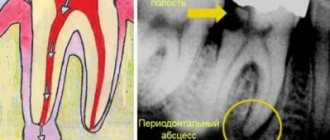
- tooth abscess;
- joint dysfunction;
- infectious diseases of the jaw;
- periodontal diseases;
- mechanical injuries.
OPTG is also necessarily prescribed when planning orthodontic correction of the dentition. A panoramic photograph allows you to see parts of the teeth that are located under the gums. With its help, the orthodontist can correctly draw up a treatment plan, predict its timing and select teeth that need to be removed (if necessary).
A panoramic photograph must be taken after sustaining jaw injuries (dislocations, fractures, etc.). Its main function is the diagnosis of dental diseases that cannot be determined solely by external signs. For this reason, OPTG is prescribed in the presence of symptoms indicating deep inflammatory and purulent processes.
The doctor gives a referral for a panoramic dental photograph after a visual examination of the oral cavity and collection of anamnesis. This takes into account the presence of pain (especially after canal filling), the nature of the discharge, dysfunction of the jaw joint and other symptoms.
A panoramic photograph is the most informative diagnostic procedure in pediatric dentistry. It is necessary for delayed teething or deviations in their formation. The picture clearly shows the rudiments of baby teeth already in the first months of a child’s life. With the help of OPTG, it is possible to predict bite pathologies and prescribe timely treatment.
With the help of OPTG, it is possible to diagnose diseases of the ENT organs, in particular, diseases of the maxillary sinuses and nasal passages. In this case, a panoramic image is prescribed for dizziness, fever, headache, and breathing discomfort.
Human teeth designation
Taking into account the anatomical structure, the teeth of an adult are divided into different groups:
- Incisors
- Normally, there are central and lateral incisors on the upper and lower jaws, only 4 upper and 4 lower. In total, the incisal group consists of 8 teeth. The physiological task of this group is to bite or cut off pieces of food.
- Fangs
- Normally, they are represented by 2 upper and 2 lower canines, for a total of 4.
- The task of the fangs is to separate the dentition when crushing and grinding food, to prevent the upper and lower dentition from abrading and rocking against each other.
- Small molars (premolars)
- Normally there are 4 upper and 4 lower ones, for a total of 8.
- The task of the premolars is the primary grinding of food for subsequent delivery to the large molars.
- Large molars (molars)
- Normally, they can be present in numbers from 4 to 6 on the upper jaw, and from 4 to 6 on the lower jaw. The last (third molars) are wisdom teeth that may not be present or may not erupt. Thus, there can be from 8 to 12 teeth in this group. The task of molars is to grind food to the state of a homogeneous food bolus, when the reflex act of swallowing becomes possible.
What diseases can be diagnosed by a panoramic dental photograph?
A panoramic photograph of teeth is an effective method for x-ray diagnosis of a number of dental diseases. Among them:
- benign and malignant neoplasms;
- fractures, cracks and dislocations;
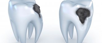
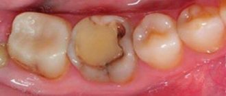
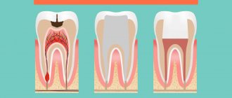
- deep caries;
- bite pathologies;
- periodontitis;
- improper teething;
- cancer of the maxillary sinuses, etc.
Preparing for a panoramic dental photograph
The procedure does not require special preparation. You can consume drinks, food and medicine - this will not affect the x-ray in any way. Immediately before the procedure, metal jewelry must be removed from the body. It is not recommended to drink alcohol.
When visiting a dentist, you must take panoramic and x-ray photographs from the last few years, as well as a medical history. With their help, the doctor will be able to track visible changes in the patient’s health status or compare treatment results.
X-ray radiation poses a possible risk to pregnant women. According to some studies, it can negatively affect the intrauterine development of the fetus. Pregnant women should tell their dentist about their pregnancy. In this case, carrying out OPTG depends on the ratio of possible risks and benefits.
Orthodontic stage: bite correction
A physiologically correct bite is one of the important criteria for a Hollywood smile. According to statistics, malocclusion occurs in 90% of patients. These can range from minor problems to serious pathologies. In any case, if you decide to get a Hollywood smile, you cannot do without consulting an orthodontist.
Getting a beautiful smile doesn't always require wearing braces. Minor deviations can be corrected by other methods. After the examination, the orthodontist will give you his recommendations on the optimal methods of treating the bite in your case.
Mouth guards for bite correction
One of the correction methods is treatment with mouth guards. A set of mouth guards is made from dental impressions, individually for each patient. The polymer is soft but elastic. The mouthguard puts pressure on the teeth, so they begin to gradually move. But unlike braces, the pressure is gentle. Creating a Hollywood smile takes place without discomfort. Due to the fact that the polymer is transparent, the aligners are not visible on the teeth even when smiling.
Treatment with mouthguards has one peculiarity. The effectiveness of the correction directly depends on how carefully you follow the orthodontist’s recommendations. If you are not ready for this, it is better to use other orthodontic methods to create a beautiful smile.
It's easy to use mouthguards:
- Wear it on your teeth daily, for a period of time calculated by the doctor when developing a bite correction plan.
- As the teeth move into the desired position, the set must be replaced with a new one.
The number of sets required to create an ideal smile will also be determined by the orthodontist at the preparatory stage.
Correction with braces
Braces are one of the most effective methods for treating malocclusion and creating a beautiful smile. It can be used even for serious pathologies. The correction period depends on the complexity of the case.
Traditional metal braces had one serious drawback - they spoiled the appearance of your smile. This caused psychological discomfort for many patients, and some refused treatment because of it. Modern braces are made from different materials. They look aesthetically pleasing on the surface of the teeth, and during long-term orthodontic treatment your smile will be beautiful, you won’t have to be embarrassed.
We offer braces from different materials: ceramic, sapphire. They will cost more than metal ones, but if a beautiful smile is important to you, it is better to opt for them.
Depending on the type of pathology, the doctor will offer you one of the types of braces:
- Lingual. They are fixed on the inner surface of the teeth, so they are absolutely invisible while wearing. Such systems are suitable for the treatment of serious pathologies.
- Vestibular. The brace lock is installed on the outside of the teeth and the arch is fixed to them.
Vestibular braces also differ in the method of correction. In ligature systems, the arch is fixed using ligatures. When the teeth move, the doctor adjusts the position of the arch. Self-ligating systems do not require correction.
Traditional metal braces are still used in orthodontics and can successfully treat malocclusions. When creating a Hollywood smile, we can recommend them if quick results are more important to you than aesthetics during treatment.
How is a panoramic dental x-ray performed?
Panoramic photography of teeth is the specialty of a dental radiologist. It is carried out using an orthopantomograph - a modern device that exposes the body to a minimal amount of x-ray radiation.
To take a photo, you need to stand or sit near the device. Next, the patient must wear a protective apron that protects the chest from radiation. When the patient is ready, the doctor starts the orthopantomograph. The entire procedure lasts no more than 15 minutes and is completely painless.
During the process you must remain stationary. After the panoramic photo is taken, you need to wait a few more minutes. During this time, the specialist will prepare the image and provide it to the patient. The entire procedure lasts no more than 15 minutes, including the time required for preparation and waiting.
Additional specialists and procedures
A patient who has received an appointment for a panoramic dental x-ray may need the help of the following specialists:
- dental surgeon;
- maxillofacial surgeon;
- oncologist;
- otolaryngologist;
- periodontist;
- orthodontist, etc.
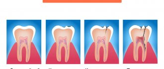
OPTG is an important, but not the final stage of diagnosis. To confirm the diagnosis and develop effective therapy, in most cases a number of additional studies are prescribed. If tumors are present, a tissue biopsy is performed. A laboratory examination of pathological discharge is also performed.
If an orthopantomogram indicates problems with the canals or subgingival part of the tooth, it is necessary to open the tooth (filling) and further treatment. In some cases, tooth extraction may be indicated (if it cannot be treated).
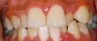
Gingivitis –
Gingivitis is an inflammation of the gingival margin, including the interdental gingival papillae (i.e., that part of the gum that is directly adjacent to the necks of the teeth). The main symptoms of this disease are periodic bleeding and soreness of the gums when brushing teeth, redness or cyanosis of the gingival margin, swelling of the gingival papillae, and often there is bad breath. Gingivitis with a predominance of such symptoms is called “catarrhal” by dentists.
Gum disease: photo of teeth with gingivitis
The cause of the development of catarrhal gingivitis is exclusively insufficient oral hygiene. As a result, soft microbial plaque accumulates at the necks of teeth, and hard dental deposits can also form (we are talking about tartar). In this case, the development of inflammation is due to the fact that pathogenic bacteria that are part of microbial plaque and tartar secrete various toxins and inflammatory mediators, which trigger a chain of inflammatory reactions in the gums.
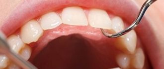
Therefore, the main treatment for inflammatory gum diseases is always aimed at eliminating the causative factor, i.e. for the removal of dental plaque (+ normalization of oral hygiene). It should be noted that gingivitis is the simplest and most quickly curable form of gum inflammation, if, of course, the treatment is carried out correctly - with regular removal of dental plaque by ultrasound (the frequency of this procedure will depend on the quality of the patient’s oral hygiene).
Important: inflammation in gingivitis affects only the mucous membrane of the gums - without destroying the dentogingival attachment or bone tissue around the teeth (which is typical for a more severe form of gum inflammation - periodontitis). But the transformation of gingivitis into periodontitis is only a matter of time if the patient focuses his treatment on antiseptic mouth rinses and gel applications on the gums, rather than on the removal of dental plaque and high-quality hygiene.
Hypertrophic form of gingivitis –
Catarrhal gingivitis is the most common form of gingivitis, but not the only form. Sometimes patients can experience a hypertrophic form of gingivitis, which is manifested by an increase in the volume of the gingival papillae or the gingival margin as a whole (usually only in the area of the front teeth). Most often, this form occurs in pregnant women and adolescents, and is associated with hormonal changes in the body. There are edematous and fibrous forms of hypertrophic gingivitis.
In the edematous form, the enlargement of the gingival papillae occurs due to their chronic persistent swelling, which is practically untreatable. This form can occur not only in pregnant women, but also in patients with long-term catarrhal gingivitis (in the absence of adequate treatment). In the fibrous form, gum enlargement occurs due to the proliferation of fibrous tissue (Fig. 4). You can read more about the forms of catarrhal and hypertrophic gingivitis and their treatment regimens at the link below.
→ Treatment of gingivitis in adults