- Surgical department
Surgical department » - Maxillofacial Surgery
Maxillofacial Surgery "
- Jaw osteotomy
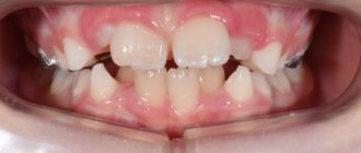
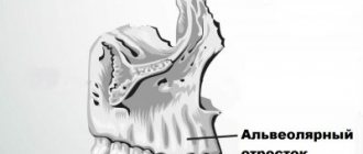
Malocclusion is a violation of the process of chewing food and swallowing it, a negative effect on the speech apparatus, as well as an aesthetic defect. With malocclusions, the upper and lower jaws can be enlarged or reduced in size, pushed forward or positioned posterior to the normal position, shifted to the side or tilted, turning clockwise or counterclockwise. In this case, the closure of the teeth is disrupted to one degree or another, they change the angle of inclination, turn around, run into each other (crowding) or diverge with the formation of cracks (treme).
What are the dangers of malocclusion?
- Violation of dental contacts makes it difficult to chew food and provokes the development of diseases of the gastrointestinal tract.
- Severe malocclusions so limit the possibilities of normal nutrition that they are a reason for deferment from the army.
- Changing the angles of inclination of the teeth affects the distribution of the load during chewing, leads to overload of the periodontium, the development of gum inflammation and, ultimately, earlier tooth loss.
- Teeth crowding, combined with misalignment, affects the natural cleaning of teeth, further leading to the development of gum disease and an increased likelihood of tooth enamel destruction. If tooth loss has already occurred, then violations of the jaw relationships can create difficulties in prosthetics.
- Malocclusion leads to a change in the trajectory of jaw movement during chewing: the temporomandibular joints are overloaded, and dysfunction of the temporomandibular joints develops.
- Some types of malocclusion are anatomical prerequisites for narrowing the airways, for the appearance of snoring and such a serious condition as sleep apnea.
To eliminate lower prognathia, O. Neuer (1962) proposed.
The technique of decortication of the mandibular ramus in the form of a narrow cavity in the horizontal direction above the mandibular foramina, leaving only small bridges of the compact layer along the posterior edge of the ramus. The operation was performed using a submandibular approach. After 2 weeks, a blow to the chin caused a posterior displacement of the lower jaw (like a subperiosteal fracture). In the postoperative period, intermaxillary fixation was performed with a rubber rod. In our opinion, this method does not guarantee against relapse.
To eliminate prognathia of the mandible or its combination with an open bite, R-Ritter (1956) proposed performing an arcuate osteotomy in the area of the lower third of the mandibular ramus, i.e., above the opening of the mandibular canal (Fig. 29).
The lower jaw moves backwards, and in the presence of an open bite, upwards, the teeth are set in an orthognathic bite. Osteosynthesis of bone fragments is carried out using a wire suture. In the postoperative period, intermaxillary fixation is performed using dental splints. O. Neuer (1962), N. Kole (1963) performed the method of partial arcuate resection of the branches of the lower jaw in the same location and for the same indications as R. Ritter.
This technique, in our opinion, is technically complex and requires great precision. It is not without the same disadvantages as horizontal osteotomy of the branches (small contact area of the wound surfaces of bone fragments, the possibility of damage to the neurovascular bundle, etc.).
Among operations on the branches of the lower jaw, oblique “sliding” osteotomy, first performed in our country in 1928 by A. A. Limberg (1924) (Fig. 30), and abroad by G. Regtes (1924), occupies a significant place.
Its essence lies in osteotomy of the jaw ramus from the middle of the semilunar notch to the posterior edge of the ramus, then the large fragment is mixed downwards, sliding the osteotomized edge over the small fragment. The operation ends with osteosynthesis with a wire suture. This surgical method has found wide application in many clinics and specialized hospitals [Kabakov B. D., Vasiliev V. S., 1966; Rudko V.F., 1966 (Fig. 31); Vasiliev V. S., 1967, 1969, 1970 (Fig. 32) and others. ] .
Jaw osteotomy surgery
Historically, the first operations were performed on the lower jaw. And the surgeon’s task was precisely to restore normal jaw relationships. But today operations on the lower jaw alone account for no more than 10% of operations performed. Because the surgeon’s tasks have changed: now it is not only the correction of the bite, but also the improvement of facial aesthetics.
Double-jaw osteotomy is an operation on the lower and upper jaw simultaneously, which allows you to achieve aesthetically favorable proportions of the face both in front and in profile, eliminate asymmetry, and make the smile brighter by eliminating dark corridors - shading of the lateral teeth of the upper jaw. We can say that surgery on one jaw is only a correction of functionality, on two – functionality and aesthetics.
Indications for the operation:
- violation of the closure of the dentition, which cannot be corrected orthodontically;
- pronounced facial disproportions that have aesthetic significance.
Contraindications:
- unpreparedness of the dentition for surgery (insufficient orthopedic or orthodontic preparation);
- diseases of internal organs that prevent surgery or anesthesia.
In the literature, this method is sometimes called “Swedish”.
In 1910, W. Babcock also used horizontal osteotomy of the branches at the level of their upper third. No bone fixation was performed. WW Babcock inserted a piece of ivory as a spacer between the fragments (Fig. 25).
In the postoperative period, the jaw was fixed using dental splints and rubber traction.
The Czech surgeon F. Kostecka in 1924 performed a horizontal osteotomy of the branches of the lower jaw without a soft tissue incision - blindly. The essence of the operation is as follows. Using a Kerger needle, an injection is made below the earlobe 5-6 mm from the posterior edge of the branch. Then, all the time feeling the medial surface of the jaw branch with the end of the needle, they move it anteriorly towards the lower edge of the zygomatic bone. At the front edge of the branch, the needle pokes out through the cheek. It is important that the needle passes above the entrance to the mandibular canal, between the medial surface of the ramus and the soft tissues containing the neurovascular bundle (inferior alveolar artery, nerve and vein). At the same time as the needle, a silk thread is passed through, held in a hole at the end of the needle. Then they do it in two ways: either the needle is removed, leaving a silk thread in the wound for further insertion of the Gigli saw, or the needle is inserted without the silk thread, and the Gigli saw is fixed to the end of the needle and brought to the level of the posterior edge of the branch. Using sawing movements in a horizontal direction (the skin at this moment must be protected from damage), the jaw branch is cut first on one side and then on the other. After this, the lower jaw is shifted posteriorly until the correct bite is established and fixed to the upper jaw using dental splints and rubber rings for a period of 2 - 3 months, and sometimes more (Fig. 26).
The main disadvantages of the Kostechka method are bleeding, the formation of salivary fistulas, damage to the facial nerve and frequent relapses (up to 50%, according to E. Reichenbach, 1955).
The operation using the Kostechka method in its initial version was used by many surgeons in our country and abroad [Kabakov B. D., Pasternak A. A., 1962; Mukhin M.V., 1963; Egiyan G. M., 1964; Klementov A.V., Vasiliev V.S., 1964; Mikhelson N. M. et al., 1965; Vasiliev V. S., Popov S. E., 1967; PopudrenkoP. I, 1970; Pichler N., 1928; Pearson W. N., 1943; Traunham V. N., 1944; Reichenbach E., 1955; Hinds E. S., 1957; Toman Y., 1957, 1968; Shira R.V., 1961; Urban F., 1961; Ullik R., 1962, etc. ] .
While maintaining the basic principle of the method, a number of surgeons made some modifications to Kostechka’s operation. Thus, A. Zindemann (1921) performed osteotomy of the branches not with a Gigli saw, but with a hacksaw. E. Skaloud (1954) performed this operation using intraoral access. S. M. Davydov (1960) proposed a protective plate to prevent injury to soft tissues and the neurovascular bundle. S. Moose (1945) performed osteotomy of the mandibular branches intraorally using a mechanical saw with a protective plate. To prevent anterior displacement of the small fragment, K. Mushka (1971) proposed fixing the fragments with a wire loop from the semilunar notch to the angle of the jaw; The author passed the wire subcutaneously using a special conductor.
Stages of treatment
Today, the method of combined treatment of malocclusions, when the orthodontist begins work first, is considered generally accepted in the world.
1) Orthodontic stage
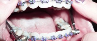
The orthodontist places braces on both jaws and begins to move the teeth so that they take the correct position in relation to the jaws. In this case, a temporary deterioration in the closure of teeth occurs. It takes about nine months to prepare. After this, the surgical stage is performed.
2) Surgical stage
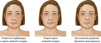
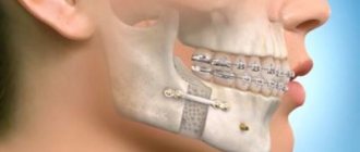
The operation is performed under general anesthesia. The intervention is performed via intraoral access, which allows it to avoid leaving scars. In accordance with the treatment plan, cuts are made on the upper and lower jaws, the fragments are mobilized and installed in the most functionally and aesthetically advantageous position. Next, the jaws are fixed with titanium miniplates, which are invisible to the body, do not ring at the airport and do not interfere with MRI. They usually remain in the body for life without causing any consequences. In rare cases, surgery may be performed before orthodontic treatment begins.
Corrective osteotomy at the Ladisten clinic
The clinic specializes in minimally invasive orthopedic surgeries. More than 6,000 patients from all over the world have already undergone osteotomy and are satisfied. Each patient is offered a tour of the facility, a separate room during rehabilitation, and 24-hour medical supervision in the first days after the procedure. The operation itself is bloodless: a small puncture is made in the leg, through which doctors correct the bone. For fixation, a unique device from Dr. Veklich is used. This is an improved design of the Ilizarov apparatus. It is less bulky, weighs little and does not involve dangerous knitting needles. Doctors at the Ladisten Clinic have been performing corrective osteotomy procedures for more than 30 years; the price varies depending on the pathology and severity of the case. To find out the exact diagnosis, consult about contraindications and discuss the cost of osteotomy, just make an appointment by calling us at: +38 +38 or Write to WHATSAPP Write to VIBER We will choose a convenient time for you to visit the clinic or online consultation.
Methodology of V. S. Vasiliev
It differs in that after osteotomy and displacement of the lower jaw posteriorly, a small fragment of the branch is superimposed on the outer surface of the branch. The negative side of this modification is the creation of unfavorable conditions for consolidation due to the contact of fragments not with a spongy, but with a compact layer.
Most surgeons perform osteotomy on the mandibular branch not in an oblique, but in a vertical direction. In 1910, WW Babekosk performed a vertical osteotomy of the branches of the lower jaw, the line of which connected the middle of the semilunar notch with the angle of the jaw. After the lower jaw was displaced upward and an orthognathic bite was established, a wedge-shaped defect formed in the area of the osteotomy; the fragments were in contact in the upper section only in a small area, where osteosynthesis was carried out with a metal wire through two holes (Fig. 33).
Too small a contact area of bone fragments after osteotomy creates unfavorable conditions for consolidation, which makes the WW Babcock method one of the imperfect ones. This method continues to be used today [Tyukalov K.V., 1967; And rzhantse in P. 3. et al., 1970; Alekseev-L, I., 1970; Y. V. Caldwell, G. S. Zettermann, 1954; M. Robinson, 1954; Toman Y., 1958; Shira R.V., 1961; Ailing G., 1965: Ginestet G., Merville L., 1965, 1966; Constantinidis Y., 1967; BerenytB., 1968; KoppW.K., 1968, etc.).
In addition to the listed methods, some surgeons use vertical wedge resection on the branches of the lower jaw. Thus, Van Zile (1955) proposed an original method of triangular resection in the vertical direction of the branch using a submandibular approach. First, he performed a vertical osteotomy of the ramus from the middle of the semilunar notch to the corner of the jaw, and then shifted the medial fragment of the jaw posteriorly (or upward when prognathism of the lower jaw was combined with an open bite) until the correct bite was established. In this case, a small fragment of the ramus was superimposed on the branch, revealing the dimensions of the excess part on the large fragment, resected by the author in the form of a triangle, the apex of which faces the angle of the jaw, and the base faces the semilunar notch. The wound surfaces of the branch were compared end-to-end, and osteosynthesis was performed with metal wire (Fig. 34).
The advantage of this technique is that it allows you to increase the height of the jaw branch, reduce the angle of the jaw and preserve the neurovascular bundle. A similar technique, but with more accurate mathematical calculations and strong fixation, was carried out in 1966 by V. F. Rudko (Fig. 35).
In our practice, we have somewhat modified the vertical “sliding” osteotomy. In December 1966, patient S. underwent bilateral vertical “sliding” osteotomy of the branches of the lower jaw for lower prognathia of the second degree and an open bite of the first degree. After osteotomy and installation of the middle fragment of the jaw into the correct position, its excess was determined, since the small fragments were displaced onto the remainder of the branches of the large fragment. To eliminate excessive layering of branch fragments and improve their contact, we removed strips of the compact layer of branches from the outer surface of the large fragment and from the inner surface of the small fragment. The width of the strips was determined by the level of layering of small fragments. Thus, the exposed surface of the spongy substance of the branches had the shape of an inverted trapezoid. Fragments of branches were compared until complete contact of the spongy substance. Fixation was performed with two wire sutures (Fig. 36).
In the postoperative period, intermaxillary fixation was carried out for 37 days using dental wire splints and rubber traction. This operation achieved good cosmetic and functional results (Fig. 37-40).
Currently, planar osteotomies in the area of the angle and lower part of the mandibular ramus are quite widely used [Arzhantsev P. 3. et al., 1970; Mitrofanov G. G., Rudko V. V., 1972; Arzhantsev P. 3., Sukachev V. A., 1974; Sukachev V. A., Gritsai N. P., 1975, 1977; Rudko V.V., 1975, 1976; Semenchenko G.I., Lozenko P.A., 1975; Dal Pont, 1961; Obwegeser H., 1968; Tsvioro F., 1972, 1978, etc.].
The most successful and promising was: