Fibrin is an essential protein. Properties, functions, fibrin and inflammation
Contents
hide
1 Fibrin is an essential protein. Properties, functions, fibrin and inflammation
2 The role of fibrin in inflammation
3 Body protection
4 Function of fibrin
5 Experiments performed
6 Enzyme treatment
7 Method of treating patients with trophic ulcers
8 Classification of wounds. Wound process 8.1 Materials used in the article:
Fibrin is a protein that is the end result of blood clotting. It is formed as a result of the effect of thrombin on fibrogen.
Fibrin is an insoluble protein produced in the body in response to bleeding. This protein is a solid element consisting of fibrous threads. The progenitor of fibrin is fibrinogen. This is a protein produced by the liver. It is in the blood. When tissue is damaged, bleeding occurs. To stop it, thrombin begins to work. It affects fibrinogen, thereby provoking its transformation into fibrin.
First, the protein molecules combine into long strands that entangle the platelet, creating a rough mass. Gradually it hardens and contracts, forming a bloody clot. The compaction process is stabilized by a fibrin-stabilizing factor.
Principles (stages) of treatment
Actions when treating a wound depend not only on the conditions and causes of its appearance, but also on the stage at which the wound process has stopped in it.
The first stage of the wound process is acute. During it, vascular changes occur caused by a reaction to damage, and the process of blood clotting is actively underway. If the wound is infected, pus is formed and toxic substances are simultaneously absorbed into the tissue.
At this stage, it is necessary, first of all, to clean the wound - remove dead tissue from it, remove foreign bodies (with the help of a doctor), drain it - remove pus and exudate from the wound. It is also necessary to prevent infection from entering the wound. It is best to wash the affected area with saline solution, as caustic disinfectant liquids can harm surrounding healthy tissue.
The second stage of the wound process is granulation. At this stage, a thin layer of connective tissue begins to form on top of the wound. The edges of the wound begin to tighten, and it closes itself. The thinnest capillaries are formed in the connective tissue layer, providing nutrition to regenerating tissues. Often chronic wounds stop at this phase, and the healing process does not proceed further; This is most often what happens with trophic ulcers.
At the granulation stage, it is important, firstly, to protect the newly formed tissue from mechanical damage, secondly, to stimulate further regeneration, and thirdly, to prevent the development of the inflammatory process - despite the fact that the wound is closed, the risk of infection and inflammation remains. To stimulate tissue regeneration, methyluracil ointment is used (you can use a ready-made Voskopran dressing with methyluracil ointment); to protect the surface of the wound, atraumatic dressings should be used that do not stick to the wound surface and do not injure it.
We should not forget about maintaining a maintenance diet and sleep schedule - this will help speed up the healing process.
The role of fibrin in inflammation
Fibrin production and inflammation are two closely related processes. Protein plays an important role in contact with deteriorating, damaged tissue. Thrombokinase released from the tissue comes into contact with fibrinogen.
When blood clots, all toxic substances become clogged in the clot. This feature of the effect of protein during the inflammatory process protects the body from the further spread of toxins and their negative effects. This reaction is called fixation. Thus, fibrin is also a protector of the body from toxins.
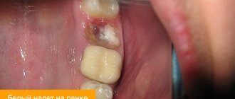
Causes of white plaque on a wound
Fibrin is a protein that normally appears on the surface of wounds during the last stage of blood clotting. If a white coating forms on the wound, this is already evidence of a pathological process. Most often, infections develop in two cases: in diabetics and patients with circulatory disorders.
In any case, the prerequisite is a weakened immune system. Therefore, the body is not able to fully resist the fibrinous process in the wound. It is necessary to begin treatment before the plaque becomes purulent.
Body protection
The formation of insoluble fibrin helps protect the body from blood loss, as well as from inflammatory processes. However, such a reaction causes pain and swelling, tissue damage, and disruption of their functionality. This is subsequently eliminated by reparative processes. At their early stage, special substances are produced that cause fibrin depolymerization. Such a reaction, even at the very beginning of the inflammatory process, can inhibit the effect of protein on the pathological focus.
How does fibrin form?
The soluble protein fibrinogen, synthesized in the liver with the participation of vitamin K, interacts during bleeding with a peptidase called thrombin, which promotes partial hydrolysis of fibrinogen molecules, transforming this protein into fibrin in the presence of calcium ions (Ca2+). In general, the formation of fibrin from fibrinogen occurs in three stages:
- The fibrinogen dimer under the influence of thrombin undergoes enzymatic cleavage, separating as a result of this process 2 peptides (fibrinopeptides A and B) - the formation of fibrin monomer occurs, which is built from two absolutely identical subunits, interconnected by disulfide bridges and consisting of three polypeptide chains ( alpha – α, beta – β, gamma – γ);
- Aggregation of fibrin monomer (the appearance of fibrin threads or fibrin aggregate - unstabilized fibrin), which occurs at the second stage of the process of formation of this substance, consists in the fact that it (fibrin monomer) without external influence (except for the participation of calcium ions) independently begins to form a bundle , the result of this reaction (polymerization) becomes soluble fibrin polymer “S”;
- The influence of the fibrin-stabilizing factor (FXIIIa), which is brought into an active state by calcium ions and thrombin, completes the reaction of formation of insoluble fibrin (“J”), it “crosslinks” the individual fibrin strands together, that is, it finally stabilizes and forms a blood clot.
Thus, fibrin threads are united molecules of this substance. They, entangling with their network (fibrin network) blood cells rushing to the accident zone (primarily platelets) or simply circulating in the bloodstream, provide a “foundation” for the construction of a spongy mass, which becomes the basis of a clot that closes a blood vessel when it is damaged . The spongy mass contracts and hardens, forming the clot itself. To ensure that the formed blood clot does not immediately collapse, at this stage a factor enters the process that stabilizes the “plug” on the wound of the vessel.
Experiments performed
Scientists conducted experiments on animals, during which it turned out that proteases introduced from outside before the inflammatory process can prevent its development completely, and in some cases the pathology was mild. Experience has shown that the use of tryptic substances in most cases stops the development of inflammatory processes at the onset of the disease.
When prophylactic doses of enzymes were administered, protein formation decreased.
Fibrin is not just a protein, but the creator of a protective barrier around the pathological focus, which protects against disease. Subsequently, this insoluble component serves to build connective tissue. It also participates in regeneration processes. The formation of scar tissue depends on the duration of storage and the amount of fibrin produced by the body.
So what is fibrin and what is it for? This substance is produced by the body's cells in such an amount that is necessary to quickly stop bleeding and which will help quickly restore damaged tissue. In some cases, fibrin is a pest. If it is produced and deposited in large quantities, the protein can cause harm to the body. As far as is known, fibrinolysis is a long process that is not able to dissolve all excess protein. Moreover, this process requires certain conditions.
To get rid of excess fibrin, special enzyme treatment is prescribed.
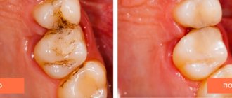
The most important medical and social problem is the treatment of long-term non-healing wounds of various etiologies. Trophic ulcers and deep skin cracks in the lower extremities, as well as chronic ulcerative pyoderma, sharply reduce the quality of life and limit the activity of patients for a long time. Local therapy occupies a special place in the treatment and prevention of these diseases, preventing the development of an infectious process in the wound and being a barrier to the spread of infection.
Modern highly effective agents for local treatment of long-term non-healing wounds have a number of medicinal qualities that allow their use taking into account the phases of the wound process. They must have anti-inflammatory and proteolytic effects, have a wide antibacterial spectrum of action, remain active in an acidic environment, not cause hypergranulation and pigmentation, have hypoallergenic qualities, be non-toxic and easy to use.
There are many combination drugs for local treatment containing antibiotics or sulfonamides in combination with each other and/or various antiseptics. Such drugs are characterized by a slower development of microbial resistance and a wider coverage of bacterial agents that cause infections.
Argosulfan cream
2% (“Elfa Farmzavod A.O.”) is a combined topical antibacterial drug that promotes healing and effective protection of wounds (trophic, purulent, burns, etc.) from infection.
silver sulfathiazole
included in its composition has a wide spectrum of antibacterial and bacteriostatic effects against gram-positive, gram-negative microflora and pathogenic anaerobes, and is also effective against fungal superinfection. The mechanism of the antimicrobial action of sulfathiazole - inhibition of the growth and reproduction of microbes - is associated with competitive antagonism with para-aminobenzoic acid and inhibition of dihydropteroate synthetase, which leads to disruption of the synthesis of dihydrofolic acid and its active metabolite - tetrahydrofolic acid, necessary for the synthesis of purines and pyrimidines of the microbial cell [1].
The silver ions present in the cream enhance the antibacterial effect of sulfonamide - they inhibit the growth and division of bacteria by binding to the deoxyribonucleic acid of the microbial cell [1]. The gradual release of silver ions suppresses the growth of pathogenic microflora over a long period of the dressing being on the wound, which is important when there is a high risk of reinfection with hospital strains and is a permanent barrier to the spread of infection.
In addition, silver ions weaken the sensitizing properties of sulfonamide. There is an opinion according to which the application of sulfonamides to the skin and mucous membranes is undesirable due to their low activity and the risk of an allergic reaction. When applying such an ointment to a burn or infected surface, burning and pain may occur [1]. Hydrophilic cream base Argosulfan
, which has an optimal pH and contains a large amount of water, provides its moisturizing and analgesic effect and leads to a decrease in wound healing time.
Due to minimal resorption Argosulfan
-cream applied even to large wound surfaces does not have a toxic effect, since silver sulfathiazole has low solubility, due to which the concentration of the drug in the wound is maintained at the same level for a long time.
A small amount of silver sulfathiazole that appears in the bloodstream undergoes acetylation in the liver and is found in the urine in the form of inactive metabolites and partially unchanged [1]. Due to the lack of systemic absorption of the drug, Argosulfan
in combination with other drugs.
Dressings with Argosulfan
containing silver ions are painless when applied to the wound surface, do not dry to the wound and are easily removed from its surface, have a mild drying effect, and penetrate well into necrotic tissue and exudate. Tissue detritus impregnated with the drug forms a “protective cushion”, under which accelerated formation of granulations and epithelization occurs.
We used Argosulfan
for long-term non-healing wounds in patients with trophic ulcers of various etiologies, necrobiosis lipoidica, chronic ulcerative and ulcerative-vegetative pyoderma, deep fissures complicating the course of tylotic eczema.
The appearance of trophic ulcers
can complicate the course of many diseases - from diseases of the circulatory system to metabolic disorders, and the development of a peptic ulcer is accompanied by both a violation of normal blood circulation and a change in innervation. That is why the recovery process may require the use of complex effects and fairly long-term treatment.
There are varicose, ischemic and neurotrophic ulcers [2-5]. The largest group (1-2% of the adult population) consists of patients with chronic venous insufficiency (CVI) of the lower extremities, which develops when venous outflow is impaired and pressure inside the capillaries increases. The main causes of CVI are varicose veins and postthrombophlebitic syndrome. When the valves of the deep veins of the leg are damaged, the function of the muscular-venous pump is disrupted and retrograde blood flow occurs. Damage to the perforating veins, which connect the superficial veins to the deep veins, aggravates venous insufficiency. Due to fibrin deposition in the perivascular space and inhibition of tissue fibrinolysis, sclerosis and obliteration of lymphatic and small blood vessels develop. Perivascular fibrosis disrupts the delivery of nutrients to the epidermis, which leads to the formation of varicose trophic ulcers [2-5].
Ischemic ulcers form in diseases of the peripheral arteries, in particular in obliterating atherosclerosis (OA). OSA is always accompanied by skin damage - from gradually increasing ischemia to infarction, i.e. necrosis, which develops when there is a sudden disruption of blood supply as a result of atheroembolism - blockage of small arteries with fragments of atherosclerotic plaques. Ischemic ulcers are localized in frequently injured areas and areas of compression, and are accompanied by severe pain. Mixed ulcers develop in patients suffering from both CVI and OSA, who clinically have features of both diseases [4, 5].
The cause of neurotrophic ulcers is damage to sensory and motor nerves, circulatory disorders and atherosclerosis in diabetes mellitus and its complications, secondary hyperparathyroidism, granulomatous inflammation in response to collagen degeneration in necrobiosis lipoidica [6, 7].
Necrobiosis lipoidica
- a rare chronic dermatosis of a vascular-metabolic nature, which is usually classified as a group of localized lipoidoses of the skin. The provoking factors in necrobiosis lipoidica in 1/3 of cases are diabetes mellitus, and in another 1/3 - impaired glucose tolerance, therefore, family history and research are necessary to identify hidden forms of diabetes mellitus. The onset of the disease is often preceded by trauma. Collagen degeneration leads to increased platelet aggregation, microangiopathies, damage to arterioles, sclerosis and obliteration of blood vessels in foci of necrobiosis. Vascular disorders lead to trophic disturbances and necrobiotic changes in the dermis with subsequent deposition of lipids in it. The disease often has a long, relapsing course, its severity does not depend on the severity of diabetes mellitus; the developing trophic ulcers are accompanied by severe pain and heal, as a rule, with the formation of a rough scar [6, 7].
Without special treatment, trophic ulcers are characterized by a low tendency to heal and a long, relapsing course [8, 9]. It is generally accepted that surgical treatment of CVI and OSA is best performed after healing of the trophic ulcer or thorough sanitation of its surface. However, conservative treatment using outdated, ineffective local medications is often complicated by dermatitis, eczema, and erysipelas, which delays the timing of vascular surgery. The choice of drug is influenced by the phase of the disease and the severity of the inflammatory reaction (phase I - pre-ulcerative state; phase II - dystrophic changes, necrosis, inflammation of the skin and adjacent tissues; phase III - ulcer cleansing and regeneration; phase IV - epithelization and scarring), complications (mycoses, eczema, pyoderma, erysipelas, recurrent thrombophlebitis, malignancy, etc.), species composition of the ulcer microflora. Numerous bacteriological studies of the qualitative composition of the microflora of the surface of trophic ulcers have revealed multidrug-resistant gram-positive and gram-negative microflora. The content of microorganisms in induratively changed tissues surrounding the ulcer sometimes reaches 107-109 microbial bodies per 1 g of wound tissue, which indicates a high risk of generalization of the infectious process [10]. As a rule, the isolated microorganisms are highly resistant not only to traditional antibacterial drugs, but also to the local agents most often used in such cases - solutions of furatsilin, chlorhexidine, syntomycin emulsion, ointments with antibiotics (tetracycline, gentamicin), etc. At the stages of granulation and epithelialization, to speed them up, as well as when wounds are complicated by dermatitis, eczema or other manifestations of allergies to traditional drugs, silver salts may become the drug of choice, given their high efficiency in suppressing both gram-positive and gram-negative microflora, better tolerability and the rarity of allergic reactions even in patients with a burdened history of allergies.
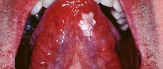
Pyoderma
(pustular skin diseases) are a group of dermatoses based on purulent inflammation of the skin and its appendages, as well as subcutaneous fat. Pyoderma accounts for 1/3 of all cases of skin diseases [4, 5]. The development of pyoderma is promoted by microtrauma, maceration and contamination of the epidermis, increased sweating, hypothermia, overheating, endocrinopathies (diabetes mellitus), hypogammaglobulinemia, insufficient protein intake, hypovitaminosis, immunity disorders and severe somatic diseases, fatigue, chronic intoxication, persistent foci of staphylococcal infection. The most common causative agents of pyoderma are Staphylococcus aureus and Staphylococcus epidermidis (80-90% of patients); in 10-15% of cases a mixed infection is detected (staphylococcus in combination with streptococcus, Pseudomonas aeruginosa, Proteus, Escherichia coli, etc.) [1, 4, 10 ].
Chronic ulcerative and ulcerative-vegetative pyoderma belong to deep streptostaphylococcal pyoderma, characterized by the formation of poorly healing ulcerative purulent lesions of the skin and underlying tissues. Often the disease is accompanied by a deterioration in general condition, increased body temperature, weakness, symptoms of intoxication, lymphangitis and lymphadenitis. Ulcerative pyoderma
(streptostaphylococcal impetigo) manifests itself as phlyctenae, ecthyma, located against the background of erythema.
The rashes, usually disseminated, cover large areas of the skin. Under the crusts, the formation of deep ulcers with flaccid granulations at the bottom and inflamed soft edges is characteristic. Streptostaphylococcal impetigo is often a complication of itchy dermatoses (eczema, scabies, atopic dermatitis, etc.). Chronic ulcerative-vegetative pyoderma
is characterized by ulcerative formations of irregular shape with pronounced vegetations in the area of the edges and bottom. Often there is a stagnant pink halo of hyperemia around the ulcer. Characterized by a chronic course with periodic exacerbations with the appearance of new ulcers or serpigenization of the main ulcer formation.
Therapy for pyoderma should be etiopathogenetic. In the treatment of chronic, recurrent and deep forms of pyoderma, antibiotics (locally and systemically), antiseptic solutions, glucocorticosteroids, immunomodulators, vitamins are used, pustules and abscesses are opened, and if necessary, vegetations and necrotic tissue are removed and scraped out.
Tylotic (horny) eczema
- a chronic persistently recurrent allergic skin disease, a type of true eczema. Tilotic eczema is manifested by hyperkeratosis of the palms and soles with the formation of rough horny and cortical layers, dry skin, as well as deep, painful, long-term non-healing, often bleeding cracks. This disease is characterized by polyvalent sensitization and autosensitization, occurs against the background of neuroendocrine changes, and is accompanied by changes in the central and autonomic nervous system, disorders of metabolic processes and tissue trophism [4, 5]. Damage to the soles and palms is usually symmetrical; cracks form against the background of erythema and peeling, especially in areas of greatest pressure and load, including on the lateral surfaces of the fingers. Inflammation can be expressed in a limited area or occupy the entire sole and/or palm; pain in the area of the cracks can be more intense than itching. The tendency for cracks to deepen is associated with the patient’s age, duration of the disease, cold season and the presence of underlying diseases.
Pyogenic infection can easily penetrate into cracks on the soles, and erysipelas or pyoderma can develop. Tilotic eczema is resistant to treatment, prone to recurrence, improvement of the condition is achieved by frequent and strong moisturizing of the skin and immediate cessation of wearing wet shoes [11].
We observed 47 patients (13 men, 34 women) aged 44-75 years with trophic ulcers that developed against the background of CVI and obliterating endarteritis, circulatory and trophic disorders in necrobiosis lipoidica, as well as with pyoderma and tylotic eczema. The duration of the disease ranged from 2 months to 4 years. Argosulfan cream externally as part of complex therapy.
.
11 (23.4%) patients had varicose ulcers, 5 (10.6%) had mixed trophic ulcers, 8 (17%) had trophic ulcers due to necrobiosis lipoidica; 8 (17%) had chronic ulcerative and chronic ulcerative-vegetative pyoderma, 15 (32%) had tylotic eczema with deep cracks on the palms and soles.
In 22 (46.8%) patients, dermatoses occurred against the background of endocrine diseases: in 4 (8.4%) - diabetes mellitus, in 2 (4.2%) - impaired glucose tolerance, in 5 (10.6%) - nodular goiter, 1 (2.1%) - hypothyroidism, 1 (2.1%) - adrenal pathology. Chronic bronchitis was detected in 2 (4.2%) patients, chronic gastritis in 3 (6.3%), chronic pancreatitis in 5 (10.6%), and viral hepatitis C in 2 (4.2%) , 10 (21.3%) had hypertension, 9 (1%) had coronary heart disease, 2 (4.2%) had uterine fibroids.
Bacteriological studies of the qualitative composition of microflora from the surface of lesions (ulcers, cracks) revealed Staphylococcus aureus and Staphylococcus epidermidis in 29 (61.7%) patients, mixed infection (staphylococci in combination with streptococcus, Escherichia coli, bacteroides, Proteus, yeast fungi) in the amount of 104-105 microbial bodies per 1 g of wound tissue.
All patients were prescribed a protective regimen. It was proposed to limit stay in an upright position in order to reduce the static load, and to place the affected limb in an elevated position. It was recommended to reduce dietary intake of salt and extractive products; patients with tylotic eczema were prescribed a hypoallergenic diet; patients with necrobiosis lipoidica were prescribed a diet with a limited carbohydrate-fat load.
As part of complex therapy, all patients received vascular drugs and venoprotectors (pentoxifylline, detralex, aescusan), antiplatelet agents (curantil), desensitizing agents (calcium gluconate, sodium thiosulfate), trophic improvers (solcoseryl, actovegin) and antihistamines (suprastin, tavegil, loratadine) drugs. Patients with trophic ulcers, necrobiosis lipoidica, pyoderma were treated with cephalosporin antibiotics, immunomodulators (methyluracil, viferon, cycloferon), vitamins (milgamma), with tylotic eczema - vitamin A preparations (Aevit, retinol palmitate), with necrobiosis lipoidica - lipoic acid preparations (berlition).
Argosulfan cream as an external remedy.
.
After cleansing with a 0.1% aqueous solution of chlorhexidine and/or surgical treatment of the wound surfaces, the drug was applied in a layer of 2-3 mm daily, 2 times a day, to the lesions on cracks or ulcers from the middle to the edges until completely absorbed. For larger lesions, Argosulfan
was applied daily under an occlusive dressing at night. During treatment, the wound was completely covered with the drug; if part of it was opened, additional cream was applied. The course of complex treatment was 25-30 days.
Pain, as well as local and general allergic reactions when using Argosulfan
was not observed.
Argosulfan cream
in the local treatment of long-term non-healing wounds, it allowed to achieve improvement in all patients.
As a result of complex therapy, complete epithelization of trophic ulcers in patients with CVI, OSA and necrobiosis lipoidica occurred in 10 (41.7%) cases, partial regression (reduction in size, inflammatory and dystrophic changes in the skin, the appearance of mature granulations and marginal epithelialization with minimally pronounced pigmentation ) - in 14 (58.3%). The symptoms of pyoderma resolved in 7 (87.5%) patients and decreased significantly (with a sharp slowdown in microflora growth to 102 microbial bodies per 1 g of wound tissue) in 1 (12.5%). Argosulfan
cream contributed to the formation of soft, mobile, weakly pigmented scars. Cracks in the area of the palms and soles against the background of resolution of tilotic eczema were epithelialized in all 15 (100%) patients.
Because Argosulfan
- a drug for long-term use, which is due to the slow restoration of physiological processes in the epidermis and dermis; for patients with trophic ulcers and pyoderma, we recommended using it according to the previously used scheme on an outpatient basis for 1-2 months after the end of the course of treatment in the hospital (until complete healing or skin transplantation ).
Thus, 2% cream Argosulfan
in the complex therapy of patients with long-term non-healing wounds, it turned out to be highly effective both at the stage of formation of trophic ulcers and during the period of filling deep wounds with granulation tissue, which in many cases led to their independent healing under a bandage. The drug was well tolerated, easy to use, and made it possible to significantly reduce the cost of treating a patient in a hospital and successfully continue treatment on an outpatient basis.
Enzyme treatment
Recently, special attention has been paid to the properties of enzymes. This is especially true for proteases. To treat fibrin enzymes, substances of this type are used. They help dissolve excess protein, thereby preventing serious complications such as blood clots.
The properties of proteolytic enzymes are different. They are able to have fibrinolytic and immunomodulatory effects on the body, as well as improve blood circulation and work as decongestants and anti-inflammatory substances.
Since thrombus formation is based on the production of fibrin, a protease is needed that causes the breakdown of this substance. Without such an enzyme, it is impossible to break the protein into molecules, therefore, there will be no improvement in blood microcirculation.
With local exposure to protease, it is possible to remove necrotic plaque, resolve fibrinous formation, and liquefy viscous secretions.
Treatment of alveolitis
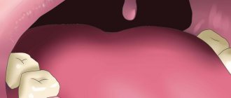
If alveolitis occurs after tooth extraction, you need to undergo dental treatment aimed at:
- elimination of the infectious focus;
- preventing possible complications;
- maintaining the integrity of the rest of the dentition.
First, the dentist cleans the socket and rinses it. It is important to wash away all purulent masses and dead tissue from it. Antiseptics and hydrogen peroxide work well for this purpose.
To reduce painful symptoms, which deprive the patient of the opportunity to fully rest and work, analgesics are used. It is better to use them by applying applications. Compresses are changed every half hour. It is also possible to take drugs orally, that is, in the form of painkillers and anti-inflammatory tablets.
If the patient has concomitant diseases or reduced immunity, the doctor may decide to prescribe antibiotics. They quickly relieve inflammation and speed up the healing of damaged tissue.
If you seek qualified dental care in a timely manner, the prognosis for alveolitis is favorable. After 3-5 days, the symptoms begin to subside, and the person’s well-being improves. Residual socket pain may persist for another 2-3 weeks, but it does not pose a threat.
Method for treating patients with trophic ulcers
Owners of patent RU 2578382:
The invention relates to medicine and can be used to treat patients with trophic ulcers. To do this, the surface of the ulcer is treated with a swab moistened with saline solution. After this, cryotherapy is carried out with an applicator maximally cooled with liquid nitrogen to a temperature of -180°C with different diameters of the flat working surface. The exposure time is no more than 5 seconds. The method ensures bloodless, painless and rapid transformation of a chronic wound into an acute one and active closure of the defect, including by removing the necrotic component, fibrin films, biofilms from the surface of the ulcer, reducing colonization and contamination of microorganisms, removing phenotypically altered cells of the edge and base of the wound. 1 salary f-ly, 2 ill., 1 pr.
The invention relates to medicine, in particular to clinical and experimental medicine, and can be used in the treatment of patients with trophic ulcers and non-healing wounds.
Trophic ulcers, regardless of ethology, attract the close attention of various specialists. This is due to both the enormous material costs of treatment and the high social significance of this issue. Regardless of wound etiology, the principles of local management are largely similar. Adequately selected local treatment plays an important role. Currently, new high-tech dressing materials have appeared, the use of which, without a doubt, helps accelerate the healing of chronic wounds. However, the use of these agents without preliminary treatment of the wound is ineffective, since in all cases at the bottom of the ulcer there is fibrin, purulent-necrotic masses and single flaccid granulations in greater or lesser quantities (RU2388418, A61B 17/00, 2010; RU2340305, A61B 18/02 , 2008; RU2278689, A61K 38/17, 2006; RU2203061, A61K 31/47, 2003; Chernetskaya Yu.G. Application dosage form of hydrogels with miramistin and chymotrypsin. Man and Medicine, 2004, p. 848).
To clean the wound and prepare it for active closure of the defect by skin grafting or the use of collagen-containing preparations, the following treatment methods are used: surgical, autolytic, chemical and physical methods.
Classic surgical treatment should include removal of necrotic tissue, fibrin film, and excision of wound edges containing phenotypically altered cells. Surgical treatment transforms a chronic wound into an acute one, shortens the time to epithelialization, reduces exudation, and reduces the risk of infectious complications. It should be noted that full surgical treatment is impossible in the presence of soft necrosis and neuro-ischemic lesions on the extremities (ABI). The group of inventions relates to the pharmaceutical industry, namely to the use of pterostilbene (PTER), or pterostilbene in combination with quercetin (QUER), or any acceptable salt thereof for the prevention of skin cancer or for the prevention and/or treatment of skin diseases selected from psoriasis, atopic dermatitis and allergic dermatosis, by topical administration.
The infection must be suppressed
To combat infection in a wound, it is best to use ACTICOAT and ACTICOAT7 dressings. These are mesh absorbent dressings that allow unimpeded drainage of exudate from the wound and fight infection. These dressings contain silver. The antibacterial activity of silver has been known for many years and its effectiveness is beyond doubt. The activity of silver is not affected by the presence of blood and pus in the wound. All causative agents of wound infections, including antibiotic-resistant bacteria, are not resistant to silver. Silver has a universal, wide spectrum of action, including against mixed microflora containing various bacteria, fungi, and yeast. The use of dressings with silver leads to rapid suppression of the inflammatory reaction in the wound, and at the same time the amount of discharged exudate decreases.
It is important that the silver in ACTICOAT and ACTICOAT7 dressings begins to be released immediately as soon as the dressing is applied to the wound, and quickly, within the first 30 minutes, the necessary concentration of silver is achieved in the wound to fight infection, and this concentration of silver is maintained at a constant level throughout long-term - up to 3 days when using ACTICOAT dressings and up to 7 days when using ACTICOAT7 dressings, thereby ensuring long-term antibacterial activity of the dressings, so the ACTICOAT dressing can be on the wound for up to 3 days, and the ACTICOAT7 dressing up to 7 days, with external absorbent dressings (gauze or Allevyn sponge) must be changed daily.
Use an acticoate dressing to effectively fight infection.
Classification of wounds. Wound process
A wound is any tissue damage accompanied by a violation of the integrity of the integument of the body. Such damage can be caused by various factors: mechanical, thermal, chemical, radiation. Combined wounds occur when several types of damaging factors are simultaneously exposed. Simultaneous or time-limited exposure to external damaging factors leads to the formation of acute wounds. Chronic wounds are the result of long-term, ongoing adverse effects on tissue. The nature of this effect is often endogenous, for example, disturbances of arterial or venous blood supply, innervation, or constant leakage of exudate from the purulent cavity. Another common cause of chronic wounds is prolonged local pressure on tissue.
Wounds are one of the most common traumatic injuries. Victims are injured in domestic, industrial, combat and criminal conditions. It is characteristic that most of the victims are of young working age. In addition, wounds are caused during surgical interventions. In the latter case, the main feature is the ability to create conditions that minimize the risk of wound complications. What is common to all wounds except post-operative wounds is that they are always contaminated with microorganisms and are often complicated by the development of infection.
Wound process
The main clinical signs of wounds immediately after their application are the presence of a defect in the skin or mucous membranes, bleeding and pain. Subsequently, the clinical picture corresponds to the phases of the wound process.
The formation of any wound is accompanied by a sequence of local and general reactions of the body. General reactions are more pronounced in the formation of acute wounds. They consist in the typical manifestations of stress syndrome - an increase in all vital processes under the influence of the sympathetic nervous system and hormones, an increase in basal metabolism and catabolism. When tissue breakdown products and microbial toxins are absorbed into the bloodstream, stimulating leukocytes to release cytokines, systemic inflammatory reactions may occur: fever, an increase in the number of leukocytes, tachycardia, and others. In the absence of complications, these phenomena completely stop after 4-5 days.
Local reactions of the body are aimed at restoring the integrity of damaged tissues. The healing of wounds of various organs and tissues has its own characteristics, depending on their morphological structure. The wound process can vary significantly in duration, but is always accompanied by the formation of a connective tissue scar. Without scar formation, only superficial wounds heal without damaging the germ layer of the skin. There are 3 phases of the wound process (Fig. 11. 1).
The inflammation phase begins immediately after injury and, in the absence of complications, lasts an average of 4-5 days. It is characterized by typical vascular reactions - vasoconstriction followed by vasodilation, exudation with the release of plasma proteins, migration and release of blood cells into the damaged area, fibrin loss with delimitation of the damaged area, edema and infiltration of surrounding tissues. Subsequently, fibrin undergoes fibrinolysis, and the wound is cleansed of necrotic tissue and microorganisms with the participation of leukocytes and their enzymes.
The regeneration or proliferation phase lasts on average 2-4 weeks. Regeneration processes begin as early as 1 day after injury, and their duration depends on the size of the wound defect and the morphology of the damaged tissue. Migration of fibroblasts, formation of collagen and ground substance, new formation of blood vessels with the development of granulation tissue at the site of the tissue defect occurs. Exudation and swelling gradually decrease, granulation tissue from the bottom of the wound fills the entire defect. Granulation tissue contains many newly formed vessels and is practically not innervated. During the normal course of the wound process, the surface of granulating wounds is bright, “juicy”; when dressings, high contact bleeding and slight pain are characteristic.
The phase of epithelialization and reorganization of the scar, depending on the morphology of the tissue, lasts from several weeks to a year. Epithelization begins from the edges of the wound simultaneously with the formation of granulation tissue. This process is regulated by the action of epidermal cheilon, which is a contact inhibitor of proliferation. Immediately after the formation of the scar, its restructuring begins: elastic fibers are formed and a new fibrous network develops, and the water content in the scar tissue decreases.
The course of the wound process is influenced by various general and local factors. Common factors include: age, nutritional status and immune status of the patient, various disorders of homeostasis, concomitant diseases, including diabetes mellitus, taking anti-inflammatory drugs, cytostatics, massive antibiotic therapy. Local factors influencing the course of the wound process include the state of the blood supply and the degree of tissue trauma in the damaged area, the level of microbial contamination of the wound and the quality of wound care.
According to the degree of contamination and the presence of signs of infection, all wounds are divided into 3 types: aseptic, contaminated and infected.
· Only surgical wounds with “clean” surgical procedures are aseptic.
· Contaminated are wounds contaminated with microflora, but without signs of suppuration. These include all accidental wounds after they are inflicted and some surgical wounds.
· Infected are purulent wounds, that is, wounds with signs of an infectious-inflammatory process. They are divided into primary infected - formed after operations for acute purulent processes, and secondary infected - wounds that festered during the healing process.
The wound process ends with wound healing. The following types of wound healing are distinguished:
· Healing by primary intention – healing without suppuration. It occurs with the development of a linear scar without the formation of visible interstitial tissue. Such healing is typical for wounds with smooth viable edges, separated from each other by no more than 1 cm, and with microbial contamination of tissues below a critical level. Surgical wounds usually heal by primary intention when primary sutures are applied.
· Delayed primary - healing according to the type of primary intention. This is healing without suppuration with delayed closure of the wound with sutures.
· Healing by secondary intention – healing through suppuration and granulation with the development of a rough scar and the formation of visible interstitial tissue. Occurs with extensive tissue defects that do not allow primary comparison of the wound walls and with the development of wound infection.
· Healing under a scab – healing without a scar. Occurs with superficial wounds without damage to the germ layer of the skin. Under the scab, consisting of fibrin and blood cells, rapid regeneration of the epidermis occurs.
The course of the wound process described above is typical for wounds that were the result of a one-time exposure to a damaging factor. With long-term or constant adverse effects leading to disruption of the functioning of the integument of the body and complicating healing, which occurs in the case of the formation of chronic wounds, it is characteristic that there are signs of all three of its phases at once. The bottom of a chronic wound is simultaneously covered with fibrin and granulations; there may be areas of necrosis and purulent discharge. Granulations are sluggish, pale. The edges of the wound and the surrounding tissue are compacted. Marginal epithelialization often occurs. Typically, palpation of the damaged area is not painful.
The surrounding tissues have changes characteristic of the disease that caused the formation of a chronic wound. If the damaging factors are not eliminated, chronic wounds, even with intensive treatment, do not heal for a long time, and after healing they recur - in the same or in a neighboring area. With long-standing chronic wounds there is a risk of their malignancy.
Excess exudate
If there are no crusts or necrosis in the wound, then the technique of local negative pressure can be used for active outflow of exudate. For these purposes, the PICO wound treatment device is used, which creates a constant outflow of purulent exudate from the wound, which is accompanied by a decrease in tissue swelling, a decrease in the number of bacteria in the wound, as a result, the inflammatory reaction subsides, and the growth of new tissues begins, the vessels that fill the wound begin the epithelium grows, the edges of the wound gradually come closer together and the wound heals. When using this device, there is no need to carry out dressings daily. It is enough to change the dressings on average once every three days.
For passive drainage of exudate from the wound, absorbent dressings of your choice are used:
- Carbonet. This multi-layer dressing perfectly absorbs viscous purulent exudate, and, thanks to a layer of activated carbon, absorbs the odor that often accompanies purulent wounds. Since Carbonet does not contain antimicrobial components, dressings on purulent wounds must be changed daily.
- NEOFIX polyurethane sponge dressings with silver - NEOFIX FibroSorb AG and NEOFIX FibroSorb AG Sacrum. Silver in dressings provides antimicrobial effect.
Sponge dressings absorb liquid exudate and retain it in their structure even under pressure, for example, under compression bandages or underwear (stockings, stockings, tights), which are one of the main attributes of the treatment of trophic venous ulcers. NEOFIX FibroSorb AG sponge dressing
without adhesive edge is indicated for wounds where the skin around which is damaged.
Such a bandage requires additional fixation with a bandage or adhesive roll bandages NEOFIX ROLL, or OpSite Flexifix, always only around the perimeter, without covering its central part with adhesive patches. This is necessary to prevent maceration of the skin and wound. NEOFIX FibroSorb AG Sacrum bandage
with a sticky edge, specially shaped for fixation in the sacrum area, while the skin around the wound should be dry and healthy, without any disturbances. The bandage protects the wound from the ingress of urine, feces, as well as from the penetration of external liquids during hygiene procedures.
When using NEOFIX FibroSorb AG and NEOFIX FibroSorb AG Sacrum dressings for wound treatment, there is no need to change dressings daily. It is recommended to change the dressing once every 2-3 days, depending on the degree of filling of the dressing with exudate.
Deviation from the norm
The following symptoms indicate that an infection is developing:
- aching or throbbing pain that appears spontaneously;
- swelling near fibrinous plaque;
- redness next to the damage at a distance of one to two cm;
- increase in body temperature – more than 37 degrees.
This indicates that the process has begun to spread throughout the body. The clinical picture of fibrinous plaque on a wound may be supplemented by general disorders, for example, weakness or nausea, and the appearance of dizziness.
With aggravated purulent infection, the human body will react to changes very hard. Immediately after the appearance of swelling, phlegmon reactions only intensify. The most striking symptom is a feverish state. It is associated with deterioration of the patient's condition and increasing pain.
In addition, during diagnosis, changes in the structure of the blood may be noted. For example, increasing the leukocyte ratio or protein production. It is better to save photographs of tests and copies of data for further examinations for fibrinous plaque.
Treatment
In order for the wound to heal as quickly as possible, it is necessary to suppress the infection. At the initial stage it is important:
- Ensure drainage of pus. If it is present under a crust formed on the wound, it must be soaked in hydrogen peroxide. Then the upper fibrinous layer is removed with a bandage, which is applied for 30 minutes. It is also soaked in peroxide or other antiseptics.
- Get rid of pus accumulated under the skin and fibrin plaque. To do this, it is squeezed out of the hole, which is made along the edges where areas of the skin have dried.
- Be sure to treat the problem area with hydrogen peroxide every day. The pus is disposed of as it accumulates, so that the wound does not become covered with fibrin again.
IMPORTANT! It is recommended to use Levomekol. This is an effective remedy that promotes healing of fibrinous plaque. It will be useful to use a bandage with it every day.
Medicines
If indicated, the patient is prescribed a number of medications. Most often these are analgesics, antihistamines and detoxification compounds. During the treatment of fibrinous plaque, immune stimulants are used. It is necessary to take into account that:
- if there is a threat of infection spreading, surgeons prescribe antibiotics;
- physicians must constantly monitor treatment, monitor the recovery process and adaptation during the postoperative period;
- attention is paid to how severe the inflammatory processes are in the wound itself, and whether it has become covered with plaque again.
It is impossible to do without studying material from the problem area, the patient’s blood. It is important to timely study the microbial spectrum in fibrinous plaque. In this case, even with a severe infection, treatment will be successful and without critical consequences.
Video: fibrin threads under a microscope
Did you find useful information for yourself? Want to read more about this topic? Like ♥, subscribe to our channel and you will be one of the first to know about new publications!
And if you have something to share, leave your comments! Your feedback is very important to us!
Source: netnaleta.ru