Photo: phlegmon of the submandibular region
Phlegmon of the submandibular region is a diffuse purulent-inflammatory lesion of the subcutaneous tissue and muscles in the area of the lower jaw. This disease is characterized by the intensive spread of the pathological process, which from the very beginning has a diffuse course.
In dental practice, it is customary to distinguish between several types of phlegmon. These can be odontological, adenophlegmon and osteophlegmon. According to statistics, the ratio of abscesses and phlegmons in the maxillofacial area is 4:1.
Causes and pathogenesis of abscesses and phlegmons
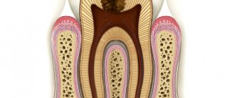
Most purulent lesions of the maxillofacial area are odontogenic in nature. This means that the disease is formed due to the spread of infection from the inflamed root or periodontium. In such cases, streptococci and staphylococci from the primary lesion penetrate through the lymphatic vessels into the deep layers of the soft tissues of the face.
The state of immunity is of great importance in the development of the purulent process. A decrease in the body's protective abilities is a powerful predisposing factor for tissue suppuration.
Reasons for formation
Due to their development, there are two types of perimaxillary abscesses:
- Odontogenic. The cause of development is damaged dental elements.
- Non-odontogenic. Not caused by the condition of the dentofacial apparatus.
The most common causes of anomalies are dental diseases:
- Advanced carious lesions of units, pulpitis.
- Deep periodontal pockets.
- Periodontitis.
- Pericoronitis.
- Infected and impacted molars and premolars.
- Root cyst.
The development of a lesion can be triggered by penetration of infection under the periosteum from a diseased unit.
Reasons for the development of non-odontogenic foci of pus:
- Traumatic lesions of the jaws, elements or soft membranes of the oral cavity with infection.
- Nasopharyngeal and ear infections.
- Complications of unqualified dental treatment.
- Spread of infection to the jaw from other organs and systems.
The development of an abscess depends critically on the general health and immunity of the patient.
Clinical picture of the disease
In the initial stages, abscesses and phlegmons adjacent to the lower jaw are manifested by compaction and progressive swelling of the soft tissues of the face. The skin over the purulent focus is often hyperemic. The key symptom of suppuration is fluctuation - the feeling of fluid in a confined space.
The course of phlegmon is accompanied by general intoxication, in which the patient presents the following complaints:
malaise and feeling of chronic fatigue; increase in body temperature; fatigue and joint pain.
Abscesses, as a rule, do not cause such symptoms due to the limited nature of the pathological process.
Abscess, phlegmon of the maxillofacial area in children is characterized by an acute and severe course of the disease. Diffuse purulent lesions of soft tissues are formed as a result of the imperfection of the children's immune system.
What is a gum abscess, definition of the disease
Let us consider in more detail what an abscess on the gum is and the mechanism of its occurrence. A purulent focus occurs as a result of long-term untreated inflammation of periodontal tissues. The pathogenesis of the disease is associated with the penetration of pathogenic microorganisms (mainly Bacteroides forsythus and Porphyromonas gingivalis) into the periodontal tissues. Possible routes of penetration of microbes:
- from the outside: through a carious cavity, chips, cracks on the surface of the segment;
- from the inside (hematogenously, lymphogenously): in the presence of other septic foci in the body.
During their life, bacteria produce toxic substances that lyse tissue cells, resulting in the formation of a cavity filled with pus. The abscess under the tooth is surrounded by a connective tissue capsule, which separates it from the intact periodontal tissues.
Microbial metabolic products have a neurotoxic effect on nerve receptors located in the pulp of the segment, which causes pain. In addition, they increase the permeability of the capillary walls, resulting in swelling of the surrounding tissues.
Diagnosis of the disease
Establishing a diagnosis for acute odontogenic infections includes the following measures:
Collection of medical history. The doctor finds out the patient’s complaints and the general condition of the patient. External examination of the maxillofacial area and palpation of regional lymph nodes. Most inflammatory and purulent processes cause enlargement and pain of the lymph nodes. Instrumental examination of the oral cavity, during which the doctor detects chronic foci of odontogenic infection. X-ray in frontal and lateral projection. Laboratory blood test, in which there is an increase in SOE, leukocytes and a decrease in the concentration of red blood cells and hemoglobin.
Anatomy
Rice.
1. Schematic representation of the boundaries of the submandibular region and adjacent areas of the neck: 1 - fossa retromandibularis, 2 - lateral area of the face, 3 - submandibular region, 4 - trigonum submentale, 5 - carotid triangle, 6 - scapulotracheal triangle, 7 - sternocleidomastoid region (the solid line indicates the boundaries of the submandibular region, the dotted line indicates the neck region). From above, the submandibular region (submandibular triangle, T.; trigonum submandibulare) is limited by the edge of the lower jaw (mandibula), on the sides - by two bellies of the digastric muscle (m. digastricus). By. easily accessible for examination with the head thrown back and turned to the side (Fig. 1). Above P. o. borders on the areas of the face - chin, buccal and parotid-masticatory (regiones mentalis, buccalis et parotideomasseterica); below - with triangles of the neck - trigonum submentale in front and sleepy (trigonum carotieum) in the back; above and behind it continues into fossa retromandibularis.
Rice. 2. Schematic representation of the topography of the submandibular region (head thrown back, front view): 1 - bed of the submandibular gland, lined with the superficial plate of the cervical fascia, 2 - v. submentalis, 3 - mylohyoid nerve, 4 - a. submentalis, 5 - mylohyoid muscle, 6 - trigonum submentale tissue, 7 - anterior belly of the digastric muscle, 8 - lower jaw, 9 - facial artery, 10 - facial vein, 11 - submandibular gland. Sublingual region, sublingual gland, submandibular region, submandibular gland. Rice. 1. Topography of the submandibular and sublingual areas (the right half of the lower jaw is removed, the skin and muscles are retracted). Rice. 2. Topography of the sublingual area (the tongue is raised, part of its mucous membrane is removed). Rice. 3. Topography of the submandibular region (the subcutaneous muscle of the neck and the superficial plate of the cervical fascia have been removed): 1 - tongue, 2 - anterior lingual salivary gland, 3 - deep artery of the tongue, 4 - submandibular duct, 5 - sublingual gland, 6 - lower jaw (partially removed ), 7 - mylohyoid muscle, 8 - geniohyoid muscle, 9 - submandibular gland, 10 - facial artery, 11 - facial vein, 12 - sublingual - lingual muscle, 13 - submandibular lymph node, 14 - upper root of the cervical loop , 15 - sternocleidoid - mastoid muscle, 16 - submandibular vein, 17 - internal jugular vein, 18 - internal carotid artery, 19 - external carotid artery, 20 - deep cervical lymph nodes, 21 - hypoglossal nerve, 22 - posterior belly of the digastric muscles, 23 - stylohyoid muscle, 24 - parotid gland, 25 - masticatory muscle, 26 - deep vein of the tongue, 27 - branch of the mandible (partially removed), 28 - lingual nerve, 29 - sublingual papillae, 30 - sublingual fold, 31 - frenulum of the tongue, 32 - anterior belly of the digastric muscle, 33 - subcutaneous muscle of the neck, 34 - superficial plate of the cervical fascia, 35 - base of the mandible, 36 - marginal branch of the mandible of the facial nerve.
Skin P. o. thin, in men partially covered with hair, fused with the subcutaneous muscle of the neck (platysma). Small arteries and veins run under the skin. The cervical branch of the facial nerve (n. colli n. facialis) and the branches of the transverse nerve of the neck (n. transversus colli) pierce the subcutaneous muscle of the neck, innervating it and the skin of the neck. Under this muscle there may be significant accumulations of subcutaneous tissue, causing the presence of the so-called. double chin. The superficial plate of the cervical fascia in the P. region, moving away from the hyoid bone, splits into a denser superficial and thinner deep plate, which form the fascial sac (capsule) of the submandibular gland (saccus gl. submandibularis) and the sheath of the digastric and stylohyoid muscles (Fig. .2). Behind the angle of the lower jaw, both plates again merge into a thickened cord - the stylomandibular ligament (lig. stylomandibulare), the edges are separated by the Po. from the mandibular fossa. The facial vein (v. facialis) runs through the superficial plate of the cervical fascia. Between the superficial and deep plates, in addition to the submandibular gland (see), there are the facial artery (a. facialis) with the submental artery (a. submentalis) extending from it, as well as the submental vein (v. submentalis), mylohyoid nerve (n. mylohyoideus), lymph, nodes and fiber (color. Fig. 3). The deep plate also covers the muscles that form the bottom of the P. o. - mylohyoid and hypoglossal (mm. mylohyoideus et hyoglossus), as well as the lingual vein (v. lingualis), hypoglossal and lingual nerves (nn. hypoglossus et lingualis) , lying on the outer surface of the hyoglossus muscle. The facial artery (a. lingualis) is adjacent to the inner surface of the hyoglossus muscle. In the depths of the Po., at its apex, there is Pirogov’s triangle, bounded below (behind) by the tendon of the digastric muscle, above by the hypoglossal nerve, and in front by the posterior edge of the mylohyoid muscle.
Fiber P. o. communicates through the gap between the mylohyoid and mylohyoid muscles with the fiber of the floor of the mouth (see Mouth, oral cavity) and the peripharyngeal space; along the facial artery and vein, it communicates with the fiber of the lateral area of the face and the carotid triangle. Along the fiber, inflammatory processes from the submandibular region can spread to the face (see) and to the deep parts of the neck (see).
Blood supply to the skin and muscles of P. o. carried out by the branches of the facial artery, mainly the submental artery; The deep muscles are also supplied with blood by the lingual artery (a. lingualis). Venous outflow occurs through the veins of the same name. Lymph is collected in the submandibular (Submandibular, T.) and deep cervical lymph nodes (nodi lymphatici submandibulares et seg-vicales profundi).
Skin P. o. innervates the transverse nerve of the neck from the cervical plexus, muscles - cranial (cranial, T.) nerves: trigeminal (n. trigeminus) - anterior belly of the digastric and mylohyoid muscle, facial - subcutaneous muscle of the neck, posterior belly of the digastric and stylohyoid muscle; hypoglossal nerve - hyoglossus and styloglossus muscles.
General principles of treatment of the disease
The doctor who treats phlegmon of the maxillofacial area is an oral and maxillofacial surgeon.
Each doctor, when starting to treat an odontogenic process in the maxillofacial area, is guided in his actions by the following principles:
The tooth that caused the development of phlegmon is removed. Timely diagnosis is very important due to the characteristics of this area, from where the infection can spread, causing severe consequences (in particular, mediastinitis). It is necessary to eliminate the spread of the infection, that is, promptly open the lesion and eliminate the inflammatory exudate, relieve tissue tension. The rate of subsidence of inflammation depends on the quality of evacuation of all decay products from the wound, that is, careful postoperative treatment of the wound is necessary. Comprehensive treatment of pathology using all means and methods available in this medical institution. Friendly management of the patient with colleagues from other areas, timely appointment of consultations with all specialists for combined lesions. Visible external changes often occur, which are not always pleasant for the patient, which is especially painful in the face area. Therefore, there is an urgent need to organize work with patients by a medical psychologist in the early stages after surgery, especially if there is a significant visible defect. Correct stage-by-stage treatment, organization of medical rehabilitation, referral to the appropriate department with complete information about the patient. Fully informing relatives and the patient about the violation of significant functions of the maxillofacial area, if any. Upon leaving the hospital, the patient should have clear further recommendations, especially if it is necessary to continue treatment on an outpatient basis.
Symptoms of perimandibular abscess
The first sign of the formation of a perimaxillary abscess is toothache, which intensifies when biting in the affected area. Subsequently, the clinical picture of the disease is supplemented by the appearance of edema and the formation of a hyperemic, painful compaction in it. Some patients experience severe facial asymmetry.
Without treatment, the patient's condition worsens. A spontaneous opening of the abscess occurs, the bacterial flora begins to actively multiply in the oral cavity, leading to a chronic process. Chronic suppuration appears from the formed fistula canals, and intoxication of the sick person’s body with decay products is observed.
Complications of abscesses and phlegmon
Purulent lesions of the maxillofacial area can be complicated by the following pathologies:
Sepsis is a serious condition of the body that is caused by the penetration of a bacterial infection into the circulatory system. Treatment of such a complication is difficult due to the development of body resistance to antibiotic therapy. Sepsis is often the cause of death. Mediastenitis in the form of purulent inflammation of the tissue of the mediastinum, where the heart, lungs and bronchi are located. Meningitis . Inflammatory damage to the meninges develops as a result of the spread of purulent infection through the lymphatic and blood vessels of the head.
Forecast and prevention of the disease
The prognosis of odontogenic abscesses and phlegmons is usually favorable. A positive treatment result is observed with timely indication of full surgical care. In such cases, the patient must be hospitalized in a specialized medical hospital.
Lethal outcomes from purulent lesions of the soft tissues of the maxillofacial area are associated with late presentation of the patient and systemic suppression of his immunity.
Prevention of the disease is achieved in the following ways:
sanitation of the oral cavity, during which the dentist treats all carious, pulpitic and periodontitis teeth; strict adherence by patients to the rules of personal hygiene and regular brushing of teeth; undergoing regular preventive examinations at the dentist, at least twice a year; timely consultation with a doctor if symptoms of dental diseases are detected. Every person should remember that the cost of prevention is much lower than the cost of treatment. And in some cases, sanitation of the oral cavity can prevent the development of severe complications, which are accompanied by high mortality in patients.
